Dear Editor,
Severe coronavirus disease 2019 (COVID‐19) has been shown to have a wide spectrum of clinical presentations. Fever, dry cough, asthenia, headache, myalgia, anosmia, diarrhea, and a great recent number of skin manifestations are widely referenced. 1 , 2 , 3 Some patients do not present these symptoms on their initial presentation, which complicates a correct diagnosis. COVID‐19 seems to be a systemic multiorgan viral invasion. Previous histological lung and skin COVID‐19 analysis demonstrated obliterative microangiopathy with an increased vascular permeability. 4 Moreover, postmortem lung analysis identified pulmonary inflammation and swelling. 5 Due to the physiopathological mechanisms observed in COVID‐19, edema could affect other tissues.
Spain has been one of the hardest‐hit countries with COVID‐19, and we have accumulated vast knowledge of clinical manifestations. Description of clinical expressions observed could help an early diagnosis and contribute to better management of the disease. Herein, we describe a new clinical feature commonly observed in patients during the COVID‐19 pandemic.
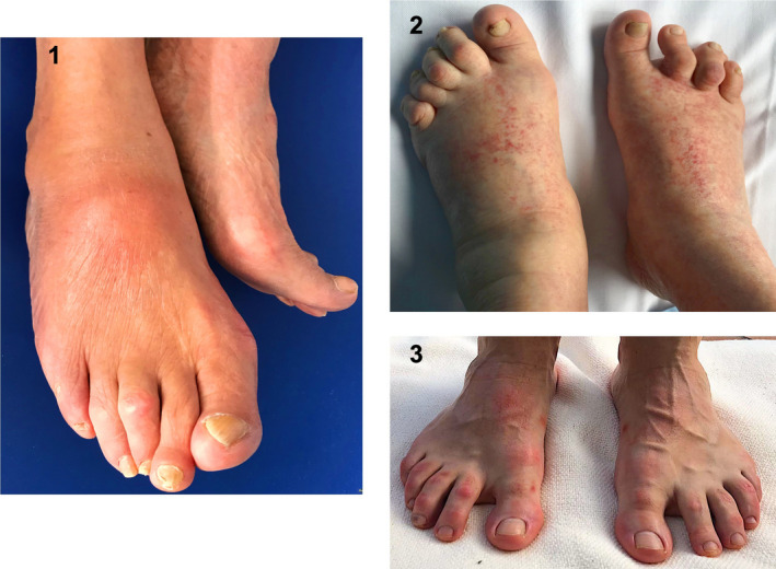
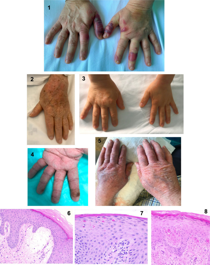
Our observations showed unexplained acute‐onset, painful acral edema across many patients during the COVID‐19 pandemic (a representative sample of eight cases is shown in Figs. 1 and 2). Some of them had confirmed severe acute respiratory syndrome coronavirus‐2 (SARS‐CoV‐2) infection, and others presented previous COVID‐19 symptoms or had contact with someone who was infected. Previous diarrhea and/or light asthenia were reported among all the patients. Most of them presented good overall condition when edema was manifested. None of them had previous similar acral history, recent new drug therapy, or sun exposure reported.
Figure 1.

Foot edema and associated skin lesions during the COVID‐19 pandemic. (1) Right dorsal foot edema with an erythematous skin area. (2) Symmetrical feet edema with multiple micropacular purpuric lesions in the dorsum area. (3) Both feet edema more evident in right foot with chilblain‐like lesions
Figure 2.

Edema in hands and associated skin lesions during the COVID‐19 pandemic. (1) Edema in both hands, more evident in left hand, with intense chilblain‐like lesions. (2) Left hand edema with an erythematous skin area. (3) Right hand edema. (4) Left hand edema with multiple dark purple micro‐petechiae on the acral areas of the whole left hand fingers. (5) Symmetrical hand edema, more evident in the right hand, that included dryness, slight peeling, and erythema. (6) Histological edematous skin features: Significant serum and erythrocyte extravasation in the superficial dermis (hematoxylin and eosin, ×40). (7 and 8) Extravasated erythrocytes in the epidermis (erythro transepidermal diapedic) (hematoxylin and eosin, ×63 and ×40, respectively)
Physical examination revealed painful edema in the feet (Fig. 1) or hands (Fig. 2) with a mean duration of 6 days. Edema was associated with an intense numbness across all the patients. The overlaying skin presented non‐bleachable purpuric/erythematous areas, chilblain‐like lesions, and/or epidermal component. Distal pulses were preserved, and no cellulitis signs were observed in any patient. Markers of bacterial infection showed normal values. Complementary test did not find a relationship with any other disease.
Histological findings revealed significant serum and erythrocyte extravasation in both the superficial dermis and the epidermis (Figs. 2.6–2.8).
Increased incidence of acute, painful acral edema was observed during the COVID‐19 pandemic, and it was not associated with any other disease. The diagnosis of COVID‐19 may be hindered by the diversity in symptoms at the time of presentation and the lack of accuracy of the available diagnostic tests at certain clinical stages. Skin COVID‐19 expressions are becoming a common manifestation of the disease 2 , 3 and could provide extensive information. As far as we are concerned, acute acral edema related to COVID‐19 has not been reported. Nevertheless, lung COVID‐19 postmortem studies found inflammation and edema. 5 Moreover, skin living COVID‐19 tissue analysis demonstrated an obliterative microangiopathy with increased vascular permeability. 4 This endothelial cell distortion generated vascular lumen obliteration and a strike erythrocyte and serum extravasation. 4 It could provoke a tissular conflict with the consequent associated numbness.
According to our observations, acral edema possibly related to COVID‐19 may be symmetric or asymmetric, located in hands and/or feet. Numbness could be a common accompanying symptom. The overlaying skin could present erythematous or purpuric lesions probably due to a hematic extravasation.
Suspicion of COVID‐19 when unexplained acute, painful acral edema is present may help clinicians make an earlier diagnosis and thus have better management of the disease. Further reports are required to analyze this association in depth.
Conflict of interest: None.
Funding source: None.
References
- 1. Huang C, Wang Y, Li X, et al. Clinical features of patients infected with 2019 novel coronavirus in Wuhan, China. Lancet 2020; 395: 497–506. [DOI] [PMC free article] [PubMed] [Google Scholar]
- 2. Landa N, Mendieta‐Eckert M, Fonda‐Pascual P, Aguirre T. Chilblain‐like lesions on feet and hands during the COVID‐19 Pandemic. Int J Dermatol 2020; 59(6): 739–743. [DOI] [PMC free article] [PubMed] [Google Scholar]
- 3. Galván Casas C, Català A, Carretero Hernández G, et al. Classification of the cutaneous manifestations of COVID‐19: a rapid prospective nationwide consensus study in Spain with 375 cases. Br J Dermatol 2020. 10.1111/bjd.19163 [DOI] [PMC free article] [PubMed] [Google Scholar]
- 4. Valtueña J, Martínez‐García G, Ruiz‐Sánchez D, et al.Vascular Obliteration Due To Endothelial And Myointimal Growth In COVID‐19, 29 May 2020, Preprint (Version 1) available at Research Square [+ 10.21203/rs.3.rs-32241/v1+]. [DOI] [PubMed]
- 5. Zhang H, Zhou P, Wei Y, et al. Histopathologic changes and SARS‐CoV‐2 Immunostaining in the lung of a patient with COVID‐19. Ann Intern Med 2020; 172(9): 629–632. [DOI] [PMC free article] [PubMed] [Google Scholar]


