Abstract
Accurate anatomical atlases are recognized as important tools in brain-imaging research. They are widely used to estimate disease-specific changes and therefore, are of great relevance in extracting regional information on volumetric variations in clinical cohorts in comparison to healthy populations. The use of high spatial resolution magnetic resonance imaging and the improvement in data preprocessing methods have enabled the study of structural volume changes on a wide range of disorders, particularly in neurodegenerative diseases where different brain morphometry analyses are being broadly used in an effort to improve diagnostic biomarkers. In the present dataset, we introduce the Cerebrum Atlas (CerebrA) along with the MNI-ICBM2009c average template. MNI-ICBM2009c is the most recent version of the MNI-ICBM152 brain average, providing a higher level of anatomical details. Cerebra is based on an accurate non-linear registration of cortical and subcortical labelling from Mindboggle 101 to the symmetric MNI-ICBM2009c atlas, followed by manual editing.
Subject terms: Diagnostic markers, Diagnostic markers, Brain imaging, Brain
| Measurement(s) | neuroantomical mapping • brain atlas • brain segmentation |
| Technology Type(s) | computational modeling technique |
| Sample Characteristic - Organism | Homo sapiens |
Machine-accessible metadata file describing the reported data: 10.6084/m9.figshare.12472070
Background & Summary
Brain atlases are widely recognized as important tools in research for the analysis of neuroimages. High spatial resolution magnetic resonance imaging (MRI) and improved data preprocessing have enabled the study of structural volume changes in a wide range of disorders. Anatomical atlases are central to the understanding of the brain anatomy and are the best resources to bring prior knowledge about anatomy into any computer vision methodology involved in various types of brain imaging analyses. Anatomical atlases can also be used to investigate potential disease-specific changes that occur in clinical cohorts compared with healthy populations, by providing information on region locations for various regions of interest. Analysis of fMRI data also routinely involves registration to a template and extraction of the average signal within various regions of interest within the corresponding anatomical atlases1.
The MNI-ICBM152 brain template2, from the Montreal Neurological Institute (MNI) is a crucial tool in neuroimage analysis. This multi-contrast atlas including T1w, T2w and PDw contrasts, was built recruiting brain scans from 152 young adults at 1.5 T. The 2009 edition uses group-wise non-linear registration for better alignment of cortical structures between subjects. The MNI-ICBM152 non-linear model has many advantages. It was created from a large number of subjects; hence it represents the average anatomy of the population and is not biased unlike single-subject models. In addition, the left-right symmetric version enables interpretation of asymmetries that might be found in an analysis.
Mindboggle-101 is the largest, publicly available set of manually labelled human brain images created from 101 human scans, labelled according to a surface-based cortical labelling protocol (DKT- Desikan-Killiany-Tourville labelling protocol)3,4. For the creation of the Mindboggle-101 dataset, developed to serve as brain atlas for use in labelling other brains, 101 T1-weighted (T1w) brain MRI images were selected and segmented based on a modification of the DKT cortical parcellation atlas4. These labels were then manually edited in agreement with the DKT protocol. Labelling was performed on the surface, yet, topographical landmarks visible in the folded surface were used to infer label boundaries. In addition, Mindboggle used non-cortical labels that were converted from Neuromorphometrics BrainCOLOR subcortex labels4.
The Cerebrum Atlas (CerebrA) includes co-registration of the Mindboggle atlas3 to the symmetric version of MNI-ICBM 2009c2 average template (at a resolution of 1 × 1 × 1 mm3) in addition to manual editing of cortical and subcortical labels. In the present dataset, we introduce an accurate non-linear registration of cortical and subcortical labelling from Mindboggle 101 to the symmetric MNI-ICBM2009c atlas followed by manual editing.
Methods
MNI-ICBM152 template
This section summarizes the details on generation of the nonlinear MNI-ICBM2009c average template. Further methodological details can be found in the original paper by Fonov et al.2. Within the ICBM project, MRI data from 152 young normal adults (18.5–43.5 years) were acquired on a Philips 1.5 T Gyroscan (Best, Netherlands) scanner at the Montreal Neurological Institute. The T1w data were acquired with a spoiled gradient echo sequence (sagittal acquisition, 140 contiguous 1 mm thick slices, TR = 18 ms, TE = 10 ms, flip angle 30°, rectangular FOV of 256 mm SI and 204 mm AP). The Ethics Committee of the Montreal Neurological Institute approved the study, and informed consent was obtained from all participants2.
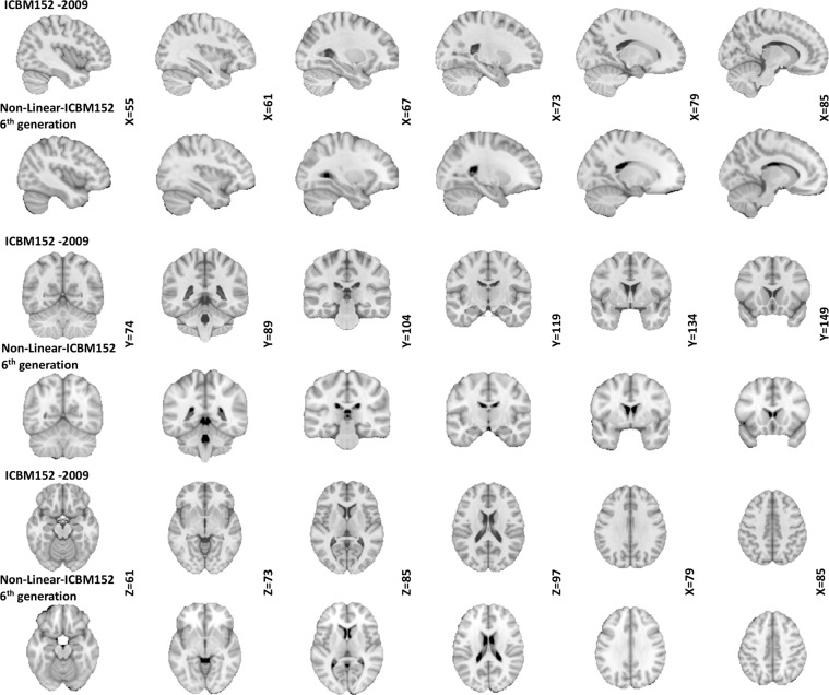
The following preprocessing steps were applied to all MRI scans prior to building the atlas: (1) N3 non-uniformity correction5; (2) linear normalization of each scan’s intensity to the range [0–100] by a single linear histogram scaling6; (3) automatic linear (nine parameters) registration to the ICBM 152 stereotaxic space7; and (4) brain mask creation8. Only the voxels within the brain volume after linear mapping into stereotaxic space were used for the nonlinear registration procedure described. The template described is generated through a hierarchical nonlinear registration procedure, with diminishing step sizes in each iteration until convergence and relies on the nonlinear registration using Automatic Nonlinear Image Matching and Anatomical Labelling (ANIMAL)9. The nonlinear versions of MNI-ICBM2009 (http://nist.mni.mcgill.ca/?p=904) have many advantages over widely used previous versions (i.e. MNI-ICBM non-linear 6th generation; http://nist.mni.mcgill.ca/?p=858, https://fsl.fmrib.ox.ac.uk/fsl/fslwiki/Atlases). Besides, anatomical variability still remains after linear transformation to stereotaxic space, therefore sulci and gyri remain blurred in previous versions10 (Fig. 1).
Fig. 1.
Comparison between the two versions of MNI-ICBM152 template. Rows 1, 3, and 5 show sagittal, coronal, and axial slices of the MNI-ICBM2009c template, respectively. Rows 2, 4, and 6 show sagittal, coronal, and axial slices of non-linear 6th generation template, respectively. Note the improved tissue contrast and cortical definition of the new template compared to the older 6th generation version.
Mindboggle-101
This section summarizes the generation of the original Mindboggle-101 atlas and additional methodological details can be found in the original paper by Klein and Tourville 20123. The authors started with publicly accessible T1-w MRI scans selected from 101 healthy participants. Scanner acquisition and demographic information can be found in Klein 20123 and are also available on the http://mindboggle.info/data website. The data sets that comprise the Mindboggle-101 include the 20 test–retest subjects from the “Open Access Series of Imaging Studies” data11, the 21 test–retest subjects from the “Multi-Modal ReproducibilityResource”12, with two additional subjects run under the same protocol in 3 T and 7 T scanners, 20 subjects from the “Nathan Kline Institute Test–Retest” set, 22 subjects from the “Nathan Kline Institute/Rockland Sample”, the 12 “Human Language Network” subjects13, the Colin Holmes 27 template14, two identical twins, and one brain imaging colleague.
T1-w MRI volumes were preprocessed and segmented and then, cortical surfaces were generated using FreeSurfer’s standard recon-all image processing pipeline15,16. FreeSurfer then automatically labelled the cortical surface using its DKT cortical parcellation atlas4,17. Vertices along the cortical surface are assigned a given label based on local surface curvature and average convexity, prior label probabilities, and neighbouring vertex labels. FreeSurfer automatically labelled the cortical surface using its DKT cortical parcellation atlas for 54 of the brains in the Mindboggle-101 data set. The region definitions of the labelling protocol represented by the DKT atlas are described by Desikan et al.4. These labels were then manually edited in agreement with the DKT protocol with 31 cortical regions per hemisphere as described by Klein and Tourville3. Then, the first 40 brains that labelled were selected to train a new FreeSurfer cortical parcellation atlas representing the DKT protocol (see http://surfer.nmr.mgh.harvard.edu/fswiki/FsTutorial/GcaFormat; S’egonne et al.17; Desikan et al.4 for details regarding the algorithm that generates the atlas and how it is implemented). The resulting atlas was named “DKT40 classifier atlas” which then automatically generated the initial set of cortical labels for the remaining 47 brains in the data. Finally, Mindboggle data includes non-cortical labels that were converted from the Neuromorphometrics BrainCOLOR subcortex labels (i.e., http://Neuromorphometrics.com/). Details on the original labels included in Mindboggle-101 can be found in https://mindboggle.readthedocs.io/en/latest/labels.html. OASIS-30_Atropos_template and OASIS-TRT-20_jointfusion_DKT31_CMA_labels_in_OASIS-30_v2 were the template and atlas files that were used for registration to the MNI-ICBM2009 template and manual correction.
Atlas registration and manual label editing
The Mindboggle-101 average template was first linearly and then non-linearly registered to the symmetric version of MNI-ICBM (MNI-ICBM2009c) template. In both registrations, the Mindboggle-101 template was used as the source image, and the MNI-ICBM2009c template was used as the target image. The linear registration was performed with 9 parameters (-lsq9, 3 for translation, 3 for rotation, and 3 for scaling), using bestlinreg_s2 pipeline from the MINCTools18. The resulting image was then nonlinearly registered to MNI-ICBM2009c template using the ANTs diffeomorphic registration pipeline19, providing both source and target masks. Using the obtained nonlinear transformation, the Mindboggle-101 atlas labels were also resampled and registered to MNI-ICBM2009c template, using the label resampling option from itk_resample tool (i.e.,–label, applying a nearest neighbor interpolator for discrete labels). The quality of the registration was visually assessed by overlaying the registered Mindboggle-101 and MNI-ICBM2009c templates as well as the registered Mindboggle-101 atlas to ensure accurate transformation of the labels to the MNI-ICBM2009 template. Any remaining inaccuracies were manually corrected on the right hemisphere using the interactive software package Display, part of the MINC Tool Kit, developed at the McConnell Brain Imaging Center of the Montreal Neurological Institute (https://www.mcgill.ca/bic/software/visualization/display, https://github.com/BIC-MNI). The program allows simultaneous viewing and segmentation in the coronal, sagittal and axial planes, as well as intensity thresholding, label filling, dilation, and erosion. The corrections were performed by A.M., a neurologist with 10 years of experience in reading and assessment of MRIs. Prior to performing these corrections, A.M. received additional training on performing manual segmentations using Display by an anatomist expert both in using Display and MRI segmentation. These corrections mainly involved improvements of boundaries between neighboring regions, addition of missing voxels in some structures (detailed in Table 1) and improving the continuity of voxels within each region. Afterwards, labels were flipped onto the left hemisphere and then visual inspection on each structure was performed. In detail, thickness and boundaries of all 51 cortical and subcortical labels from each hemisphere were improved using intensity thresholds with manual painting using MNI Display. Details on the significant edits that were made for particular structures are provided in Table 1.
Table 1.
Original label numbers from Mindboggle with new label numbers.
| MB ID | Label Name | CerebrA ID | Notes | Kappa | |
|---|---|---|---|---|---|
| RH Labels | LH Labels | ||||
| 2002 | Caudal Anterior Cingulate | 30 | 81 | 0.79 | |
| 2003 | Caudal Middle Frontal | 42 | 93 | Improved distinction from Precentral | 0.73 |
| 2005 | Cuneus | 43 | 94 | 0.67 | |
| 2006 | Entorhinal | 36 | 87 | Improved delimitation | 0.78 |
| 2007 | Fusiform | 24 | 75 | 0.77 | |
| 2008 | Inferior Parietal | 10 | 61 | 0.75 | |
| 2009 | Inferior temporal | 3 | 54 | Removed dorsal part MT | 0.72 |
| 2010 | Isthmus Cingulate | 33 | 84 | 0.79 | |
| 2011 | Lateral Occipital | 34 | 85 | 0.76 | |
| 2012 | Lateral Orbitofrontal | 7 | 58 | 0.8 | |
| 2013 | Lingual | 12 | 63 | 0.75 | |
| 2014 | Medial Orbitofrontal | 15 | 66 | 0.72 | |
| 2015 | Middle Temporal | 28 | 79 | Added dorsal part | 0.72 |
| 2016 | Para hippocampal | 18 | 69 | 0.86 | |
| 2017 | Paracentral | 16 | 67 | 0.77 | |
| 2018 | Pars Opercularis | 32 | 83 | 0.77 | |
| 2019 | Pars Orbitalis | 44 | 95 | 0.8 | |
| 2020 | Pars Triangularis | 22 | 73 | 0.76 | |
| 2021 | Pericalcarine | 6 | 57 | 0.6 | |
| 2022 | Postcentral | 13 | 64 | 0.82 | |
| 2023 | Posterior Cingulate | 47 | 98 | 0.8 | |
| 2024 | Precentral | 35 | 86 | 0.84 | |
| 2025 | Precuneus | 31 | 82 | 0.8 | |
| 2026 | Rostral Anterior Cingulate | 8 | 59 | 0.72 | |
| 2027 | Rostral Middle Frontal | 1 | 52 | Improved delimitation from CMF | 0.74 |
| 2028 | Superior Frontal | 38 | 89 | 0.82 | |
| 2029 | Superior Parietal | 9 | 60 | Improved delimitation from Precuneus and IP | 0.72 |
| 2030 | Superior Temporal | 45 | 96 | Added dorsal part limiting with IP and Supramarginal | 0.87 |
| 2031 | Supramarginal | 51 | 102 | 0.81 | |
| 2034 | Transverse Temporal | 14 | 65 | 0.85 | |
| 2035 | Insula | 23 | 74 | 0.88 | |
| 16 | Brainstem | 11 | 62 | Completed filling, removed labelled voxels out of actual brainstem and removed CWM labels in brainstem area. | 0.65 |
| 14 | Third Ventricle | 29 | 80 | 0.68 | |
| 15 | Fourth Ventricle | 37 | 88 | Missing label. Manually delimited using CSF threshold. | 0.39 |
| 85 | Optic Chiasm | 17 | 68 | Almost inexistent label and out of place in original labelling Completed OC and tracts (originally labelled as Ventral Diencephalon) | 0 |
| 43 | Lateral Ventricle | 41 | 92 | Improved continuity of labelled voxels | 0.89 |
| 44 | Inferior Lateral Ventricle | 5 | 56 | 0.12 | |
| 45 | Cerebellum Gray Matter | 46 | 97 | Completed filling using threshold for CGM, removed cerebellum labels out of area (within brainstem and vermis area) | 0.83 |
| 46 | Cerebellum White Matter | 39 | 90 | Improved according threshold for CWM, removed labels in brainstem and vermis. | 0.73 |
| 49 | Thalamus | 40 | 91 | 0.97 | |
| 50 | Caudate | 49 | 100 | Completed filling using threshold | 0.84 |
| 51 | Putamen | 21 | 72 | Corrected uniformity using threshold | 0.87 |
| 52 | Pallidum | 27 | 78 | Improved delimitation between putamen and pallidum | 0.83 |
| 53 | Hippocampus | 48 | 99 | 0.69 | |
| 54 | Amygdala | 19 | 70 | 0.64 | |
| 58 | Accumbens Area | 4 | 55 | 0.76 | |
| 60 | Ventral Diencephalon | 26 | 77 | 0.93 | |
| 92 | Basal Forebrain | 25 | 76 | 0.82 | |
| 630 | Vermal lobules I-V | 50 | 101 | Improved delimitation with other vermal lobules and cerebellar hemispheres | 0.66 |
| 631 | Vermal lobules VI-VII | 2 | 53 | Improved delimitation with other vermal lobules and cerebellar hemispheres | 0.38 |
| 632 | Vermal lobules VIII-X | 20 | 71 | Improved delimitation with other vermal lobules and cerebellar hemispheres | 0.44 |
Table is showing specific corrections that were made to some structures for CerebrA and the agreement between the two labelling methods (Dice Kappa coefficient).
Abbreviations: MB: Mindboggle-10; Vol: volume; MT: middle temporal; CMF: caudal middle frontal; IP: inferior parietal; CWM: cerebellar white matter; CSF: cerebrospinal fluid; OC: optic chiasm; CGM: cerebellar grey matter.
Data Records
CerebrA probabilistic atlas, including the corresponding T1w template, as well as segmentations of labels are available at G-Node (10.12751/g-node.be5e62)20, TemplateFlow (https://github.com/templateflow/tpl-MNI152NLin2009cSym) and on http://nist.mni.mcgill.ca/?p=904. All imaging data are in compressed MINC21,22 and NIfTI formats. The registration and resampling scripts, the obtained transformations and the final Mindboggle-101 atlas labels registered to the MNI-ICBM2009 template are also available at 10.12751/g-node.be5e62. We invite contributions by other researchers, in terms of alternative opinions on labeling of included structures.
The template can be downloaded from TemplateFlow either with datalad:
$ datalad install -r ///templateflow $ cd templateflow/tpl-MNI152NLin2009cSym/ $ datalad get -r *
or python:
$ pip install templateflow
from templateflow import api api.get(‘MNI152NLin2009cSym’)
Technical Validation
Comparison between atlases
Dice Kappa similarity index was used to assess the degree of agreement between the CerebrA labels and the original Mindboggle-101 labels, after registration to the MNI-ICBM2009 template. The label agreement before and after manual correction is included to demonstrate the improvement achieved by manual correction. Dice Kappa measures the proportion of the number of voxels that are common between the two masks, over the total number of voxels within the masks, and is defined as:
where κ denotes the Dice Kappa coefficient, and V1 and V2 denote the two volumes under comparison. A Dice Kappa of 1 implies perfect agreement, whereas a Dice Kappa of 0 implies no overlap between the two masks.
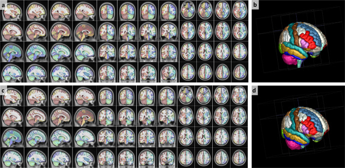
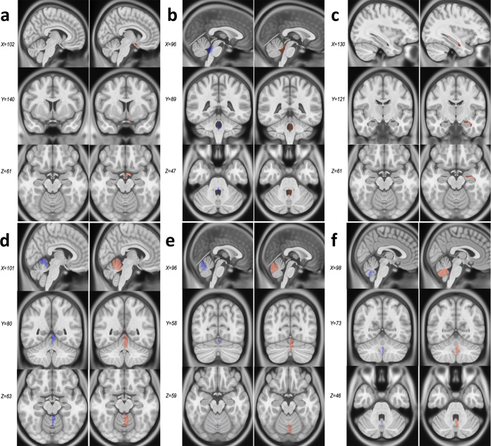
When comparing CerebrA to original labels from Mindboggle-101 (Fig. 2) registered to ICBM152, the average Dice Kappa value was κ = 0.73 ± 0.18 (Table 1). The structures with relatively lower Dice Kappa (κ < 0.6) corresponded to the structures that needed the most correction such as the optic chiasm, inferior lateral ventricles, fourth ventricle and cerebellar vermis. The optic chiasm label was barely found in the original Mindboggle-101 registered to ICBM152 and most of it was misaligned with regards to the actual structure. To ensure that this inaccuracy was not caused by the nonlinear registration process, we further inspected the original Mindboggle-101 template and label atlas and found similar issues. For CerebrA, the optic chiasm label was redefined trying to achieve continuity amongst optic chiasma itself and optic tracts (Fig. 3, panel a). Then, the inferior lateral ventricles and fourth ventricle boundaries were improved using a threshold to differentiate CSF from parenchyma (Fig. 3, panels b and c). And finally, cerebellar vermis labels were redefined for right and left side (Fig. 3, panels d–f).
Fig. 2.
Warped CerebrA atlas (a,b) and Mindboggle-101 atlas (c,d) overlaid on the ICBM152 non-linear 2009 symmetric average MRI template. Note the improved label alignment on the cortical structures.
Fig. 3.
Comparison between Mindboggle-101 and CerebrA for structures with Dice Kappa < 0.6. Panel a. Optic chiasm. Panel b. Fourth ventricle. Panel c. Inferior lateral ventricle. Panels d–f. Cerebellar vermis lobules. For each structure, the column on the left (blue) represents the original labels from Mindboggle-101, warped onto the ICBM152 symmetric template, and the right column (pink) represents CerebrA’s right sided corresponding labels, on the same template.
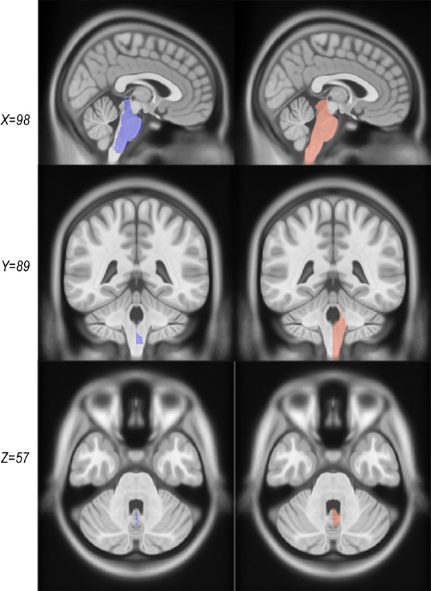
Another significant change in CerebrA from the original warped labels was the brainstem label definition. The brainstem area was manually redefined for the right side and then flipped in the same procedure as all the labels considering the symmetrical feature of the ICBM152 2009c2 template. In addition, boundaries between brainstem and fourth ventricle were carefully defined using the CSF intensity threshold, cerebellar white matter labels within the brainstem area were removed and rostral brainstem delimitation was improved (Fig. 4).
Fig. 4.
Comparison between warped Mindboggle-101 labels and CerebrA labels for the brainstem. The column on the left (purple) represents the original labels from Mindboggle-101 and the right column (coral) represents CerebrA’s right sided corresponding labels.
Inter-rater and intra-rater variability assessments
To assess intra-rater variability, the rater repeated the process of manual correction for 10 randomly selected regions, and the results were compared against the previously corrected masks. The mean Dice-Kappa values between the two masks were 0.88 ± 0.03. To assess inter-rater variability, the same 10 selected regions were rated a second time by another independent rater and these results were also compared against the previously corrected masks, yielding a mean inter-rater Dice-Kappa of 0.83 ± 0.05.
Volumes of cortical and subcortical structures
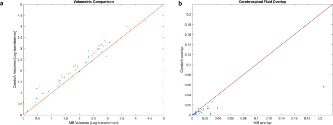
Region volumes were calculated for all cortical and subcortical structures before and after performing the manual correction by summing up the number of voxels within each label (in CCs). These volumes were then log-transformed to achieve normal distribution to enable comparisons between the two sets of volumes. Figure 5a shows the correlation plot between the log-transformed CerebrA and Mindboggle volumes. Although the volumes were strongly and significantly correlated (R = 0.9657, P value < 0.001), overall volumes estimated with CerebrA were larger than those estimated with Mindboggle-101 (Fig. 5a). The volumes estimated per structure using Mindboggle and CerebrA segmentation are listed in Table 2.
Fig. 5.
(a) Correlation plot between CerebrA and Mindboggle-101 volumes. For better visualization and to achieve a normal distribution, the volumes have been log-transformed. Correlation coefficient R = 0.9657, P value < 0.001. (b) Plot showing the proportion of overlap (the number of voxels in the specific mask overlapping with the CSF mask/the total number of voxels inside the specific mask) between the atlas labels and CSF for both atlases. MB: Mindboggle-101. CSF: CerebroSpinal Fluid.
Table 2.
Volume per structure using Mindboggle-101 and CerebrA segmentations.
| Label Name | Volume (cc) | Label Name | Volume (cc) | ||
|---|---|---|---|---|---|
| MB | CerebrA | MB | CerebrA | ||
| Caudal Anterior Cingulate | 2.75 | 3.65 | Superior Parietal | 12.97 | 19.33 |
| Caudal Middle Frontal | 9.1 | 13.22 | Superior Temporal | 20.25 | 25.35 |
| Cuneus | 4.97 | 8.07 | Supramarginal | 12.2 | 16.03 |
| Entorhinal | 2.56 | 3.21 | Transverse Temporal | 1.45 | 1.88 |
| Fusiform | 10.48 | 13.47 | Insula | 8.33 | 9.52 |
| Inferior Parietal | 17.73 | 26.19 | Brainstem | 9.17 | 17.01 |
| Inferior temporal | 15.1 | 16.22 | Third Ventricle | 0.6 | 1.1 |
| Isthmus Cingulate | 3.27 | 4.43 | Fourth Ventricle | 0.51 | 0.77 |
| Lateral Occipital | 14.97 | 22.81 | Optic Chiasm | 0 | 0.77 |
| Lateral Orbitofrontal | 11.1 | 15.04 | Lateral Ventricle | 8.16 | 9.57 |
| Lingual | 9.13 | 12.36 | Inferior Lateral Ventricle | 0.09 | 1.06 |
| Medial Orbitofrontal | 5.28 | 8.88 | Cerebellum Gray Matter | 75.85 | 71.23 |
| Middle Temporal | 16.77 | 28.29 | Cerebellum White Matter | 17.86 | 13 |
| Para hippocampal | 2.36 | 2.71 | Thalamus | 10.48 | 10.04 |
| Paracentral | 5.94 | 8.24 | Caudate | 4.28 | 5.64 |
| Pars Opercularis | 5.79 | 7.67 | Putamen | 5.4 | 6.63 |
| Pars Orbitalis | 2.61 | 3.64 | Pallidum | 1.93 | 1.7 |
| Pars Triangularis | 5.29 | 8.2 | Hippocampus | 4.64 | 4.69 |
| Pericalcarine | 2.8 | 5.03 | Amygdala | 1 | 1.65 |
| Postcentral | 13.1 | 18.19 | Accumbens Area | 0.41 | 0.55 |
| Posterior Cingulate | 3.99 | 5.41 | Ventral Diencephalon | 6.39 | 6.1 |
| Precentral | 16.13 | 21.39 | Basal Forebrain | 0.16 | 0.19 |
| Precuneus | 13.79 | 18.77 | Vermal lobules I-V | 3.05 | 4.5 |
| Rostral Anterior Cingulate | 2.52 | 3.83 | Vermal lobules VI-VII | 0.61 | 2.32 |
| Rostral Middle Frontal | 13.77 | 21.74 | Vermal lobules VIII-X | 0.75 | 2.57 |
| Superior Frontal | 38.87 | 52.82 | |||
Abbreviations: MB: Midboggle-101.
Overlap with CSF
Using the CSF mask of the MNI-ICBM152 template, the number of voxels within each label that overlapped with the CSF was calculated to assess which template had a lower overlap with CSF. The four ventricular regions (i.e. lateral ventricles, inferior lateral ventricles, 3rd and 4th ventricles) were excluded from this analysis. Figure 5b shows the proportion of overlap of each of the labels with the CSF for each atlas; i.e. the number of voxels in the specific mask overlapping with the CSF mask divided by the total number of voxels inside the specific mask. Overall, Mindboggle-101 showed greater overlap of cortical and subcortical structures with CSF (Fig. 5b). Cerebellar vermal lobule regions (630 and 631) from Mindboggle-101 had the highest degree of overlap (20%) with the CSF, due to a combination of misalignment and over-segmentation errors (see Fig. 2c).
Usage Notes
Atlases are sometimes used to compare individual subjects. Such comparisons, made based on average templates and corresponding atlases, are by nature prone to errors caused by registration of a subject’s brain to the template. Such errors are dependent on the individual scans, and are generally greater in presence of pathologies such as tumors, lesions, severe atrophy, etc.18. Therefore, these types of errors might introduce systematic biases in the findings, and great care should be taken to assess registration accuracy when performing such analyses.
Acknowledgements
We would like to acknowledge funding from the Famille Louise & André Charron.
Author contributions
Ana L. Manera: Study concept and design, manual correction of the labels, analysis and interpretation of the data, drafting and revision of the manuscript. Mahsa Dadar: Study concept and design, analysis and interpretation of the data, revising the manuscript. Vladimir Fonov: Generation of the MNI-ICBM2009c average template, revising the manuscript. D. Louis Collins: Study concept and design, interpretation of the data, revising the manuscript.
Code availability
The scripts used to perform both the linear and nonlinear registrations (including the ANTs code with all the selected registration parameters), the obtained transformations that were used to register the DKT atlas to the MNI-ICBM2009c template, the code for resampling the labels based on these transformations, as well as the registered DKT atlas in the MNI space, after applying the transformations are available at https://gin.g-node.org/anamanera/CerebrA/src/master/.
Competing interests
The authors declare no competing interests.
Footnotes
Publisher’s note Springer Nature remains neutral with regard to jurisdictional claims in published maps and institutional affiliations.
These authors contributed equally: Ana L. Manera, Mahsa Dadar.
References
- 1.Mateos-Perez JM, et al. Structural neuroimaging as clinical predictor: A review of machine learning applications. Neuroimage Clin. 2018;20:506–522. doi: 10.1016/j.nicl.2018.08.019. [DOI] [PMC free article] [PubMed] [Google Scholar]
- 2.Fonov V, et al. Unbiased average age-appropriate atlases for pediatric studies. Neuroimage. 2011;54:313–327. doi: 10.1016/j.neuroimage.2010.07.033. [DOI] [PMC free article] [PubMed] [Google Scholar]
- 3.Klein A, Tourville J. 101 labeled brain images and a consistent human cortical labeling protocol. Front Neurosci. 2012;6:171. doi: 10.3389/fnins.2012.00171. [DOI] [PMC free article] [PubMed] [Google Scholar]
- 4.Desikan RS, et al. An automated labeling system for subdividing the human cerebral cortex on MRI scans into gyral based regions of interest. Neuroimage. 2006;31:968–980. doi: 10.1016/j.neuroimage.2006.01.021. [DOI] [PubMed] [Google Scholar]
- 5.Sled JG, Zijdenbos AP, Evans AC. A nonparametric method for automatic correction of intensity nonuniformity in MRI data. IEEE Trans Med Imaging. 1998;17:87–97. doi: 10.1109/42.668698. [DOI] [PubMed] [Google Scholar]
- 6.Nyul, L. G. & Udupa, J. K. On standardizing the MR image intensity scale. Magn Reson Med42, 1072–1081, doi:10.1002/(sici)1522-2594(199912)42:6<1072::aid-mrm11>3.0.co;2-m (1999). [DOI] [PubMed]
- 7.Collins DL, Neelin P, Peters TM, Evans AC. Automatic 3D intersubject registration of MR volumetric data in standardized Talairach space. J Comput Assist Tomogr. 1994;18:192–205. doi: 10.1097/00004728-199403000-00005. [DOI] [PubMed] [Google Scholar]
- 8.Eskildsen SF, et al. BEaST: brain extraction based on nonlocal segmentation technique. Neuroimage. 2012;59:2362–2373. doi: 10.1016/j.neuroimage.2011.09.012. [DOI] [PubMed] [Google Scholar]
- 9.Evans AC. D. L. C. a. Animal: validation and applications of nonlinear registration-based segmentation. International Journal ofPattern Recognition and Artificial Intelligence. 1997;11:1271–1294. doi: 10.1142/S0218001497000597. [DOI] [Google Scholar]
- 10.Collins, M. P. D. L. in Atlas of the Morphology of the Human Cerebral Cortex on the Average MNI Brain (ed Academic Press) 17-22 (Natalie Farra, 2019).
- 11.Marcus DS, et al. Open Access Series of Imaging Studies (OASIS): cross-sectional MRI data in young, middle aged, nondemented, and demented older adults. J Cogn Neurosci. 2007;19:1498–1507. doi: 10.1162/jocn.2007.19.9.1498. [DOI] [PubMed] [Google Scholar]
- 12.Landman BA, et al. Multi-parametric neuroimaging reproducibility: a 3-T resource study. Neuroimage. 2011;54:2854–2866. doi: 10.1016/j.neuroimage.2010.11.047. [DOI] [PMC free article] [PubMed] [Google Scholar]
- 13.Morgan VL, Mishra A, Newton AT, Gore JC, Ding Z. Integrating functional and diffusion magnetic resonance imaging for analysis of structure-function relationship in the human language network. PLoS One. 2009;4:e6660. doi: 10.1371/journal.pone.0006660. [DOI] [PMC free article] [PubMed] [Google Scholar]
- 14.Holmes CJ, et al. Enhancement of MR images using registration for signal averaging. J Comput Assist Tomogr. 1998;22:324–333. doi: 10.1097/00004728-199803000-00032. [DOI] [PubMed] [Google Scholar]
- 15.Dale AM, Fischl B, Sereno MI. Cortical surface-based analysis. I. Segmentation and surface reconstruction. Neuroimage. 1999;9:179–194. doi: 10.1006/nimg.1998.0395. [DOI] [PubMed] [Google Scholar]
- 16.Fischl B, Sereno MI, Dale AM. Cortical surface-based analysis. II: Inflation, flattening, and a surface-based coordinate system. Neuroimage. 1999;9:195–207. doi: 10.1006/nimg.1998.0396. [DOI] [PubMed] [Google Scholar]
- 17.Fischl B, et al. Automatically parcellating the human cerebral cortex. Cereb Cortex. 2004;14:11–22. doi: 10.1093/cercor/bhg087. [DOI] [PubMed] [Google Scholar]
- 18.Dadar M, Fonov VS, Collins DL. & Alzheimer’s Disease Neuroimaging, I. A comparison of publicly available linear MRI stereotaxic registration techniques. Neuroimage. 2018;174:191–200. doi: 10.1016/j.neuroimage.2018.03.025. [DOI] [PubMed] [Google Scholar]
- 19.Avants BB, Epstein CL, Grossman M, Gee JC. Symmetric diffeomorphic image registration with cross-correlation: evaluating automated labeling of elderly and neurodegenerative brain. Med Image Anal. 2008;12:26–41. doi: 10.1016/j.media.2007.06.004. [DOI] [PMC free article] [PubMed] [Google Scholar]
- 20.Manera A, Dadar M, Fonov V, Collins DL. 2020. CerebrA: Accurate registration and manual label correction of the Mindboggle-101 atlas for the MNI-ICBM152 template. G-Node. [DOI] [PMC free article] [PubMed]
- 21.Neelin, P., MacDonald, D., Collins, D. L. & Evans, A. C. The minc file format: From bytes to brains (1998).
- 22.Vincent RD, et al. MINC 2.0: A Flexible Format for Multi-Modal Images. Front Neuroinform. 2016;10:35. doi: 10.3389/fninf.2016.00035. [DOI] [PMC free article] [PubMed] [Google Scholar]
Associated Data
This section collects any data citations, data availability statements, or supplementary materials included in this article.
Data Citations
- Manera A, Dadar M, Fonov V, Collins DL. 2020. CerebrA: Accurate registration and manual label correction of the Mindboggle-101 atlas for the MNI-ICBM152 template. G-Node. [DOI] [PMC free article] [PubMed]
Data Availability Statement
The scripts used to perform both the linear and nonlinear registrations (including the ANTs code with all the selected registration parameters), the obtained transformations that were used to register the DKT atlas to the MNI-ICBM2009c template, the code for resampling the labels based on these transformations, as well as the registered DKT atlas in the MNI space, after applying the transformations are available at https://gin.g-node.org/anamanera/CerebrA/src/master/.


