
Figure 2. Case 1 glottis images.
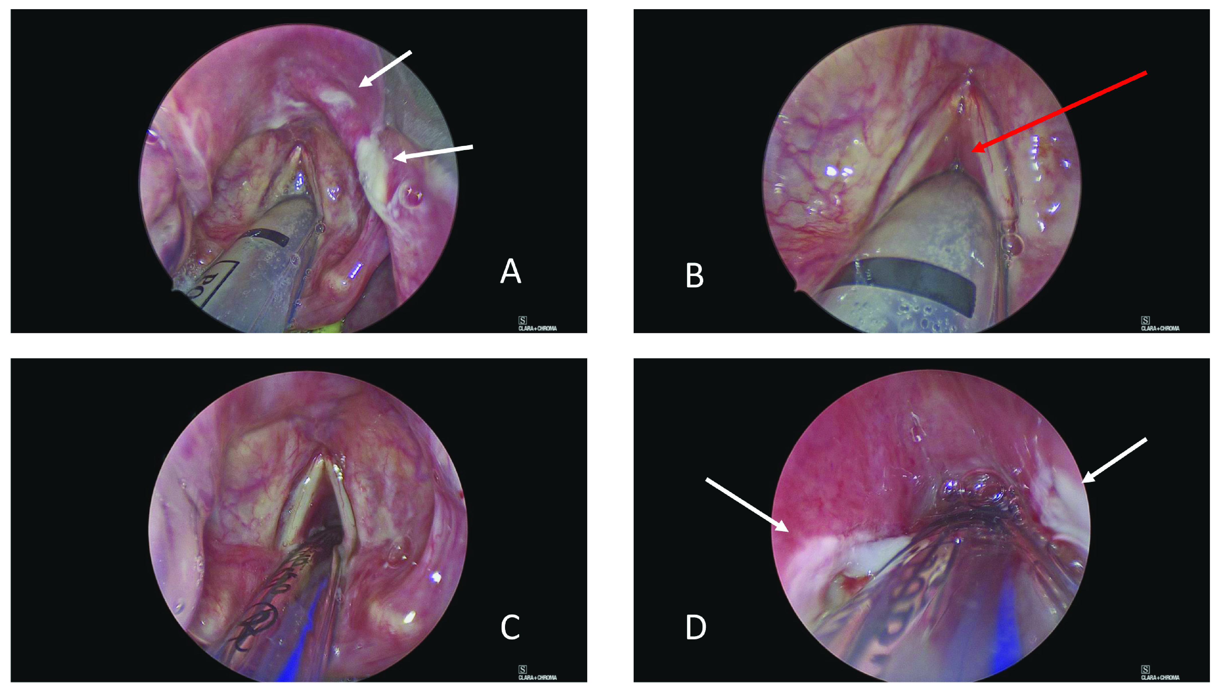
( A) View of supraglottis showing ulcerated epiglottis. ( B) Glottis showing relative sparing of vocal cords and false cords, but profound subglottic oedema. ( C) Following change to size 6 endotracheal tube, there is some anterior glottic airway. ( D) However, the subglottis is also ulcerated and oedematous mucosa prevents rigid bronchoscopy (0 o Hopkins’ rod) beyond the third tracheal ring. White arrows indicate areas of ulceration and red arrow subglottic oedema.