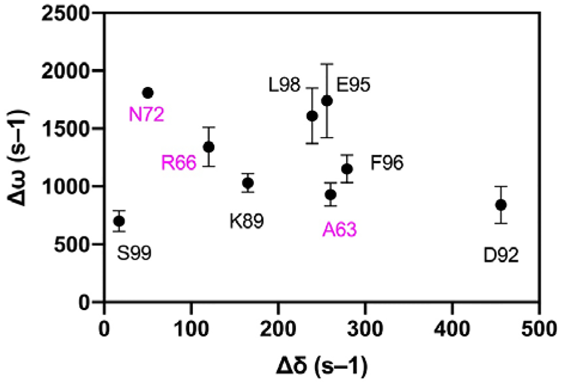
Figure 8.

Chemical shift differences. Dynamic chemical shift changes (Δω) in apo VHR from CPMG relaxation dispersion experiments plotted against chemical shift changes (Δδ) from a tungstate titration into apo VHR. Residues are labeled and colored with VI residues in magenta and acid loop residues in black. The lack of correlation between the two values indicates that loop motions in the apo enzyme are distinct from those conformational changes that occur due to ligand binding.
