Abstract
Background
English springer spaniels (ESS) show an increased risk of chronic hepatitis (CH). In a previous study of 68 ESS with CH, in which only one dog received corticosteroids, a median survival time of 189 days was noted. Some ESS with CH appear to improve with prednisolone treatment; therefore, we aimed to investigate the response to prednisolone in this breed.
Participants
ESS with histologically confirmed idiopathic CH were treated with prednisolone 1–2 mg/kg/day. Nine female and three male ESS were enrolled (median age at diagnosis of five years). Patients were monitored clinically and had biochemistry samples taken to assess markers of hepatocellular damage and function.
Results
The mean starting dose of prednisolone was 1.1 mg/kg/day. All symptomatic patients showed an initial clinical improvement. Two cases were euthanased while receiving prednisolone. The median time since diagnosis is 1715 days (range: 672–2105 days) and the remaining patients are clinically well, with seven patients still receiving a mean dose of 0.4 mg/kg prednisolone every other day. Statistical analysis demonstrated significant (P<0.05) reductions in serum alkaline phosphatase, alanine aminotransferase and bilirubin following 2–4 weeks of prednisolone treatment.
Conclusion
This study demonstrates improved clinical and biochemical parameters when some ESS with CH are managed with prednisolone and standard supportive treatments.
Keywords: hepatic disease, dogs, histopathology
Introduction
English springer spaniels (ESS) in the UK have an increased risk of chronic hepatitis (CH).1 CH is defined by the World Small Animal Veterinary Association (WSAVA) Liver Standardisation Project2 as histological evidence of hepatocellular apoptosis, necrosis, regeneration, predominant mononuclear cell infiltration and fibrosis. The reported postmortem prevalence of CH in dogs in first-opinion practice is 12 per cent, suggesting this is a common disease which likely has a range of aetiologies including nutritional, environmental, genetic and infectious.3 There are several well-documented causes of canine CH, such as copper accumulation due to a defect in copper metabolism4 and possible infectious causes due to Bartonella species,5 Leptospira species6 and Helicobacter species.7 Although previous studies have identified viral causes of CH, including canine adenovirus type I,8 there is currently no substantial evidence to suggest viral causes are a significant aetiology for canine CH.9–12 Studies have been performed which support an immune-mediated aetiology to CH in some breeds.13 14 CH has a number of well-reported breed predispositions including the Labrador retriever,15 American Cocker Spaniel,16 English Cocker Spaniel,17 ESS,1 Dalmatian,18 Doberman,19 Great Dane,20 Cairn Terrier and Samoyed;1 however, the underlying aetiology is frequently unknown and therefore treatment often remains non-specific and supportive.21
In a previous study of 68 ESS with biopsy confirmed idiopathic CH, only one was treated with prednisolone, and the median survival time of the whole cohort was 189 days (range: 1–1211 days).22 This suggested that the underlying disease process in the ESS was aggressive and rapidly fatal in most cases, in contrast to a previous study looking at 79 dogs of various breeds with histologically confirmed CH which reported mean survival times of 21.1–36.4 months when cirrhosis was not present.23 Research investigating the disease in ESS and other breeds in the UK initially concentrated on attempts to find a viral cause for the disease because of the histological similarity to human viral hepatitis and canine acidophil cell hepatitis.9 24 Corticosteroid treatment was not initially advised and cases were instead managed supportively. However, the progression of CH in these cases remained mostly rapid and short survival times were noted,22 yet some clinicians reported improved survival when these cases were given corticosteroids. Despite the widespread use of corticosteroids in the general treatment of canine CH, only two previous studies have investigated their efficacy.25 26 One of those studies was published before the WSAVA Liver Standardisation Project, which generates concern that some patients were not truly idiopathic, and both studies included a range of canine breeds. Although both studies identified some clinical and biochemical improvements in some cases of canine CH, it is likely that the study populations included a diverse range of underlying disease processes and therefore the results are likely difficult to interpret. There are no currently published studies investigating the response to prednisolone in canine patients with CH in a single breed.
Therefore, the authors instituted a prospective cohort study aimed at investigating the clinical and biochemical response to prednisolone and other supportive treatments in a group of ESS with histopathologically confirmed idiopathic CH.
Materials and methods
ESS being treated in first-opinion practice, or referred to the Queen’s Veterinary School Hospital (University of Cambridge) with a histological diagnosis of idiopathic CH were enrolled prospectively between 2009 and 2017. No cases had previously been involved in studies investigating CH in ESS. Cases were identified when veterinary surgeons contacted the authors for advice. A histopathological diagnosis was based on a predominantly lymphoplasmacytic, interface hepatitis and variable fibrosis, and according to WSAVA criteria for a diagnosis of CH. All liver biopsies were stained with rhodanine for qualitative copper assessment using a previously published copper grading system.27 Samples were scored as grade 1: absence or few copper-containing granules in the cytoplasm of an occasional hepatocyte; grade 2: obvious copper-containing granules in some centrilobular hepatocytes; grade 3: numerous granules in most centrilobular hepatocytes (one-third of each lobule); grade 4: presence of numerous granules in all centrilobular and midzonal hepatocytes (approximately two-thirds of the hepatocytes in all lobules); grade 5: abundant granules in more than two-thirds of the liver cells in all lobules. The histopathology specimens were examined by several board-certified pathologists and were excluded if significant copper accumulation (grade 3–5) was documented. Cases with evidence of pyogranulomatous hepatitis were also excluded. No cases had been treated with corticosteroids within six months of the study. Attending veterinary surgeons gave prednisolone 1–2 mg/kg/day and submitted haematology and biochemistry samples and progress reports to the authors. The prednisolone starting dose range was based on two previous studies that investigated the response to prednisolone in various canine breeds with CH.25 26 Prednisolone is a well-reported treatment option for canine CH, and the decision to start the patients on this therapy was at the discretion of the clinician in charge of each case. Informed owner consent was obtained for analysis of patient data. All patients had their first recheck 2–4 weeks following initiation of prednisolone therapy, which included biochemical assessment of liver parameters. Further blood samples were advised to be taken before any prednisolone dose reduction. The prednisolone dose was tapered by 25 per cent–50 per cent every 4 weeks according to the patient’s clinical and biochemical response, focusing on alkaline phosphatase (ALKP), alanine aminotransferase (ALT) and bilirubin. While all cases had these values assessed at diagnosis and at the first recheck, not all cases had these values measured each time the prednisolone dose was altered. To account for variation in instruments used to assess biochemical parameters, and corresponding variation in reference ranges, the ALKP, ALT and bilirubin have been presented as multiples of the upper limit of the reference range for the individual machine used. Due to individual patient variation in the way prednisolone was tapered and timing of blood samples, the biochemical values were plotted against the prednisolone dose at the time of sampling rather than against specific time points. Thus, the values were recorded and plotted graphically against different oral doses of prednisolone, including the values at diagnosis of CH; the values after prednisolone 1–2 mg/kg/day for 2–4 weeks; 0.5–1 mg/kg/day; 0.5–1 mg/kg/EOD (every other day); 0.25–0.5 mg/kg/EOD; after cessation of prednisolone treatment (if applicable) and after restarting prednisolone treatment if cessation of medication caused a relapse in clinical signs. Additional therapies used included combinations of S-adenosylmethionine, silybin, ursodeoxycholic acid, antibiotics and hepatic diet.
Statistical analysis
Shapiro-Wilk normality testing was performed on the data and identified a lack of normal distribution, typical of small data sets. Therefore, the non-parametric two-sided Wilcoxon test was used to demonstrate significant (P<0.05) changes in serum ALKP, ALT and bilirubin following prednisolone therapy.
Results
Sixteen cases of suspected ESS CH were evaluated during the study period. Two cases were excluded because hepatic biopsies were not performed, and two cases were excluded because their histopathology results were not consistent with the previously published WSAVA criteria for CH. Nine female and three male ESS were enrolled with a median age at diagnosis of five years (range: 11 months–10 years). All cases had been diagnosed following evaluation of wedge liver biopsies taken during laparotomy or laparoscopy. Within these cases, one dog was being treated with levothyroxine for hypothyroidism at the time of recruitment, and one case was subsequently diagnosed with protein-losing nephropathy eight months after initially presenting for hepatic disease, and its urine protein creatinine ratio improved following treatment with standard doses of benazepril (0.5 mg/kg SID PO). Four of the 12 cases showed no clinical signs of CH at diagnosis but were investigated after routine blood sampling for an unrelated reason detected elevations of liver enzymes. Seven of the 12 cases had bile culture performed and no bacteria were cultured; however, the remaining five patients did not have this evaluated. Table 1 summarises the histopathological diagnosis for each of the 12 ESS cases included in the study. None of the 12 ESS displayed significant qualitative copper accumulation (table 1) and therefore quantitative copper analysis was not performed in these cases. Furthermore, there was no histopathological evidence of significant biliary tract inflammation in any of the evaluated samples from the 12 cases. The mean prednisolone starting dosage was 1.1 mg/kg/day (range: 1.0–2.0 mg/kg/day). Symptomatic patients showed a subjective improvement clinically within four weeks according to the owners. Table 2 reports the clinical abnormalities reported for each case, both at enrolment and at the first recheck appointment, as well as additional treatments provided to each patient at the time of diagnosis. Two out of the 12 cases were euthanased due to CH-related signs while receiving prednisolone, with survival times of 122 and 741 days from diagnosis. Clinical signs that prompted euthanasia for these two patients included hepatic encephalopathy, melaena, jaundice and lethargy. The remaining 10 patients are alive and clinically well at the time of manuscript submission, with seven patients still receiving a mean dosage of 0.4 mg/kg prednisolone EOD (range: 0.25 mg/kg/EOD–1 mg/kg/day). Three patients stopped prednisolone therapy without a concurrent elevation in liver parameters, while three cases stopped and needed to be restarted on prednisolone due to recurrence of CH-related signs or an elevation in liver parameters. At the time of submission, four patients are currently in the process of having their prednisolone dose reduced with the aim to stop and monitor for recurrence of clinical signs. The median time since diagnosis for the 10 remaining cases is 1715 days (range: 672–2105 days).
Table 1.
Histopathological diagnosis for each of the 12 English springer spaniels included in the study
| Patient | Histopathological diagnosis |
| 1 | Hepatitis, interface, lymphocytic, plasmacytic and neutrophilic, chronic, mild, with moderate to marked hepatocyte apoptosis. Grade 1 copper |
| 2 | Hepatitis, periportal, lymphocytic and neutrophilic, chronic, marked, with moderate porto-portal bridging fibrosis. Grade 2 copper |
| 3 | Hepatitis, lymphocytic, plasmacytic and neutrophilic, chronic, moderate, with moderate hepatocyte apoptosis and necrosis. Grade 2 copper |
| 4 | Hepatitis, interface, lymphocytic, plasmacytic and neutrophilic, chronic, severe, with mild porto-portal bridging fibrosis. Grade 2 copper |
| 5 | Hepatitis, periportal, lymphocytic and neutrophilic, chronic, marked with moderate hepatocyte apoptosis and necrosis. Grade 2 copper |
| 6 | Hepatitis, periportal, lymphocytic, plasmacytic and neutrophilic, chronic, marked, with mild porto-portal bridging fibrosis. Grade 1 copper |
| 7 | Hepatitis, interface, lymphocytic, plasmacytic and neutrophilic, chronic, severe, with mild biliary hyperplasia and hepatocyte necrosis. Grade 2 copper |
| 8 | Hepatitis, lymphocytic, plasmacytic, chronic, moderate, with moderate porto-portal bridging fibrosis. Grade 1 copper |
| 9 | Hepatitis, lymphocytic, plasmacytic, chronic, moderate, with moderate portal fibrosis and portal biliary hyperplasia. Grade 1 copper |
| 10 | Hepatitis, lymphocytic, plasmacytic, subacute, moderate. Grade 2 copper |
| 11 | Hepatitis, lymphocytic, plasmacytic and neutrophilic, subacute, moderate, with occasional pigmented histiocytes and mild hepatocyte apoptosis. Grade 2 copper |
| 12 | Hepatitis, lobular and interface, lymphocytic and neutrophilic, chronic, severe with mild portal fibrosis. Grade 1 copper |
Table 2.
Clinical signs documented for each of the 12 English springer spaniels reported in the study, both at enrolment and at the first recheck appointment (2–4 weeks after initiation of prednisolone therapy) with additional treatments
| Patient | Clinical signs at diagnosis | Clinical signs at first recheck | Additional treatments |
| 1 | Reduce appetite, vomiting, jaundice | Resolution of jaundice and no abnormal clinical signs currently reported by owner. | UDCA, SAMe, amoxicillin-clavulanate |
| 2 | PUPD | Resolution of PUPD according to owner. | UDCA, SAMe |
| 3 | Anorexia, PUPD, vomiting, weight-loss, jaundice, lethargy | Jaundice still identified during clinical examination and appetite improved but reduced compared with normal. Resolution of vomiting, PUPD and lethargy according to owner, however weight-loss continued. The patient subsequently developed neurological abnormalities with worsening jaundice, and was euthanased 122 days after initiating prednisolone therapy. | UDCA, SAMe, metronidazole |
| 4 | Lethargy, reduced appetite, vomiting, jaundice. | Jaundice not identified during clinical examination and no abnormal signs currently reported by the owner. | UDCA, SAMe |
| 5 | No abnormal clinical signs reported by owner | Still reported to be clinically normal by owner. | UDCA, SAMe, hepatic diet |
| 6 | No abnormal clinical signs reported by owner | Still reported to be clinically normal by owner. | SAMe, hepatic diet |
| 7 | No abnormal clinical signs reported by owner | Still reported to be clinically normal by owner. | SAMe |
| 8 | Jaundice, ascites, weight-loss | Resolution of jaundice and ascites during clinical examination. No abnormal signs currently reported by the owner. The patient subsequently developed neurological abnormalities and melaena, and was euthanased 741 days after initiating prednisolone therapy. | UDCA, SAMe, spironolactone |
| 9 | Reduced appetite and PUPD | PUPD still present but improved, according to owner. Appetite now normal. | UDCA, SAMe |
| 10 | No abnormal clinical signs reported by owner | Still reported to be clinically normal by owner. | |
| 11 | Vomiting, lethargy, jaundice, ascites, PUPD | Mild PUPD and polyphagia reported by owner, otherwise no abnormal clinical signs. | Hepatic diet |
| 12 | Lethargy, diarrhoea, vomiting | Resolution of vomiting and diarrhoea but owner still reported mild lethargy. | SAMe, hepatic diet, cefalexin |
PUPD, polyuria/polydipsia; SAMe, S-adenosylmethionine; UDCA, ursodeoxycholic acid.
Table 3 presents the median values for ALKP, ALT and bilirubin at diagnosis of CH, as well as the values at the patients’ first recheck 2–4 weeks after starting prednisolone. Two-sided Wilcoxon test demonstrated a significant reduction in ALKP, ALT and bilirubin at the first recheck following prednisolone treatment with P values 0.0010, 0.002 and 0.0156, respectively. Due to variability in the length of time patients remained on the tapering doses of prednisolone, the authors elected not to assess for significant changes between the remaining prednisolone doses.
Table 3.
Median values for serum ALKP, ALT and bilirubin in English springer spaniels at diagnosis of CH and at first recheck (2–4 weeks after initiation of prednisolone 1–2 mg/kg/day)
| Median value* at diagnosis (range) | Median value* at first recheck (range) | P value | |
| ALKP | 8.5 (3.7–16.2) | 2.7 (1.1–8.3) | 0.0010 |
| ALT | 10.8 (2.5–46.3) | 3.9 (0.8–14.7) | 0.0020 |
| Bilirubin | 1.4 (0.3–27.9) | 0.45 (0.1–4.1) | 0.0156 |
*The values are reported as a multiple of the upper limit of the reference range.
ALKP, alkaline phosphatase; ALT, alanine aminotransferase.
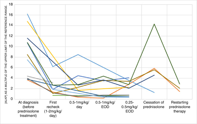
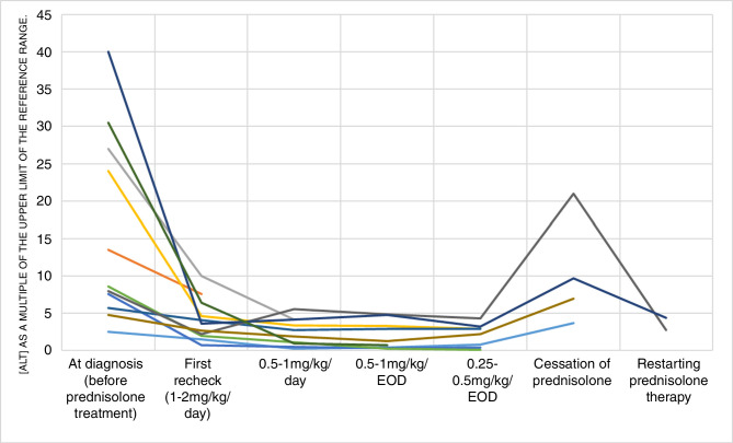
Figures 1 and 2 depict the serum values for ALKP and ALT, respectively, from the 12 ESS cases in this study. In all dogs, there were elevations in ALKP and ALT before prednisolone treatment, but following initiation of prednisolone therapy all values significantly reduced for all patients. However, in the 11 patients that had oral prednisolone reduced to 0.25–0.5 mg/kg/EOD or stopped entirely, seven (64 per cent) documented an increase in either or both ALKP and ALT. In the three cases that had ALKP measurements following restarting prednisolone after cessation of treatment, the ALKP values were subjectively decreased (figure 1), and the same was found for measured ALT (figure 2). In 5 of the 12 ESS cases, the measured serum ALKP never returned to within the reference range. Furthermore, we found that 9 of the 12 cases documented serum ALT that did not return to within the reference range, despite resolution of clinical signs.
Figure 1.
Serum (ALKP) as a multiple of the upper limit of the reference range in 12 ESS with CH, at diagnosis (before prednisolone treatment), first recheck (2–4 weeks after starting 1–2 mg/kg/day prednisolone) and at tapering doses of prednisolone. A significant difference was identified between the values at diagnosis and first recheck; however, due to variability of dosing, the authors did not assess statistical differences between the remaining prednisolone doses. Each coloured shape represents an individual patient. ALKP, alkaline phosphatase; CH, chronic hepatitis; EOD, Every Other Day; ESS, English springer spaniels.
Figure 2.
Serum (ALT) as a multiple of the upper limit of the reference range in 12 ESS with CH, at diagnosis (before prednisolone treatment), first recheck (2–4 weeks after starting 1–2 mg/kg/day prednisolone) and at tapering doses of prednisolone. A significant difference was identified between the values at diagnosis and first recheck; however, due to variability of dosing, the authors did not assess statistical differences between the remaining prednisolone doses. Each coloured shape represents an individual patient. ALT, alanine aminotransferase; CH, chronic hepatitis; EOD, Every Other Day; ESS, English springer spaniels.
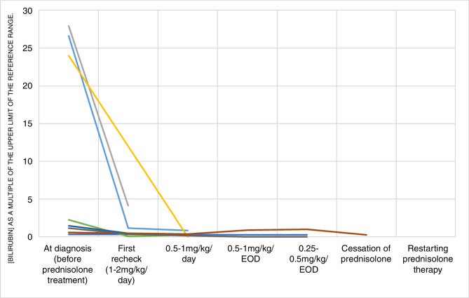
Figure 3 presents the values of serum bilirubin in the nine ESS that had these values measured. The initial values are elevated before prednisolone treatment in six patients, and there is a significant reduction in these values following initiation of prednisolone. Five of the six patients with elevated serum bilirubin showed a return to normal range, and the one patient whose elevated bilirubin did not return to normal is still early in the treatment course and has shown a substantial decrease which is approaching the reference interval.
Figure 3.
Serum (bilirubin) as a multiple of the upper limit of the reference range in nine ESS with CH, at diagnosis (before prednisolone treatment), first recheck (2–4 weeks after starting 1–2 mg/kg/day prednisolone) and at tapering doses of prednisolone. A significant difference was identified between the values at diagnosis and first recheck; however, due to variability of dosing, the authors did not assess statistical differences between the remaining prednisolone doses. Each coloured shape represents an individual patient. CH, chronic hepatitis; EOD, Every Other Day; ESS, English springer spaniels.
Discussion
This study documents that some ESS with histologically confirmed idiopathic CH show clinical and clinicopathological improvement to prednisolone 1–2 mg/kg/day, in addition to standard supportive treatments. The median time since diagnosis in our current study was 1715 days (range: 672–2105 days) and although we cannot make direct comparisons, it does appear that the ESS in our study had an improved survival compared with the previously documented median survival time of 189 days (range: 1–1211 days).22 Unfortunately, we do not have a direct control population for comparison; however, given the aggressive nature of ESS CH, and the previously published benefits of prednisolone for canine CH,25 26 we felt it was inappropriate to deny patients medication that could benefit them. As a result, we must acknowledge that the supportive treatments provided to the patients may have contributed to our results. Furthermore, our results suggest that serial measurements of alkaline phosphatase (ALKP), alanine aminotransferase (ALT) and serum bilirubin are useful for monitoring the patient’s response to prednisolone therapy, and the increase in some dogs when prednisolone therapy was stopped further supports their use. This is the first study providing evidence for the use of prednisolone in some ESS with CH and indeed the first study documenting corticosteroid response in a single rather than multiple breeds.26 This positive response was convincing in spite of the absence of a control population without treatment. These were different dogs from those described in the previous study22 and offers additional support for a female predisposition with 9 of the 12 cases being female. Interestingly, two previous studies1 22 identified a young to middle-aged onset of disease in ESS (median age at diagnosis of five years and five years seven months, respectively) which is similar to the median age at diagnosis in our current study of five years. This is younger than the overall median age of eight years in a study of 551 dogs of varying breeds with CH in the UK1. Histologically, the liver tissue from ESS CH cases shows a predominant lymphoplasmacytic inflammation with interface hepatitis, variable fibrosis that can extend between portal triads and hepatocellular apoptosis and necrosis. While it would have been interesting to assess liver histopathology following treatment with prednisolone, this was not evaluated in the current study. Table 1 summarises the histopathological diagnosis for each of our 12 ESS cases and the features identified are remarkably similar to those expected with human autoimmune hepatitis (AIH).28 Hepatic histopathology alone is not considered diagnostic for AIH in people, but instead further validation is required with response to immunosuppressive drugs and positive detection of various serum autoantibodies including non-organ-specific and organ-specific autoantibodies such as antinuclear, smooth muscle, liver cytosol type-I and liver-kidney-microsomal type-I antibodies.29 Human leucocyte antigen alleles which confer an increased risk for developing AIH have been found in affected individuals.30 These alleles have also been shown to influence progression of the disease, which is interesting in light of the previously documented association between dog leucocyte antigen and CH in the ESS.31 Regarding assessment of hepatic copper, none of the 12 cases were reported to have significant copper accumulation following qualitative copper grading, but it is possible that variation between the pathologists resulted in a degree of interobserver variation. However, all pathologists were board-certified and as such are very likely to have made the authors aware if they had concerns regarding the qualitative copper assessment of the histopathology specimens.
An unexpected finding in this study was that 4 of the 12 cases were perceived to be asymptomatic at the time of diagnosis, in contrast to a previous case series suggesting that the disease is usually aggressive and rapidly fatal.22 These patients may have been identified early in the course of their disease and it is possible they will have become clinically unwell in the future if the disease had not been investigated. Human AIH has an asymptomatic presentation in 25 per cent–34 per cent cases,32 with 26 per cent–70 per cent of these patients going on to develop clinical signs within 32 months of diagnosis. The number of asymptomatic cases in our current study is not too dissimilar from that reported in human literature which could suggest some similarities between human AIH and ESS CH. It is impossible to know in either people or dogs how long a patient may be asymptomatic before clinical presentation because patients without symptoms are not routinely blood tested. An equally unexpected finding was the significant reduction in ALKP in patients despite prednisolone therapy. It is known that corticosteroids induce ALKP activity in dogs, and therefore it is common for patients treated with prednisolone to experience increased serum concentrations of the enzyme.33 The significant reduction in serum ALKP seen in our cohort following initiation of prednisolone treatment suggests that, in these cases, the initial enzyme elevation before corticosteroid administration was primarily disease induced. Therefore, controlling the disease with prednisolone appeared to result in a corresponding, significant reduction in hepatocellular damage and cholestasis. The continued mild elevations in ALKP on treatment are consistent with steroid induction of enzymes supported by the fact that bilirubin became normal in six cases.
We recognise there are limitations to this investigation. Due to the clinical nature of this cohort study, with individual patients being managed by different veterinarians, there was some variability in the way prednisolone was tapered and timing of blood samples. Therefore, the authors did not plot the measured serum values of ALT, ALKP and bilirubin against time from initiation of treatment, but instead the values were plotted against different doses of prednisolone. Furthermore, all cases received additional supportive medications for CH which varied between patients and could have influenced results. However, this variability is inherently difficult to overcome when dealing with patients in clinical practice and also made it challenging to accurately standardise a clinical scoring system. It is also important to note that cases were enrolled at different times which resulted in patients being at different stages of their disease with some having fully recovered while others having more recently been diagnosed and started on medication at the time of manuscript submission.
Conclusion
This study documents that some ESS with histologically confirmed idiopathic CH show clinical and clinicopathological improvement to prednisolone 1–2 mg/kg/day, in addition to standard supportive dietary and medical management. Further studies are indicated to investigate potential serum markers of autoimmunity and the use of other immunosuppressive treatments in affected dogs.
Acknowledgments
The authors would like to thank the primary practitioners who contributed cases for this study, including but not limited to Dr Peter Haworth, Dr Chirag Patel, Dr Anne Wilson, Dr Gabi Habacher and Dr Will Hodge. The authors would also like to thank Dr Tim Williams for his help with statistical analysis.
Footnotes
Funding: The authors have not declared a specific grant for this research from any funding agency in the public, commercial or not-for-profit sectors.
Competing interests: None declared.
Patient consent for publication: Not required.
Provenance and peer review: Not commissioned; externally peer reviewed.
Data availability statement: All data relevant to the study are included in the article or uploaded as supplementary information.
References
- 1. Bexfield NH, Buxton RJ, Vicek TJ, et al. Breed, age and gender distribution of dogs with chronic hepatitis in the United Kingdom. Vet J 2012;193:124–8. 10.1016/j.tvjl.2011.11.024 [DOI] [PMC free article] [PubMed] [Google Scholar]
- 2. Rothuizen J, Bunch SE, Charles JE, et al. WSAVA standards for clinical and histological diagnosis of canine and feline liver diseases, 1st ED. Philadelphia: Saunders Elsevier, 2006. [Google Scholar]
- 3. Watson PJ, Roulois AJA, Scase TJ, et al. Prevalence of hepatic lesions at post-mortem examination in dogs and association with pancreatitis. J Small Anim Pract 2010;51:566–72. 10.1111/j.1748-5827.2010.00996.x [DOI] [PubMed] [Google Scholar]
- 4. Hoffmann G, van den Ingh TSGAM, Bode P, et al. Copper-associated chronic hepatitis in Labrador Retrievers. J Vet Intern Med 2006;20:856–61. 10.1111/j.1939-1676.2006.tb01798.x [DOI] [PubMed] [Google Scholar]
- 5. Gillespie TN, Washabau RJ, Goldschmidt MH, et al. Detection of Bartonella henselae and Bartonella clarridgeiae DNA in hepatic specimens from two dogs with hepatic disease. J Am Vet Med Assoc 2003;222:47–51. 10.2460/javma.2003.222.47 [DOI] [PubMed] [Google Scholar]
- 6. Adamus C, Buggin-Daubié M, Izembart A, et al. Chronic hepatitis associated with leptospiral infection in vaccinated beagles. J Comp Pathol 1997;117:311–28. 10.1016/S0021-9975(97)80079-5 [DOI] [PubMed] [Google Scholar]
- 7. Sykes JE, Hartmann K, Lunn KF, et al. 2010 ACVIM small animal consensus statement on leptospirosis: diagnosis, epidemiology, treatment, and prevention. J Vet Intern Med 2011;25:1–13. 10.1111/j.1939-1676.2010.0654.x [DOI] [PMC free article] [PubMed] [Google Scholar]
- 8. Bulut O, Yapici O, Avci O, et al. The serological and virological investigation of canine adenovirus infection on the dogs. ScientificWorldJournal 2013;2013:587024 10.1155/2013/587024 [DOI] [PMC free article] [PubMed] [Google Scholar]
- 9. Bexfield NH, Watson PJ, Heaney J, et al. Canine hepacivirus is not associated with chronic liver disease in dogs. J Viral Hepat 2014;21:223–8. 10.1111/jvh.12150 [DOI] [PMC free article] [PubMed] [Google Scholar]
- 10. Chouinard L, Martineau D, Forget C, et al. Use of polymerase chain reaction and immunohistochemistry for detection of canine adenovirus type 1 in formalin-fixed, paraffin-embedded liver of dogs with chronic hepatitis or cirrhosis. J VET Diagn Invest 1998;10:320–5. 10.1177/104063879801000402 [DOI] [PubMed] [Google Scholar]
- 11. Rakich PM, Prasse KW, Lukert PD, et al. Immunohistochemical detection of canine adenovirus in paraffin sections of liver. Vet Pathol 1986;23:478–84. 10.1177/030098588602300419 [DOI] [PubMed] [Google Scholar]
- 12. Webster CRL, Center SA, Cullen JM, et al. ACVIM consensus statement on the diagnosis and treatment of chronic hepatitis in dogs. J Vet Intern Med 2019;33:1173–200. 10.1111/jvim.15467 [DOI] [PMC free article] [PubMed] [Google Scholar]
- 13. Dyggve H, Kennedy LJ, Meri S, et al. Association of Doberman hepatitis to canine major histocompatibility complex II. Tissue Antigens 2011;77:30–5. 10.1111/j.1399-0039.2010.01575.x [DOI] [PubMed] [Google Scholar]
- 14. Dyggve H, Meri S, Spillmann T, et al. Antihistone autoantibodies in Dobermans with hepatitis. J Vet Intern Med 2017;31:1717–23. 10.1111/jvim.14838 [DOI] [PMC free article] [PubMed] [Google Scholar]
- 15. Shih JL, Keating JH, Freeman LM, et al. Chronic hepatitis in Labrador Retrievers: clinical presentation and prognostic factors. J Vet Intern Med 2007;21:33–9. 10.1111/j.1939-1676.2007.tb02925.x [DOI] [PubMed] [Google Scholar]
- 16. Kanemoto H, Sakai M, Sakamoto Y, et al. American Cocker Spaniel chronic hepatitis in Japan. J Vet Intern Med 2013;27:1041–8. 10.1111/jvim.12126 [DOI] [PubMed] [Google Scholar]
- 17. Sevelius E, Andersson M, Jönsson L. Hepatic accumulation of alpha-1-antitrypsin in chronic liver disease in the dog. J Comp Pathol 1994;111:401–12. 10.1016/S0021-9975(05)80098-2 [DOI] [PubMed] [Google Scholar]
- 18. Webb CB, Twedt DC, Meyer DJ. Copper-associated liver disease in Dalmatians: a review of 10 dogs (1998-2001). J Vet Intern Med 2002;16:665–8. [DOI] [PubMed] [Google Scholar]
- 19. Mandigers PJJ, van den Ingh TSGAM, Spee B, et al. Chronic hepatitis in Doberman pinschers. A review. Vet Q 2004;26:98–106. 10.1080/01652176.2004.9695173 [DOI] [PubMed] [Google Scholar]
- 20. Raffan E, McCallum A, Scase TJ, et al. Ascites is a negative prognostic indicator in chronic hepatitis in dogs. J Vet Intern Med 2009;23:63–6. 10.1111/j.1939-1676.2008.0230.x [DOI] [PubMed] [Google Scholar]
- 21. Poldervaart JH, Favier RP, Penning LC, et al. Primary hepatitis in dogs: a retrospective review (2002-2006). J Vet Intern Med 2009;23:72–80. 10.1111/j.1939-1676.2008.0215.x [DOI] [PubMed] [Google Scholar]
- 22. Bexfield NH, Andres-Abdo C, Scase TJ, et al. Chronic hepatitis in the English springer spaniel: clinical presentation, histological description and outcome. Vet Rec 2011;169:415 10.1136/vr.d4665 [DOI] [PMC free article] [PubMed] [Google Scholar]
- 23. Sevelius E. Diagnosis and prognosis of chronic hepatitis and cirrhosis in dogs. J Small Animal Practice 1995;36:521–8. 10.1111/j.1748-5827.1995.tb02801.x [DOI] [PubMed] [Google Scholar]
- 24. Jarrett WF, O'Neil BW, O’Neil BW. A new transmissible agent causing acute hepatitis, chronic hepatitis and cirrhosis in dogs. Vet Rec 1985;116:629–35. 10.1136/vr.116.24.629 [DOI] [PubMed] [Google Scholar]
- 25. Favier RP, Poldervaart JH, van den Ingh TSGAM, et al. A retrospective study of oral prednisolone treatment in canine chronic hepatitis. Vet Q 2013;33:113–20. 10.1080/01652176.2013.826881 [DOI] [PubMed] [Google Scholar]
- 26. Strombeck DR, Miller LM, Harrold D. Effects of corticosteroid treatment on survival time in dogs with chronic hepatitis: 151 cases (1977-1985). J Am Vet Med Assoc 1988;193:1109–13. [PubMed] [Google Scholar]
- 27. Thornburg LP, Shaw D, Dolan M, et al. Hereditary copper toxicosis in West highland white terriers. Vet Pathol 1986;23:148–54. 10.1177/030098588602300207 [DOI] [PubMed] [Google Scholar]
- 28. Tiniakos DG, Brain JG, Bury YA. Role of histopathology in autoimmune hepatitis. Dig Dis 2015;33:53–64. 10.1159/000440747 [DOI] [PubMed] [Google Scholar]
- 29. Maggiore G, Nastasio S, Sciveres M. Juvenile autoimmune hepatitis: spectrum of the disease. World J Hepatol 2014;6:464–76. 10.4254/wjh.v6.i7.464 [DOI] [PMC free article] [PubMed] [Google Scholar]
- 30. Czaja AJ, Donaldson PT. Gender effects and synergisms with histocompatibility leukocyte antigens in type 1 autoimmune hepatitis. Am J Gastroenterol 2002;97:2051–7. 10.1111/j.1572-0241.2002.05921.x [DOI] [PubMed] [Google Scholar]
- 31. Bexfield NH, Watson PJ, Aguirre-Hernandez J, et al. Dla class II alleles and haplotypes are associated with risk for and protection from chronic hepatitis in the English Springer Spaniel. PLoS One 2012b;7:e42584 10.1371/journal.pone.0042584 [DOI] [PMC free article] [PubMed] [Google Scholar]
- 32. Czaja AJ. Diagnosis and management of autoimmune hepatitis: current status and future directions. Gut Liver 2016;10:177–203. 10.5009/gnl15352 [DOI] [PMC free article] [PubMed] [Google Scholar]
- 33. Ginel PJ, Lucena R, Fernández M. Duration of increased serum alkaline phosphatase activity in dogs receiving different glucocorticoid doses. Res Vet Sci 2002;72:201–4. 10.1053/rvsc.2001.0541 [DOI] [PubMed] [Google Scholar]


