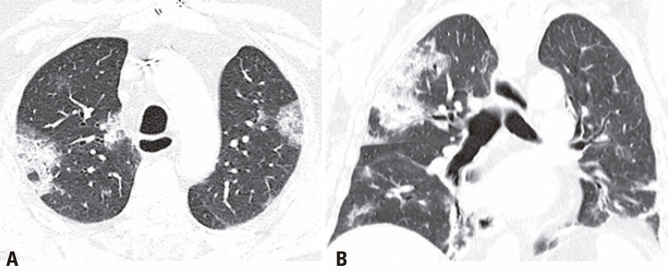
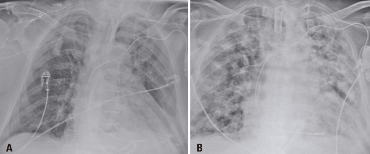
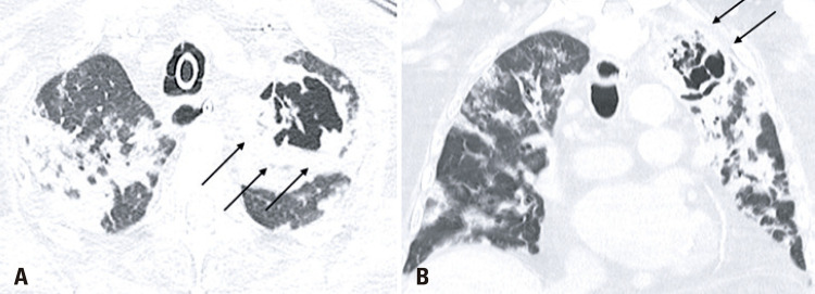
An 86-years-old male patient was admitted to the emergency department with a 1-day history of fever, dyspnea, and cough. After an assessment, there were no criteria that justified the patient’s hospitalization, therefore, he was dismissed and instructed to continue treatment at home. One week after the onset of symptoms, the patient returned with worsening dyspnea and persistent fever, which prompted his hospitalization. A chest computed tomography (CT) demonstrated typical coronavirus disease (COVID-19) findings(1,2) (Figure 1), and the real-time polymerase chain reaction (rt-PCR) test confirmed the diagnosis. After 13 days of hospitalization, the patient experienced clinical worsening, and nosocomial pneumonia caused by Enterococcus faecalis was diagnosed (Figure 2). The following CT 10 days after the diagnosis of the nosocomial infection showed multiple bilateral areas of ground-glass opacities, accompanied by septal thickening and consolidation areas, possibly related to COVID-19. There was also an excavated new lesion in the left upper lobe (Figure 3). A new rt-PCR test was still positive for COVID-19 on the same day of this last chest CT scan.
Figure 1. Axial and coronal chest computed tomography shows multiple and bilateral ground-glass opacities with areas of consolidation and septal thickening, typical findings of COVID-19.
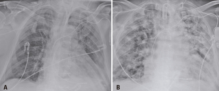
Figure 2. Bedside anteroposterior chest X-ray images demonstrate the radiographic evolution of the nosocomial infection with multiple areas of consolidation in both lungs.
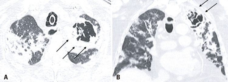
Figure 3. Axial and coronal chest computed tomography images show diffuse areas of consolidation, ground-glass opacities and a cavitating lesion in the superior lobe of the left lung (black arrows).
Recent studies have demonstrated that pulmonary co-infection caused by other agents is not uncommon in the COVID-19 context.(3,4) Complications such as necrotizing pneumonia and subsequently excavated lung lesions become possible, and their imaging presentation is considered an atypical finding in COVID-19.(5)
The persistent positive COVID-19 rt-PCR seen in this case did not allow us to differentiate whether findings of pulmonary excavation were determined by the bacterial co-infection or were related to the COVID-19 infection alone.
Even though many clinical and imaging aspects of COVID-19 have already been elucidated, some cases of the novel coronavirus continue to surprise the medical team. Further studies are still needed to develop a better understanding of this new virus.
REFERENCES
- 1.1. Ding X, Xu J, Zhou J, Long Q. Chest CT findings of COVID-19 pneumonia by duration of symptoms. Eur J Radiol. 2020;127:109009. [DOI] [PMC free article] [PubMed]; Ding X, Xu J, Zhou J, Long Q. Chest CT findings of COVID-19 pneumonia by duration of symptoms. Eur J Radiol. 2020;127:109009. doi: 10.1016/j.ejrad.2020.109009. [DOI] [PMC free article] [PubMed] [Google Scholar]
- 2.2. Chate RC, Fonseca EK, Passos RB, Teles GB, Shoji H, Szarf G. Presentation of pulmonary infection on CT in COVID-19: initial experience in Brazil. J Bras Pneumol. 2020;46(2):e20200121. [DOI] [PMC free article] [PubMed]; Chate RC, Fonseca EK, Passos RB, Teles GB, Shoji H, Szarf G. Presentation of pulmonary infection on CT in COVID-19: initial experience in Brazil. J Bras Pneumol. 2020;46(2):e20200121. doi: 10.36416/1806-3756/e20200121. [DOI] [PMC free article] [PubMed] [Google Scholar]
- 3.3. Nowak MD, Sordillo EM, Gitman MR, Paniz Mondolfi AE. Co-infection in SARS-CoV-2 infected Patients: Where Are Influenza Virus and Rhinovirus/Enterovirus? J Med Virol. 2020; April 30. doi: 10.1002/jmv.25953. [Epub ahead of print]. [DOI] [PMC free article] [PubMed]; Nowak MD, Sordillo EM, Gitman MR, Paniz Mondolfi AE. Co-infection in SARS-CoV-2 infected Patients: Where Are Influenza Virus and Rhinovirus/Enterovirus? 10.1002/jmv.25953J Med Virol. 2020 Apr 30; doi: 10.1002/jmv.25953. Epub ahead of print. [DOI] [PMC free article] [PubMed] [Google Scholar]
- 4.4. Kim D, Quinn J, Pinsky B, Shah NH, Brown I. Rates of Co-infection Between SARS-CoV-2 and Other Respiratory Pathogens. JAMA. 2020;323(20):2085-6. [DOI] [PMC free article] [PubMed]; Kim D, Quinn J, Pinsky B, Shah NH, Brown I. Rates of Co-infection Between SARS-CoV-2 and Other Respiratory Pathogens. JAMA. 2020;323(20):2085–2086. doi: 10.1001/jama.2020.6266. [DOI] [PMC free article] [PubMed] [Google Scholar]
- 5.5. Farias LP, Strabelli DG, Fonseca EK, Loureiro BM, Nomura CH, Sawamura MV. Thoracic tomographic manifestations in symptomatic respiratory patients with COVID-19. Radiol Bras. 2020; May 7. [Epub ahead of print]. [DOI] [PMC free article] [PubMed]; Farias LP, Strabelli DG, Fonseca EK, Loureiro BM, Nomura CH, Sawamura MV. Thoracic tomographic manifestations in symptomatic respiratory patients with COVID-19. Radiol Bras. 2020 May 7; doi: 10.1590/0100-3984.2020.0030. Epub ahead of print. [DOI] [PMC free article] [PubMed] [Google Scholar]


