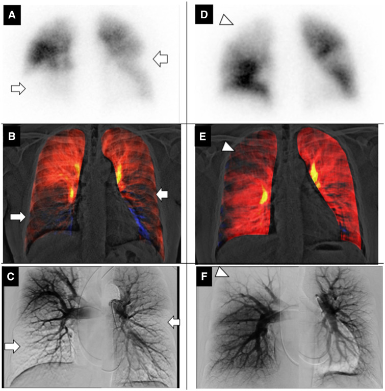
A 46-year-old man with chronic thromboembolic pulmonary hypertension (CTEPH) was referred to our hospital to undergo pulmonary endarterectomy (PEA). Tc-99m MAA lung perfusion scintigraphy showed multiple perfusion defects in the bilateral lung field (Panel A, arrows). Right heart catheterization including pulmonary angiography revealed pulmonary hypertension (mean PAP 31 mmHg and PVR 5.2 Woods Unit) and multiple stenoses/obstructions of pulmonary arteries. Pulmonary circulation image created from dynamic digital radiography captured by a flat-panel detector (AeroDR fine, KONICAMINOLTA, Japan) and a conventional X-ray system (RADspeed Pro, SHIMADZU, Japan) demonstrated lung perfusion defects (Panel B and Supplementary material online, Videos S1), which are similar to the lung perfusion scintigraphy and pulmonary angiography (Panels A and C). After PEA, his symptoms and pulmonary hypertension were improved (mean PAP 15 mmHg and PVR 0.8 Woods Unit). Post-operative pulmonary circulation image (Panel E and Supplementary material online, Videos S2) demonstrated improvement of lung perfusion in bilateral lower lung fields with a relative decrease in right upper lung field, which is so-called ‘vascular steal’ (Panels D–F, arrowheads).
Dynamic digital radiography is a cineradiography, based on conventional X-ray technology. It can be performed along with chest radiography and is simple in operation. Pulmonary circulation image can visualize small changes in pixel value representing the changes in pulmonary circulation during a breath-holding without the contrast media. It takes just 7–10 s for scanning and 1 min for image analysis using a prototype workstation (KONICAMINOLTA, Japan). Dynamic digital radiography has great potential to be an easy and non-invasive diagnostic tool for CTEPH. Furthermore, required radiation exposure is one-fifth when compared with lung perfusion scintigraphy (0.15 mSv vs. 0.8 mSv). This is the first demonstration of dynamic digital radiography for pulmonary circulation analysis in CTEPH.
Supplementary material is available at European Heart Journal online.
Supplementary Material
Associated Data
This section collects any data citations, data availability statements, or supplementary materials included in this article.


