

Abstract
Background:
The authors present three cases of high-level athletes with successful return to competitive collegiate athletics following distal femoral osteotomy for knee lateral compartment overload.
Conclusion:
Distal femoral varus osteotomy (DFO) is used to treat valgus knee malalignment and to offload the lateral knee compartment in the setting of symptomatic cartilage or meniscus pathology. DFO can be considered a viable treatment for collegiate athletes, with satisfactory outcomes and ability to return to sport participation at pre-injury functional levels.
Level of Evidence: IV
Keywords: sport, athletes, athlete, collegiate, youth, adolescent, DFO, distal femoral varus osteotomy, valgus, knee, malalignment, medial, tear, meniscus, menisectomy, cartilage, lateral compartment, osteotomy, outcomes, alignment, return to sport
Introduction
Lateral meniscus tears are more common among youth athletes than medial tears and often result in partial to subtotal meniscectomy. This can occur with, or subsequently result in, injury or degeneration of the articular cartilage in the lateral compartment.1 In the young active patient this can be a difficult clinical situation. Meniscus transplant is generally not recommended in patients attempting to return to sport2,3 and articular cartilage restoration in the athlete is challenging.4 It is generally agreed that mechanical axis malalignment accentuates knee symptoms related to meniscus and cartilage pathology. Correcting malalignment can offload a symptomatic knee compartment and potentially enhance the ability of an athlete to return to higher level activities. DFO is indicated for symptomatic valgus knee malalignment or to offload the lateral compartment in conjunction with cartilage restoration surgery.5,6
However, there is a paucity of literature reporting return to sport (RTS) following DFO for the treatment of isolated lateral compartment symptoms related to cartilage and meniscus pathology with valgus deformity. To our knowledge, no prior study has reported return to high-level collegiate sports following DFO.7,8 We present a case series of three patients who returned to collegiate contact sports following DFO at preoperative performance levels. All DFO surgeries were performed to shift the weight bearing axis out of the lateral compartment to the medial tibial spine.
The patients were informed that data regarding these cases would be collected and submitted for publication, and all provided consent.
Clinical Case Summaries
Case 1
A 17-year-old high-level high school basketball player presented for evaluation of right knee pain six months following a partial lateral meniscectomy for a radial meniscal tear sustained during basketball practice. He complained of lateral knee pain, swelling, catching and locking, exacerbated with activity. Standing radiographs revealed right lower extremity valgus alignment of 8° (Figure 1). MRI demonstrated a recurrent lateral meniscal tear with propagation. The patient was indicated for repeat diagnostic arthroscopy, partial meniscectomy, and DFO.
Figure 1.

Long Leg AP showing the right mechanical axis measures 8° valgus and the left mechanical axis measuring 4° valgus.
At the time of surgery, diagnostic arthroscopy revealed diffuse synovitis, several small cartilaginous loose bodies and a degenerative appearing horizontal tear in the mid-body of the lateral meniscus in the same area as his prior partial meniscectomy. There was also grade 2 chondromalacia of the lateral femoral condyle directly above the area of the meniscal injury and grade 1-2 early chondromalacia of the lateral tibial plateau. A partial synovectomy and chondroplasty of the lateral femoral condyle were performed with partial meniscectomy to stable and viable meniscus tissue. A DFO was then performed using a 15-mm opening wedge Arthrex Puddu plate, fixed with bicortical 4.5 mm fully threaded screws proximally, and 6.5 mm screws distally, with femoral head allograft placed into the opening wedge defect.
Postoperatively, the patient was placed in a hinged knee brace locked in full extension when upright and allowing 0-90° motion while seated. He was maintained non-weightbearing for six weeks (Figure 2) and then progressed to weightbearing as tolerated (WBAT) in the brace until 10 weeks. He continued formal physical therapy for five months, and at 10 months, returned to high-level basketball to conclude his high school basketball career as an elite-level recruit. He obtained a scholarship to play Division I collegiate basketball as a starter, completing two full seasons without limitations, and continues to play Division I collegiate basketball. After 1-year of clinical follow-up, the patient reported no further symptoms.
Figure 2.

Long Leg AP of R leg showing neutral alignment after distal femoral osteotomy 6 weeks postoperatively.
Case 2
A 19-year-old Division I collegiate women’s basketball player with history of prior ACL reconstruction with a hamstring autograft and two previous partial lateral meniscectomies presented 2 years following ACL reconstruction with knee pain. Examination demonstrated a moderate effusion with lateral joint line tenderness and a stable ligamentous exam. MRI revealed further lateral meniscus tear with a displaced fragment in the lateral gutter, and a small focal chondral injury on the lateral femoral condyle; ACL graft was found to be intact. Standing radiographs demonstrated valgus alignment with early arthritic changes in the lateral compartment. The patient tried conservative management with use of a lateral unloader brace for three months, but continued to experience recurrent lateral knee pain, locking, and swelling. She was indicated for arthroscopy and DFO.
Diagnostic arthroscopy revealed only 25% of the lateral meniscus remained with grade II chondromalacia on the tibial plateau and lateral femoral condyle; debridement of the meniscal tear was done and microfracture was performed on the lateral femoral condyle. Surgeons then proceeded with DFO, using a 10-mm opening wedge Arthrex Puddu plate and femoral head allograft. The procedure was complicated by fracture propagation through the medial cortex of the femur, and an additional 3.5mm locking plate was placed at the distal medial femoral cortex (Figure 3) to augment fixation.
Figure 3.

Left lower extremity before (left) and 6 weeks after (right) distal femoral osteotomy, with left mechanical axis demonstrating neutral alignment.
Postoperatively, the patient was maintained non-weight bearing for six weeks in a hinged knee brace locked in extension for ambulation and 0-90° while seated. She then progressed to toe-touch weight bearing for two weeks until progressing to WBAT in the brace until three months postoperatively. The patient was cleared for full return to activity without the brace at nine months postoperatively. She played Division I basketball during her freshman season prior to undergoing DFO. She subsequently underwent hardware removal at nine months postoperatively and was released to full sporting activities without limitations. The patient transferred schools and was able to complete a season of Division II collegiate basketball as a starting player (Figure 4). After 4-years of clinical follow-up, radiographs demonstrated stable alignment and the patient was asymptomatic.
Figure 4.

Long Leg AP and Lateral Left X-ray 4 obtained four years after left distal femur opening wedge osteotomy and hardware removal.
Case 3
A 21-year-old woman’s collegiate Division I basketball player with a history of two prior left knee arthroscopies with partial lateral menisectomy presented with 1.5 years of left lateral knee pain and swelling nonresponsive to conservative measures. Standing radiographs demonstrated 8° of valgus alignment (Figure 5) and MRI revealed a chondral defect on the lateral femoral condyle. The patient was indicated for a DFO.
Figure 5.
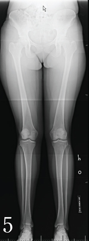
Long Leg AP. There is 3° of valgus angulation of the mechanical axis on the right and 2° of valgus angulation on the left.
DFO was performed using a 7.5-mm Puddu plate (Arthrex) for fixation, with Osferion bone grafting. The patient was placed in a hinged knee brace postoperatively, locked in extension while up, with 0-90° degrees motion seated, and was maintained non-weight bearing for five weeks. She progressed to WBAT at six weeks postoperatively and began formal physical therapy. At six months, the patient returned to Division I collegiate basketball without restrictions. She continued her career as a collegiate basketball player, and returned to play the second half of her final collegiate season (Figure 6). After 4-years of clinical follow-up, radiographs demonstrated stable alignment and the patient was asymptomatic.
Figure 6.

Long Leg AP and Lateral Left obtained four years after left distal femur opening wedge osteotomy showing stable alignment.
Discussion
Lateral meniscectomy in the setting of valgus knee alignment in young athletes poses a risk for subsequent lateral knee compartment articular cartilage degeneration.9 The three clinical cases presented in this report all involved prior lateral meniscectomy in the setting of valgus malalignment of the knee. Although not always feasible depending on the type of meniscus tearing present, meniscal repair should be considered whenever it is a viable alternative to lateral meniscectomy, especially among youth athletes.10 Repair with meniscus healing can provide long term cartilage protection, with reported failure rates of 6-28%,11 often less in adolescent populations.12 Lateral meniscectomy is often the appropriate treatment given the meniscus tear pattern. However, this predisposes the knee to higher risk of articular cartilage wear and damage, especially when valgus mechanical alignment is present.
DFO is a treatment option for younger and more active patients with a symptomatic lateral knee compartment.13 DFO offers the opportunity to offload damaged cartilage and meniscus.14 In the setting of valgus alignment and lateral meniscus insufficiency, DFO is one of the few treatment options for the athletic patient who, due to high activity level, is not indicated for meniscus transplant and may also be performed with cartilage restoration surgery.3 Limited data exists delineating rates of return to high-level athletics following DFO.
Return to sport has previously been assessed after high tibial osteotomy (HTO), demonstrating RTS rates of 86-90%.15,16 However, high impact activities such as soccer, jogging, and tennis had a reduced RTS compared to low-impact activities such as swimming or cycling. In a systematic review of 19 studies that examined return to work and sport after HTO, 13 studies reported return to the same or higher level of sport in 378 patients, with 7/13 patients (54%) returning to sport at an elite level.17
Active patients with valgus deformity have been shown to benefit from DFO to decrease pain and regain function.13 Drextler et al. documented survivorship for distal femoral varus osteotomy combined with fresh osteochondral allograft, with results of good or excellent outcomes for most patients and low conversion rates to TKA.14 RTS following DFO has been reported at rates of 58-80%, at a mean of 11 months postoperatively.7,18 Previous literature has reported a 77% overall RTS rate, with 50% RTS by 15 weeks and 71% RTS by 6 months.8 However, few studies in the literature have assessed RTS at the elite or collegiate level.
Conclusion
Return to sport at the collegiate level following DFO is feasible with mid-term follow-up. Further studies are warranted to assess RTS in additional sports and contact activities using standardized measure of sport level, intensity, frequency, and duration. Meniscal preservation should be attempted for repairable lateral meniscus injuries in young athletes, especially those with pathologic overloading of the lateral compartment.
References
- 1.Lee CR, Bin SI, Kim JM, Lee BS, Kim NK. Arthroscopic partial meniscectomy in young patients with symptomatic discoid lateral meniscus: an average 10-year follow-up study. Archives of Orthopaedic and Trauma Surgery. 2018;138(3):369–376. doi: 10.1007/s00402-017-2853-1. [DOI] [PubMed] [Google Scholar]
- 2.Lee YS, Lee OS, Lee SH. Return to Sports After Athletes Undergo Meniscal Surgery: A Systematic Review. Clinical Journal of Sport Medicine. 9000;Publish Ahead of Print. [DOI] [PubMed]
- 3.Samitier G, Alentorn-Geli E, Taylor DC, et al. Meniscal allograft transplantation. Part 2: systematic review of transplant timing, outcomes, return to competition, associated procedures, and prevention of osteoarthritis. Knee Surgery, Sports Traumatology, Arthroscopy. 2015;23(1):323–333. doi: 10.1007/s00167-014-3344-3. [DOI] [PubMed] [Google Scholar]
- 4.Murray IR, Benke MT, Mandelbaum BR. Management of knee articular cartilage injuries in athletes: chondroprotection, chondrofacilitation, and resurfacing. Knee surgery, sports traumatology, arthroscopy: official journal of the ESSKA. 2016;24(5):1617–1626. doi: 10.1007/s00167-015-3509-8. [DOI] [PubMed] [Google Scholar]
- 5.Jannik Frings MK, Ralph Akoto, Peter Wohlmuth, Karl-Heinz Frosch. Combined distal femoral osteotomy (DFO) in genu valgum leads to reliable patellar stabilization and an improvement in knee function. Knee Surg Sports Traumatol Arthros. 2018. pp. 1–10. [DOI] [PubMed]
- 6.Rosso F, Margheritini F. Distal femoral osteotomy. Current reviews in musculoskeletal medicine. 2014;7(4):302–311. doi: 10.1007/s12178-014-9233-z. [DOI] [PMC free article] [PubMed] [Google Scholar]
- 7.Voleti PB, Wu IT, Degen RM, Tetreault DM, Krych AJ, Williams RJ. Successful Return to Sport Following Distal Femoral Varus Osteotomy. CARTILAGE. 2017. p. 1947603517743545. [DOI] [PMC free article] [PubMed]
- 8.Hoorntje A, van Ginneken BT, Kuijer PPFM, et al. Eight respectively nine out of ten patients return to sport and work after distal femoral osteotomy. Knee surgery, sports traumatology, arthroscopy : official journal of the ESSKA. 2018. [DOI] [PMC free article] [PubMed]
- 9.Beaufils P, Pujol N. Management of traumatic meniscal tear and degenerative meniscal lesions. Save the meniscus. Orthopaedics & Traumatology: Surgery & Research. 2017;103(8, Supplement):S237–S244. doi: 10.1016/j.otsr.2017.08.003. [DOI] [PubMed] [Google Scholar]
- 10.Nepple JJ, Dunn WR, Wright RW. Meniscal repair outcomes at greater than five years: A systematic literature review and meta-analysis. Journal of Bone and Joint Surgery - Series A. 2012;94(24):2222–2227. doi: 10.2106/JBJS.K.01584. [DOI] [PMC free article] [PubMed] [Google Scholar]
- 11.Pujol N, Lorbach O. Meniscal repair: Results. Surgery of the Meniscus. 2016:343–355. [Google Scholar]
- 12.Tagliero AJ, Desai VS, Kennedy NI, et al. Seventeen-Year Follow-up After Meniscal Repair With Concomitant Anterior Cruciate Ligament Reconstruction in a Pediatric and Adolescent Population. The American Journal of Sports Medicine. 2018;46(14):3361–3367. doi: 10.1177/0363546518803934. [DOI] [PubMed] [Google Scholar]
- 13.Backstein D, Morag G, Hanna S, Safir O, Gross A. Long-Term Follow-Up of Distal Femoral Varus Osteotomy of the Knee. The Journal of Arthroplasty. 2007;22(4, Supplement):2–6. doi: 10.1016/j.arth.2007.01.026. [DOI] [PubMed] [Google Scholar]
- 14.Drexler M, Gross A, Dwyer T, et al. Distal femoral varus osteotomy combined with tibial plateau fresh osteochondral allograft for post-traumatic osteoarthritis of the knee. Knee Surgery, Sports Traumatology, Arthroscopy. 2015;23(5):1317–1323. doi: 10.1007/s00167-013-2828-x. [DOI] [PubMed] [Google Scholar]
- 15.Faschingbauer M, Nelitz M, Urlaub S, Reichel H, Dornacher D. Return to work and sporting activities after high tibial osteotomy. International orthopaedics. 2015;39(8):1527–1534. doi: 10.1007/s00264-015-2701-2. [DOI] [PubMed] [Google Scholar]
- 16.Saragaglia D, Rouchy RC, Krayan A, Refaie R. Return to sports after valgus osteotomy of the knee joint in patients with medial unicompartmental osteoarthritis. International orthopaedics. 2014;38(10):2109–2114. doi: 10.1007/s00264-014-2435-6. [DOI] [PubMed] [Google Scholar]
- 17.Ekhtiari S, Haldane CE, de SA D, Simunovic N, Musahl V, Ayeni OR. Return to Work and Sport Following High Tibial Osteotomy: A Systematic Review. JBJS. 2016;98(18):1568–1577. doi: 10.2106/JBJS.16.00036. [DOI] [PubMed] [Google Scholar]
- 18.de Carvalho LH, Temponi EF, Soares LFM, Gonçalves MBJ, Costa LP. Physical activity after distal femur osteotomy for the treatment of lateral compartment knee osteoarthritis. Knee Surgery, Sports Traumatology, Arthroscopy. 2014;22(7):1607–1611. doi: 10.1007/s00167-012-2316-8. [DOI] [PubMed] [Google Scholar]