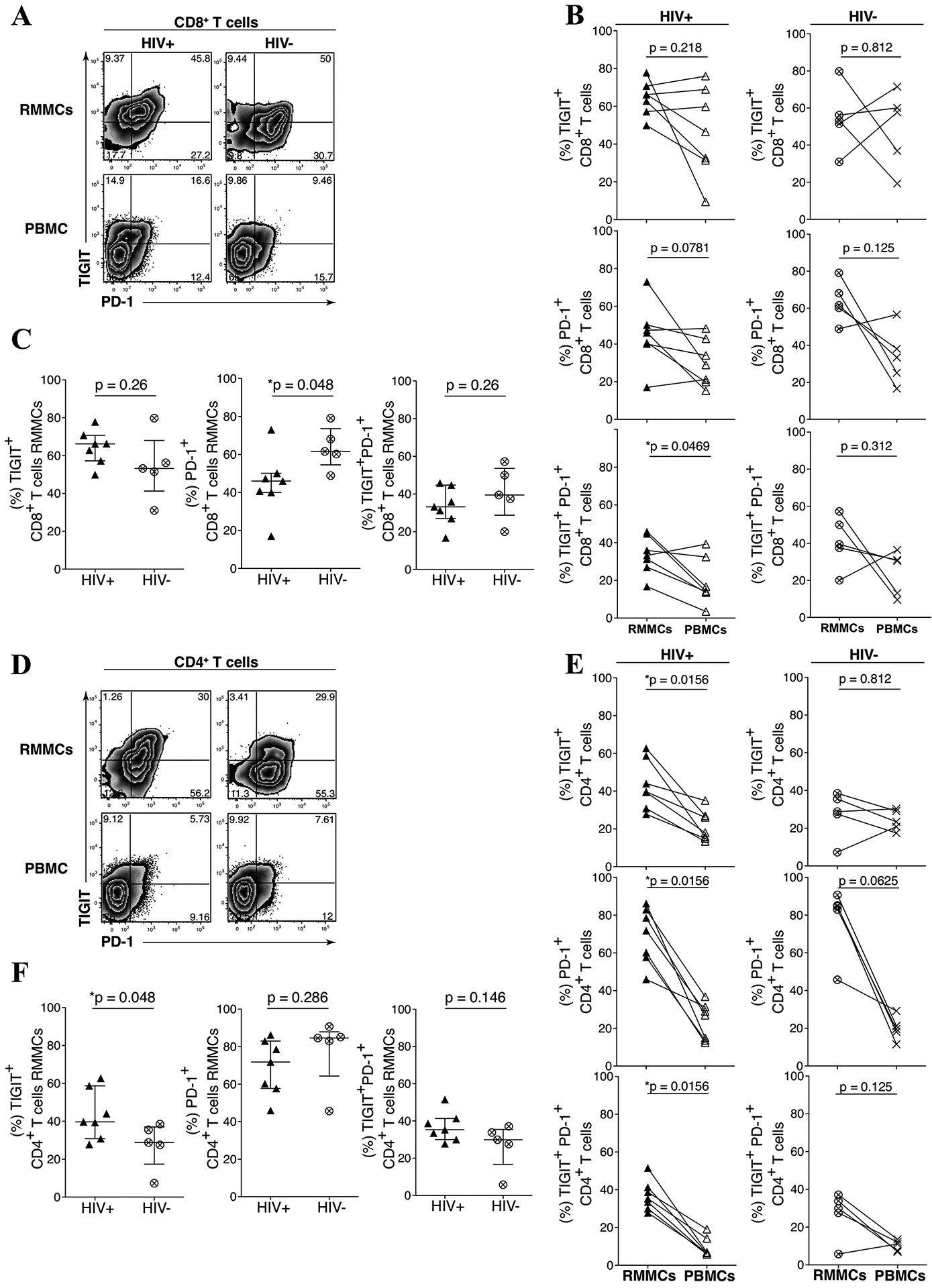
Figure 2. TIGIT and PD-1 are upregulated on mucosal mononuclear T cells derived from rectosigmoid biopsies.

Rectosigmoid mucosal mononuclear cells (RMMCs) and peripheral blood mononuclear cells (PBMCs) from cART-suppressed UH HIV-infected and HIV-uninfected individuals were stained for viability and antibodies against CD3, CD4, CD8, TIGIT and PD-1. Representative flow cytometry plots of matched RMMCs and PBMCs gated on live CD3+ lymphocytes, from representative UH HIV-infected (A) and HIV-uninfected (D) individual. (B) Compiled data comparing the differences of TIGIT and PD-1 expression on CD8+ T cells from RMMCs and matched PBMCs of UH HIV-infected (n = 7) and HIV-uninfected (n = 5) individuals. (C) Compiled data of PD-1+, TIGIT+, and PD-1+ TIGIT+ CD8+ T cells from RMMCs from UH HIV-infected and HIV-uninfected individuals. (E) Compiled data comparing the differences of TIGIT and PD-1 expression on CD4+ T cells from RMMCs and matched PBMCs of UH HIV-infected (n = 7) and HIV-uninfected (n = 5) individuals. (F) Compiled data of PD-1+, TIGIT+, and PD-1+ TIGIT+ CD4+ T cells from RMMCs from UH HIV-infected and HIV-uninfected individuals. The p-values were calculated using Wilcoxon matched-pairs signed ranked test for matched pairs and Mann-Whitney U test for group comparison.
