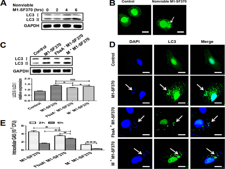
FIG 2.
Evaluation of autophagy induction by M1 GAS strain SF370 proteins. (A and B) Hep2 cells were infected with nonviable M1 GAS strain SF370 for the indicated times, and LC3 expression was assessed by Western blotting (A) or confocal laser scanning microscopy (B). Scale bar, 25 μm. (C to E) Hep2 cells were infected with WT M1 GAS strain SF370, FbaA−M1 GAS strain SF370, or M−M1 GAS strain SF370. (C) LC3II expression was analyzed by Western blotting. (D) The colocalization of fluorescent EGFP-LC3 puncta and M1 GAS strain SF370 (DAPI staining) was observed by confocal laser scanning microscopy. Scale bar, 25 μm. (E) Numbers of CFU of WT M1 GAS strain SF370, FbaA−M1 GAS strain SF370, and M−M1 GAS strain SF370 within Hep2 cells were counted after infection for the indicated times. *, P < 0.05; **, P < 0.01; ***, P < 0.001.

