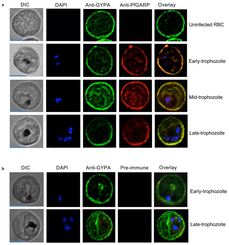
Extended Data Figure 6 ∣. PfGARP co-localizes with glycophorin A to the exofacial surface of trophozoite infected RBC membranes.
a) Uninfected and infected RBCs were probed with rabbit anti-glycophorin A (green) and recombinant DNA vaccine immunized mouse anti-PfGARP (red) and counterstained with 4′,6′-diamidino-2-phenylindole (DAPI) to label parasite nuclei. PfGARP is detected only in trophozoite-infected RBCs and co-localizes with human glycophorin A on the RBC membrane. Scale bars for all the images are 5 μm. b) Neither early nor late-trophozoite-infected RBCs label when probed with pre-immune mouse sera. Scale bar for all the images is 5 μm. Panels A and B are representative of 3 independent experiments.

