Abstract
Methemoglobinemia is a rare disorder of the blood in which there is an increase in methemoglobin, which occurs when hemoglobin is present in the oxidized form. Methemoglobin impairs hemoglobin’s ability to transport oxygen, produces functional anemia, and leads to tissue hypoxia. We report the successful management of a case of refractory hypoxia due to acutely acquired methemoglobinemia in a patient undergoing treatment for coronavirus disease 2019 (COVID-19) pneumonia. The cause of methemoglobinemia in this patient remains unknown. Hypoxia and methemoglobinemia did not respond to methylene blue and required administration of packed red blood cell transfusions.
Methemoglobinemia is a rare condition in which the iron in hemoglobin is present in the ferric (Fe3+), not the ferrous (Fe2+) state of normal hemoglobin, which renders red blood cells unable to release oxygen to tissues, produces functional anemia, and leads to tissue hypoxia.1 Methemoglobinemia is present if methemoglobin concentrations in the blood exceed the normal physiological levels of 1%–2%.2
Methemoglobinemia may be congenital due to deficiencies of methemoglobin reductase or structural abnormalities of hemoglobin. Methemoglobinemia may be acquired, usually secondary to exposure to drugs or chemicals that oxidize hemoglobin and occasionally from pathologic conditions, such as sepsis and sickle cell crisis. Medications rarely produce clinically significant methemoglobinemia when given to a healthy adult in therapeutic doses. Some oxidizing drugs require a biochemical transformation in the liver to toxic metabolites after exposure, and methemoglobinemia occurs several hours later.2 The risk of methemoglobinemia associated with oxidizing drug use increases in elderly patients with medical comorbidities such as renal failure, anemia, and human immunodeficiency virus.3 Novel coronavirus proteins can alter hemoglobin structure which may interfere with the red blood cell’s ability to carry oxygen.4
We report a case of refractory hypoxia from acquired methemoglobinemia in a critically ill patient with coronavirus disease 2019 (COVID-19), which was not managed successfully with only methylene blue but required packed red blood cell transfusion. The Health Insurance Portability and Accountability Act authorization was obtained from the patient for publication of this case report. This manuscript adheres to the applicable enhancing the quality of and transparency of health research network guidelines.
CASE DESCRIPTION
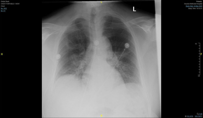
A 74-year-old retired African American man with a medical history significant for prostate cancer, hypertension, and hyperlipidemia, presented to our facility with a 7-day history of fevers, nonproductive cough, and progressively worsening shortness of breath. He had never traveled to China nor had contact with a known COVID-19 patient. On examination, he had tachypnea with oxygen saturation measured by pulse oximetry (Spo2) of 90% on room air. Laboratory testing revealed a negative respiratory pathogen panel and nasopharyngeal swab positive for severe acute respiratory syndrome coronavirus-2 on polymerase chain reaction assay. The chest radiograph (Figure) demonstrated bilateral perihilar and right lower lobe opacities. The patient was admitted to the floor with a diagnosis of COVID-19–associated pneumonia and treated with supplemental oxygen, oral azithromycin, and hydroxychloroquine (Table). His respiratory status worsened on hospital day 5 with increased oxygen requirements. He was then transferred to an intensive care unit (ICU) with acute hypoxic respiratory failure and acute respiratory distress syndrome for which he required intubation and mechanical ventilation. His treatment regimen included lopinavir-ritonavir, ribavirin, and tocilizumab (Table), and he was not a candidate for our hospital’s remdesivir therapy trial. Corynebacterium pneumonia, acute renal failure, septic shock, and cytokine release inflammatory syndrome further complicated his hospital course. The patient intermittently required renal replacement therapy. He was treated with appropriate intravenous antibiotics, a second dose of tocilizumab, thiamine, hydrocortisone, ascorbic acid (vitamin C), and a brief norepinephrine infusion. Over subsequent days, his respiratory status improved with a normal arterial oxygen tension (Pao2) on adaptive support ventilation mode of ventilator with positive end-expiratory pressure (PEEP) of 8 mm Hg and 40% fraction of inspired oxygen (Fio2). He cleared his pneumonia, and his renal function began to recover. Due to excessive secretions and frequent need for suctioning, extubation was not indicated.
Table.
Timeline of COVID-19 Treatmentsa
| Medication | Dosage | Duration of Treatment (d) | HD Completed (d) |
|---|---|---|---|
| Hydroxychloroquine | 400 mg twice a day | 5 | 7 |
| Ribavirin | 400 mg twice a day | 10 | 14 |
| Lopinavir-ritonavir | 400–100 mg twice a day | 10 | 13 |
| Tocilizumab | 400 mg once | 1 (2 treatments) | 4, 17 |
Abbreviations: COVID-19, coronavirus disease 2019; HD, hospital day.
The Table compares the date of diagnosis of methemoglobinemia at HD 15 and COVID-19 treatment duration and completion.
Figure.

Chest radiograph of the patient on initial presentation showing bilateral perihilar and right lower lobe opacities.
On hospital day 15, he developed severe hypoxia with Spo2 80%–90% with appropriate waveforms on the monitor. Hypoxia did not improve despite the transition to assist control ventilation with PEEP of 12 mm Hg and 100% Fio2. Arterial blood gas analysis revealed elevated Pao2 of 311 mm Hg and arterial oxygen saturation (Sao2) of 100%, which were discordant with Spo2 80%–90%. The chest radiograph was unchanged from the previous day. Methemoglobin and carboxyhemoglobin were checked by cooximeter and were elevated (6.3% and 3.2%, respectively). Our ICU pharmacy staff reviewed the patient’s medication list extensively, and he was not receiving any agents known for inducing methemoglobinemia. The patient was already on intravenous ascorbic acid, and the dose was continued (1500 mg every 8 hours). Intravenous hydroxocobalamin (5 g) and intravenous methylene blue (1.5 mg/kg) were given with minimal improvement in Spo2. The carboxyhemoglobin concentration normalized, but the methemoglobin concentration continued to rise with a peak at 15.9% (normal 0%–1%). Another dose of intravenous methylene blue (1.5 mg/kg) was administered. Hematology consultants recommended the continuation of methylene blue as well as red blood cell transfusion with consideration of red blood cell exchange if hypoxia persisted. Fortunately, the Spo2 improved to >90% with 2 units of packed red blood cells. Methemoglobin levels declined to 2%–4%, and hemoglobin increased from 7.8 to 10.4 g/dL.
After a prolonged ICU course (29 days), the patient slowly recovered. He was transitioned from continuous renal replacement therapy to intermittent hemodialysis on hospital day 23. He was extubated on day 27 and discharged to a long-term acute care facility 4 days later.
DISCUSSION
Methemoglobinemia is a rare condition characterized by increased methemoglobin levels, which can lead to tissue hypoxia and anoxia.1 The most common cause of acquired methemoglobinemia is exposure to oxidizing agents.2 Drugs reported to cause methemoglobinemia include nitrate derivatives (nitrates salt, nitroglycerin), nitrite derivatives (nitroprusside, amyl nitrite, nitric oxide), sulfonamides, dapsone, phenacetin, phenazopyridine, and some local and topical anesthetics (lidocaine, prilocaine).3 Methemoglobin levels may be elevated in patients with sepsis due to release of nitric oxide, which converts to nitrate and subsequently to methemoglobin.5 However, in that specific report, patients did not have methemoglobinemia above 2%. Moreover, patients with underlying cardiac, pulmonary, and hematologic disease, cirrhosis, human immune deficiency virus infection, and renal failure on hemodialysis are more susceptible to methemoglobinemia.3
The etiology of methemoglobinemia, in this case, remains unknown. Several medications (acetaminophen, hydroxychloroquine, tocilizumab, and norepinephrine) could have been the cause. However, the timing of administration of these medications does not coincide with the onset of symptoms (Table). His episode of septic shock due to corynebacterium pneumonia had also resolved with the completion of the antibiotic course. The patient had been on renal replacement therapy for almost 2 weeks before his diagnosis of methemoglobinemia, which makes renal failure an unlikely culprit. He had no history of sickle cell disease, which can cause methemoglobinemia. In this case of methemoglobinemia, we were unable to identify the cause, which raised the possibility of coronavirus-related hemoglobin changes that may have contributed. There is no clinical evidence in the literature to support this possibility. However, a recent nonclinical and nonbiological study from China elucidates this point and explains that some of the proteins of the severe acute respiratory syndrome coronavirus-2 bind to the porphyrin of hemoglobin to change its oxygen-binding capacity and decrease oxygen release in the tissues.4
Clinical features of methemoglobinemia are variable based on the concentration of methemoglobin and more severe in patients with preexisting conditions (eg, anemia, respiratory and cardiovascular diseases that compromise oxygenation of tissues).2,3 In a healthy person, the clinical features are pallor, fatigue, weakness, tachycardia, tachypnea, and cyanosis, which may be clinically evident with methemoglobin as low as 10%. As the percentage of methemoglobinemia approaches 20%, the patient may experience anxiety, light-headedness, and headaches. At methemoglobin concentrations of 30%–50%, there may be tachypnea, confusion, and loss of consciousness. If methemoglobin approaches 50%, patients are at risk for seizures, dysrhythmias, metabolic acidosis, and coma.1,6 Since our patient was intubated and sedated, the only clinical indicator was hypoxia refractory to maximum oxygen therapy.
The Pao2 measures by arterial blood gas analysis may remain remarkably normal and may falsely be elevated in methemoglobinemia.7 As methemoglobin increases from 2% to 30%, Spo2 falls from normal (around 98%) to around 85%, but no further fall in oxygen saturation occurs if methemoglobin rises above 30%–35%.8 Cooximetry is the only reliable method of measuring methemoglobin concentration to confirm a diagnosis of methemoglobinemia.9 Most modern blood gas analyzers have an incorporated cooximeter, which allows arterial blood to be examined at multiple wavelengths spectrophotometrically. All hemoglobin species have characteristic absorbance spectra. Therefore, cooximetry identifies and quantifies all hemoglobin species, including methemoglobin. Cooximetry also calculates oxygen saturation and is a more reliable method of assessing oxygen saturation in patients with methemoglobin than either pulse oximetry or blood gas analysis.9
Treatment of methemoglobinemia includes removal of the inciting agent and consideration of treatment with an antidote. Recommendations include methylene blue (1.5 mg/kg), ascorbic acid (1500 mg every 8 hours), and high-flow oxygen.10 In rare cases of refractory hypoxia from methemoglobinemia, red blood cell transfusions and red blood cell exchange have been successful in some case reports.11
In conclusion, critically ill patients with COVID-19 may develop severe refractory hypoxia from an unexplained cause of methemoglobinemia, which can be challenging to diagnose. One needs to have a high index of suspicion for methemoglobinemia in a critically ill patient with COVID-19 with persistent hypoxia and discrepancies between Spo2, Pao2, and Sao2. The mechanism of methemoglobinemia in COVID-19 disease needs further research, but we demonstrated that successful management might require red blood cell transfusions.
DISCLOSURES
Name: Hina Faisal, MD, MRCS.
Contribution: This author is a primary and corresponding author who identified this case, performed an extensive literature review, obtained consent from a patient for publication, helped with the case description, gathering clinical information, writing drafts of the paper and revised manuscript critically for valuable intellectual content. This author is also responsible for communication with the journal during the manuscript submission, peer review, and publication process.
Name: Alexi Bloom, MD.
Contribution: This author helped as a coauthor in the necessary literature review, helped with gathering clinical information, writing the manuscript draft, and revision.
Name: A. Osama Gaber, MD, FACS.
Contribution: This senior author helped write and edit the original draft of the manuscript.
This manuscript was handled by: BobbieJean Sweitzer, MD, FACP.
GLOSSARY
- COVID-19
- coronavirus disease 2019
- Fio2
- fraction of inspired oxygen
- HD
- = hospital day
- ICU
- intensive care unit
- Pao2
- arterial oxygen tension
- PEEP
- positive end-expiratory pressure
- Sao2
- arterial oxygen saturation
- Spo2
- oxygen saturation measured by pulse oximetry
Funding: None.
The authors declare no conflicts of interest.
REFERENCES
- 1.Wright RO, Lewander WJ, Woolf AD. Methemoglobinemia: etiology, pharmacology, and clinical management. Ann Emerg Med. 1999; 34:646–656 [DOI] [PubMed] [Google Scholar]
- 2.Skold A, Cosco DL, Klein R. Methemoglobinemia: pathogenesis, diagnosis, and management. South Med J. 2011; 104:757–761 [DOI] [PubMed] [Google Scholar]
- 3.Alanazi MQ. Drugs may be induced methemoglobinemia. J Hematol Thrombo Dis. 2017; 270:1–5 [Google Scholar]
- 4.Wenzhong L, Li H. COVID-19: attacks the 1-beta chain of hemoglobin and captures the porphyrin to inhibit human heme metabolism. ChemRxiv. 2020. Preprint. 10.26434/chemrxiv.11938173.v7 [DOI]
- 5.Ohashi K, Yukioka H, Hayashi M, Asada A. Elevated methemoglobin in patients with sepsis. Acta Anaesthesiol Scand. 1998; 42:713–716 [DOI] [PubMed] [Google Scholar]
- 6.Wilkerson RG. Getting the blues at a rock concert: a case of severe methaemoglobinaemia. Emerg Med Australas. 2010; 22:466–469 [DOI] [PubMed] [Google Scholar]
- 7.Haymond S, Cariappa R, Eby CS, Scott MG. Laboratory assessment of oxygenation in methemoglobinemia. Clin Chem. 2005; 51:434–444 [DOI] [PubMed] [Google Scholar]
- 8.Watcha MF, Connor MT, Hing AV. Pulse oximetry in methemoglobinemia. Am J Dis Child. 1989; 143:845–847 [DOI] [PubMed] [Google Scholar]
- 9.Brunelle JA, Degtiarov AM, Moran RF, Race LA. Simultaneous measurement of total hemoglobin and its derivatives in blood using CO-oximeters: analytical principles; their application in selecting analytical wavelengths and reference methods; a comparison of the results of the choices made. Scand J Clin Lab Invest Suppl. 1996; 224:47–69 [DOI] [PubMed] [Google Scholar]
- 10.Rehman HU. Methemoglobinemia. West J Med. 2001; 175:193–196 [DOI] [PMC free article] [PubMed] [Google Scholar]
- 11.Pritchett MA, Celestin N, Tilluckdharry N, Hendra K, Lee P. Successful treatment of refractory methemoglobinemia with red blood cell exchange transfusion. Chest. 2006; 130:294S [Google Scholar]


