Abstract
Necrotizing fasciitis (NF) is a rapidly progressing soft tissue infection with a mortality rate as high as 30% to 50%. However, the incidence rate of NF after liposuction is extremely low. In the current case report, we describe a woman with NF who developed multiple organ dysfunction syndrome (MODS) after fat acquisition. The aim of this paper is to summarize the management of these patients. After debridement and drainage, correction of multiple organ failure, and plastic surgery, the patient’s organ and lower limb functions improved to a normal level. Early diagnosis, early operative treatment, and correction of systemic abnormalities are the keys to successful recovery of patients with NF complicated with MODS after liposuction.
Keywords: Liposuction, breast augmentation, necrotizing fasciitis, multiple organ dysfunction syndrome, mortality, case report
Introduction
Necrotizing fasciitis (NF) is a rapidly progressing soft tissue infection involving mainly superficial fascia and subcutaneous tissues.1,2 The disease is life-threatening and severely painful and has a mortality rate as high as 30% to 50%.3–6 NF can occur after minor surgically induced trauma or slight abrasions or at the base of chronic wounds, and it can even occur spontaneously in normal skin. NF can affect elderly individuals, who often have many other diseases, and can also occur in the young and healthy population.7
The incidence rate of NF after liposuction is extremely low,8,9 and very few cases have been reported worldwide, including in China. We treated a young woman with NF who developed multiple organ dysfunction syndrome (MODS) after fat acquisition and herein present the details of this case.
Case presentation
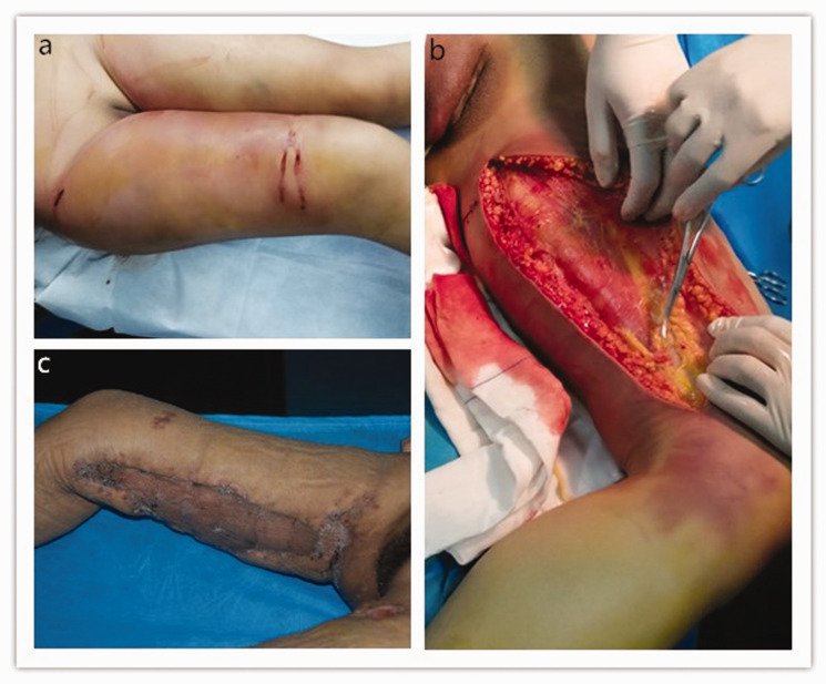
A 30-year-old woman underwent bilateral liposuction on 15 May 2018. Ten hours after the operation, she experienced unbearable pain in the bilateral thigh liposuction sites. Erythema with blisters occurred in the same area. The patient was thereafter admitted to our hospital on 16 May. On physical examination, she was febrile (38.2°C) and had a heart rate of 128 beats/minute. Small scattered incisions from the liposuction process were observed in the bilateral waist and thigh regions. The local skin over the bilateral thighs exhibited swelling and tenderness (Figure 1(a)). Blisters were also observed in the posterior thigh region. After admission, the sutures in the bilateral thigh regions were immediately removed. Additionally, the patient’s urine was black. Initial laboratory analysis revealed leukocytosis (25 × 109 cells/L) and an elevated C-reactive protein level (35 mg/dL). Her serum creatinine level (250 U/L) and blood urea nitrogen level were also elevated. Fasciotomy and drainage of the bilateral thighs were performed in an emergency unit on the same day. She was additionally treated with vancomycin and meropenem as anti-infection measures during the operation. However, her vital signs were unstable, and she was diagnosed with sepsis, renal insufficiency, and shock. She was then transferred to the surgical intensive care unit for continuous renal replacement therapy, anti-infection treatment, blood transfusion, and shock correction.
Figure 1.
(a) On 16 May, the local skin of the bilateral thighs exhibited swelling and tenderness. (b) Degeneration and necrosis of the subcutaneous fat, fascia, and muscle were observed during the operation. (c) The patient was discharged after complete healing of the wound.
On 17 May, a large amount of exudation and local swelling developed in both thighs. Thus, extensive fasciotomy, osteofascial compartment incision, and vacuum sealing drainage (VSD) of the bilateral thigh regions were performed. Degeneration and necrosis of the subcutaneous fat, fascia, and muscle were observed during the operation (Figure 1(b)). The patient’s high-sensitivity troponin level was 0.71 ng/mL. On 24 May, she developed bilateral iliolumbar local skin edema, an obvious inflammatory reaction, and subcutaneous effusion. A thigh necrotizing fasciitis incision, dilatation, and VSD were performed in an emergency unit. The exudate obtained during the operation was sent for bacterial culture testing. Candida dubliniensis was detected in the subcutaneous effusion culture, and Klebsiella pneumoniae was detected in the blood culture. Two weeks after the operation (7 June), the skin necrotizing area in the bilateral iliolumbar regions and thighs tended to be stable. With the patient under general anesthesia, the bilateral iliolumbar and thigh regions were then thoroughly debrided. The fascia and subcutaneous fat exhibited obvious necrosis, and the local tissue lacked the elasticity of normal tissue. After full debridement, the wounds were washed with hydrogen peroxide and normal saline and treated with VSD. The wounds of the bilateral iliolumbar and thigh regions remained stable without further enlargement. Interestingly, growth of relatively fresh granulation tissue was observed.
Debridement and skin grafting of the bilateral iliolumbar regions and thighs were performed on 21 June and 11 July, respectively. The skin grafts survived and the function of both lower limbs recovered well (Figure 1(c)).
The patient eventually developed systemic MODS after fat acquisition. Multiple operations, active treatment in the surgical intensive care unit, and plastic surgery were performed. The patient’s organ function recovered to normal levels after healing.
Discussion
NF is a rare complication after liposuction. Although its incidence is extremely low, it develops rapidly once it has occurred, and its mortality rate is high. The pathophysiology of NF involves the rapid spread of infection in the subcutaneous fat, superficial or deep fascial planes, and overlying skin; this spread is facilitated by bacterial toxins and enzymes, causing vascular occlusion, ischemia, and necrosis.10 Several case reports have described NF following liposuction,10–12 including a few reports published in China.13
NF can occur after minor surgically induced trauma or even slight scratches. It can also occur at the base of a chronic wound or under seemingly intact skin. NF often has a clear source of infectious bacteria, but its source may also be unclear; local bacterial culture is frequently negative. NF after liposuction occurs mostly within a few days after the operation. The keys to successful treatment are early detection and diagnosis, thorough debridement in a timely manner, complete removal of necrotic skin and subcutaneous tissue until viable tissue is reached, and the use of systemic broad-spectrum antibiotics to control the spread of infection.14
NF is characterized by rapidly developing progressive necrosis of the subcutaneous tissue layer. In most cases, it requires clinicians to draw preliminary diagnoses based on clinical manifestations, and the necessary condition for its diagnosis is that the surrounding soft tissues lack normal elasticity and resistance to blunt separation caused by extensive necrosis. Following radical debridement, closure of the remaining wound can pose significant reconstructive challenges.10 In our case, the extent of the affected subcutaneous tissue layer exceeded the necrotic skin boundaries. Repeated debridement was performed to preserve as much of the normal skin tissue as possible and reduce the scope of excision of necrotic tissue. The main purpose of such treatment is to gradually remove the necrotic subcutaneous tissue under the marginal surviving skin. In the present case, the subcutaneous tissue lacunae at the edge of the wound did not completely close and heal until the final debridement and skin grafting. After survival of the skin grafts, residual effusion was present in both the waist and thigh regions. Regular aspiration was performed. One month after the operation, there was no obvious local effusion, and the function of both lower limbs was close to normal. Although the patient was treated in a timely manner after the development of infectious NF, extensive scars remained on the bilateral waist and thigh regions.
In conclusion, when performing liposuction, the operation must be conducted under strict aseptic conditions to minimize the degree of damage to surrounding tissues and thus avoid causing unnecessary harm to the patient. Early diagnosis, early operative treatment, and correction of systemic abnormalities are the keys to a successful recovery of patients who develop NF complicated by MODS after liposuction.
Declaration of conflicting interest
The authors declare that there is no conflict of interest.
Ethics
This study was conducted with approval from the Ethics Committee of Zhengzhou First People’s Hospital and in accordance with the Declaration of Helsinki. Written informed consent was obtained from the patient.
Funding
This research received no specific grant from any funding agency in the public, commercial, or not-for-profit sectors.
ORCID iD
Yonglin Li https://orcid.org/0000-0002-0061-6185
References
- 1.Rüfenacht MS, Montaruli E, Chappuis E, et al. Skin-sparing debridement for necrotizing fasciitis in children. Plast Reconstr Surg 2016; 138: 489e–497e. [DOI] [PubMed] [Google Scholar]
- 2.Horta R, Nascimento R, Silva A, et al. The free-style gluteal perforator flap in the thinning and delay process for perineal reconstruction after necrotizing fasciitis. Wounds 2016; 28: 200–205. [PubMed] [Google Scholar]
- 3.Wladis EJ. Modified wound closure technique in periorbital necrotizing fasciitis. Ophthalmic Plast Reconstr Surg 2015; 31: 242–244. [DOI] [PubMed] [Google Scholar]
- 4.Nolff MC, Meyerlindenberg A. Necrotising fasciitis in a domestic shorthair cat - negative pressure wound therapy assisted debridement and reconstruction. J Small Anim Pract 2015; 56: 281–284. [DOI] [PubMed] [Google Scholar]
- 5.Goh T, Goh LG, Ang CH, et al. Early diagnosis of necrotizing fasciitis. Br J Surg 2013; 101: e119–e125. [DOI] [PubMed] [Google Scholar]
- 6.Vayvada H, Demirdöver C, Menderes A, et al. [Necrotizing fasciitis: diagnosis, treatment and review of the literature]. Ulus Travma Acil Cerrahi Derg 2012; 18: 507. [DOI] [PubMed] [Google Scholar]
- 7.Jones EG, El-Zawahry AM. Curative treatment without surgical reconstruction after perineal debridement of Fournier’s gangrene. J Wound Ostomy Continence Nurs 2012; 39: 98–102. [DOI] [PubMed] [Google Scholar]
- 8.Wähmann M, Wähmann M, Schütz F, et al. Severe Fournier’s gangrene-a conjoint challenge of gynaecology and plastic surgery. J Surg Case Rep 2017; 2017: rjx239. [DOI] [PMC free article] [PubMed] [Google Scholar]
- 9.Marchesi A, Marcelli S, Parodi PC, et al. Necrotizing fasciitis in aesthetic surgery: a review of the literature. Aesthetic Plast Surg 2017; 41: 1–7. [DOI] [PubMed] [Google Scholar]
- 10.Chiang IH, Chang SC, Wang CH. Management of necrotising fasciitis secondary to abdominal liposuction using a combination of surgery, hyperbaric oxygen and negative pressure wound therapy in a patient with burn scars. Int Wound J 2017; 14: 989–992. [DOI] [PMC free article] [PubMed] [Google Scholar]
- 11.Park SY, Jeong WK, Kim MJ, et al. Necrotising fasciitis in both calves caused by Aeromonas caviae following aesthetic liposuction. J Plast Reconstr Aesthet Surg 2010; 63: e695–e698. [DOI] [PubMed] [Google Scholar]
- 12.Sherman JE, Fanzio PM, White H, et al. Blindness and necrotizing fasciitis after liposuction and fat transfer. Plast Reconstr Surg 2010; 126: 1358–1363. [DOI] [PubMed] [Google Scholar]
- 13.Goh T, Goh LG, Ang CH, et al. Early diagnosis of necrotizing fasciitis. Br J Surg 2014; 101: e119–e125. [DOI] [PubMed] [Google Scholar]
- 14.Sarani B, Strong M, Pascual J, et al. Necrotizing fasciitis: current concepts and review of the literature. J Am Coll Surg 2009; 208: 279–288. [DOI] [PubMed] [Google Scholar]


