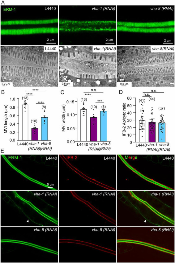
Fig. 6.

V0-ATPase silencing induces microvillus atrophy in C. elegans. (A) Visualization of microvilli by super-resolution (upper panels, 96 h RNAi, ERM-1::GFP) and TEM (lower panels, 72 h RNAi) analysis. Control animals showed the typical intestinal brush border morphology consisting of a dense array of microvilli surrounded by a glycocalyx and stabilized by the terminal web/endotube. Conversely, V0/vha-1(RNAi) worms display a microvillus atrophy, with irregular, shorter and sparse microvilli, a phenotype that is not observed in V1/vha-8-depleted worms. The terminal web/endotube appears normal in all cases. L, lumen; mv, microvilli; tw, terminal web/endotube. (B-C) Measurement of microvilli length (B) and width (C) upon 72 h V0 and V1-ATPase knockdown from TEM images. Data are mean±s.e.m., n=5 microvilli/worm for the number of worms indicated in brackets. (D-E) Effect of V-ATPase silencing on the endotube component IFB-2. V0/vha-1 or V1/vha-8 were silenced in a strain co-expressing endogenously tagged ERM-1::mNG and IFB-2::wSc for 72 h and then imaged. Arrowheads show the basolateral mislocalization of ERM-1. (D) The quantification of the apical/cytoplasmic ratio of IFB-2. Histograms are mean±s.e.m., dots represent individual worms and the total number of worms from two independent experiments is indicated in brackets. In micrographs, worms are at the L4/young adult (72 h RNAi) or adult (96 h RNAi) developmental stage. Scale bars: 0.2 µm or 0.5 µm in A; 5 µm in E. n.s., nonsignificant, ***P<0.001, ****P<0.0001.
