

Abstract
We present the first case of simultaneous development of Graves’ disease and type 1 diabetes during anti‐programmed cell death 1 therapy. A 48‐year‐old man with parotid gland adenocarcinoma and lung metastasis had received five courses of nivolumab. Fourteen days after administration of the sixth course, his casual plasma glucose and hemoglobin A1c levels were 379 mg/dL and 7.2%, respectively. Furthermore, thyrotoxicosis was detected with a blood test. Serum total ketone body and thyroid‐stimulating hormone receptor antibody levels increased, and serum C‐peptide level decreased to 0.01 ng/mL thereafter. Thus, we concluded that he simultaneously developed anti‐programmed cell death 1 therapy‐associated type 1 diabetes and Graves’ disease. Among Japanese patients with autoimmune polyglandular syndrome type III, the frequency of human leukocyte antigen‐DRB1*04:05 is higher in those with both type 1 diabetes and Graves’ disease. Our case had human leukocyte antigen‐DRB1*04:05, which might be associated with the simultaneous development of the two diseases.
Keywords: Anti‐programmed death 1 therapy, Graves’ disease, Type 1 diabetes
We present the first case of simultaneous development of Graves’ disease and type 1 diabetes during anti‐programmed cell death 1 therapy. Among Japanese participants with autoimmune polyglandular syndrome type III, the frequency of human leukocyte antigen‐DRB1*04:05 is higher in those with both type 1 diabetes and Graves’ disease. The present participant carried the human leukocyte antigen‐DRB1*04:05; which might be associated with the simultaneous development of the two diseases.
Introduction
Anti‐programmed cell death 1 (PD‐1) therapy for malignancies causes immune‐related endocrine diseases, including thyroid dysfunction and type 1 diabetes. Among thyroid dysfunctions, hypothyroidism/thyroiditis is usually reported in such patients1, although the development of hyperthyroidism as a result of Graves’ disease is virtually non‐existent2, 3, 4. Furthermore, anti‐PD‐1 therapy‐associated type 1 diabetes is also very rare1. Here, we present the first case of simultaneous development of Graves’ disease and type 1 diabetes during anti‐PD‐1 therapy for malignancies.
Case Report
A 48‐year‐old man with parotid gland adenocarcinoma and lung metastasis received five courses of the anti‐PD‐1 immunotherapy, nivolumab, at a dose of 240 mg every 2–5 weeks for approximately 13 weeks. On the day of the administration of the sixth course (113 days after the first nivolumab), casual plasma glucose and glycated hemoglobin levels were 190 mg/dL and 6.4% (46 mmol/mol), respectively, and the thyroid‐stimulating hormone (TSH) level was 0.037 μIU/mL (normal range [NR] 0.35–4.94 μIU/mL). Considering that he might be experiencing impaired glucose tolerance and thyrotoxicosis, the seventh nivolumab course was deferred. Fourteen days later (127 days after the first nivolumab), casual plasma glucose and glycated hemoglobin levels were 379 mg/dL and 7.2% (55 mmol/mol), respectively. Free triiodothyronine and free thyroxine levels increased to 3.88 pg/mL (NR 1.71–3.71 pg/mL) and 1.72 ng/dL (NR 0.7–1.48 ng/dL), respectively, and the TSH level decreased to 0.008 μIU/mL. Thus, he was immediately admitted (Figure 1; Table S1).
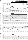
Figure 1.

Clinical time course of the development of Graves’ disease and type 1 diabetes. The clinical time course of the development of Graves’ disease and type 1 diabetes in the present patient is shown according to several clinical parameters. (a) Glycated hemoglobin (HbA1c) and serum C‐peptide levels. Filled circle and open triangle represent HbA1c and serum C‐peptide levels, respectively. (b) Free triiodothyronine (FT3) and thyroxine (FT4) levels. Dashed and solid lines represent FT3 and FT4 levels, respectively. (c) Thyroid‐stimulating hormone (TSH) level. The values are plotted with a base‐10 logarithmic scale on the y‐axis. (d) TSH receptor antibody (TRAb) level. The x‐axis of each figure shows the time course (day) before and after admission. Day 0 corresponds to the day of admission. (b–d) The gray areas show the normal ranges for each parameter.
The patient’s height, weight and body mass index were 160.4 cm, 54.9 kg and 21.3 kg/m2, respectively. He had no history of glucose intolerance and thyroid dysfunction, and no evidence of preceding acute viral infection. He also had no family history of thyroid diseases and other autoimmune diseases. Furthermore, he had no hyperglycemia‐ or thyrotoxicosis‐related symptoms before and during hospitalization. Laboratory examination at admission showed the following results: urine ketones, negative; serum creatinine, 0.78 mg/dL; islet‐associated autoantibodies (glutamic acid decarboxylase antibody, insulinoma‐associated antigen‐2 antibody, zinc transporter 8 antibody), negative; venous pH, 7.375, venous , 27.0 mmol/L; and serum total ketone body, 444.8 μmol/L (NR ≤130 μmol/L); serum acetoacetate, 164.0 μmol/L (NR ≤55 μmol/L); and serum 3‐hydroxybutyrate, 280.8 μmol/L (NR ≤85 μmol/L). His fasting serum C‐peptide level was 1.55 ng/mL at admission, which decreased to 0.01 ng/mL after 3 months and remained <0.01 ng/mL thereafter. Thus, he was diagnosed with anti‐PD‐1 therapy‐associated type 1 diabetes. He carried a homozygote of the human leukocyte antigen (HLA)‐DRB1*04:05 allele, associated with autoimmune type 1 diabetes in the Japanese population. The patient had no diabetes complications. Intensive insulin therapy was started immediately after admission; his plasma glucose levels finally improved with 7 units of insulin degludec, and 12, 8 and 5 units of insulin aspart before breakfast, lunch and dinner, respectively. He was discharged on hospitalization day 25. The seventh and subsequent nivolumab courses were resumed approximately 9 months post‐discharge. On the day after the 31st course of nivolumab treatment (835 days after the first nivolumab), computed tomography confirmed that the parotid gland tumor and lung metastasis lesions had grown larger compared with the size before the first course of nivolumab therapy. However, the tumor growth rate appeared to be extremely slow owing to nivolumab, thus suggesting the potential antineoplastic effect of nivolumab on tumors.
Meanwhile, the patient’s TSH receptor antibody titer was 3.7 IU/L (NR <2.0 IU/L), but thyroglobulin antibody and thyroid peroxidase antibody were both negative. Although thyroid ultrasonography showed no enlargement and normal vascularity of the thyroid gland (Figure S1), the patient was clinically diagnosed with mild Graves’ disease and started receiving methimazole therapy. Thereafter, his TSH level remained <0.005 μIU/mL for approximately 10 months (except for a brief period), and free triiodothyronine levels, which decreased soon after initiating methimazole, increased to the near‐upper limit of the normal range (3.47 pg/mL) at 213 days after manifesting Graves’ disease, despite using methimazole (2.5 mg/day), thereby suggesting the persistence of subclinical hyperthyroidism as a result of Graves’ disease, but not painless thyroiditis.
Written informed consent was obtained from the patient.
Discussion
Some studies have reported that 39.0–54.2% of individuals treated with anti‐PD‐1 antibodies show immune‐related adverse events1. Although the most common endocrine adverse event with anti‐PD‐1 therapy was hypothyroidism (~5.9% cases)1, there have been no reports of participants with anti‐PD‐1‐associated Graves’ disease based on correct diagnosis thus far2, 3, 4. Furthermore, type 1 diabetes was very rare, and recorded in 0.4% of cases1. Thus, the present patient with simultaneous development of Graves’ disease and type 1 diabetes after anti‐PD‐1 therapy is believed to be extremely rare and valuable. However, TSH receptor antibody and islet‐associated autoantibodies had never been measured before the manifestation of the two diseases in this patient. Hence, a limitation of this report is that the true time point of the development of the two diseases could not be completely ascertained.
The detailed mechanism for the development of anti‐PD‐1 therapy‐associated Graves’ disease remains unknown. A recent study showed that PD‐1+ CD4+ T cells were remarkably increased among intrathyroidal lymphocytes of Graves’ disease patients5. Furthermore, programmed cell death ligand‐1 (PD‐L1) was expressed in epithelial thyroid follicular cell clusters in their thyroid tissues5. These findings suggest that the PD‐1/PD‐L1 pathway plays an important role in regulating thyroid autoimmunity, including Graves’ disease. Therefore, thyroid autoimmunity arising after PD‐1/PD‐L1 blocking therapies might be because of inhibiting the PD‐1/PD‐L1 tolerance mechanism in the thyroid, leading to the development of Graves’ disease through CD4+ helper T‐cell‐associated activation of B cells and plasma cells producing TSH receptor antibody6.
Among Japanese patients with autoimmune polyglandular syndrome type III, the frequency in HLA‐DRB1*09:01 is higher in individuals with both Hashimoto’s thyroiditis and type 1 diabetes, whereas that of HLA‐DRB1*04:05 is higher in those with both Graves’ disease and type 1 diabetes7. The present patient had the HLA‐DRB1*04:05 allele, which could be associated with the simultaneous development of type 1 diabetes and Graves’ disease. Furthermore, the frequency of homozygotes with DRB1*04:05‐DQB1*04:01, the most common DRB1*04:05‐related haplotype, is approximately 2.5% in the Japanese population8, possibly explaining the very rare frequency of simultaneous development of the two diseases after anti‐PD‐1 therapy. Furthermore, non‐HLA genes, including cytotoxic T‐lymphocyte‐associated protein 4 gene, which was previously reported to be related with the concurrent development of type 1 diabetes and autoimmune thyroid disease9, might also be associated with the simultaneous development of Graves’ disease and type 1 diabetes, and this warrants further investigation. These points of discussion cannot be concluded based on a single case, and thus, accumulating similar cases might help in understanding the pathogenic mechanisms underlying the simultaneous development of thyroid dysfunction and type 1 diabetes during immune checkpoint inhibitor treatment to identify methods for preventing this side‐effect.
Disclosure
Akira Shimada has received lecture fees from Novo Nordisk Pharma Inc., Sanofi K.K. and Eli Lilly Japan K.K. Hiroshi Kagamu is a scientific advisor of ImmuniT Research Inc; has received honoraria from AstraZeneca K.K., Bristol‐Myers Squibb Co. and Ono Pharmaceutical Co.; and research funding from Boehringer Ingelheim Pharmaceuticals Inc. The other authors declare no conflict of interest.
Supporting information
Figure S1 | Ultrasound images of the thyroid gland in the current case.
Table S1 | Time course of main laboratory data in the present case.
Acknowledgments
No specific funding or grant was received for this work.
J Diabetes Investig 2020; 11: 1006–1009
References
- 1. Byun DJ, Wolchok JD, Rosenberg LM, et al Cancer immunotherapy ‐ immune checkpoint blockade and associated endocrinopathies. Nat Rev Endocrinol 2017; 13: 195–207. [DOI] [PMC free article] [PubMed] [Google Scholar]
- 2. de Filette J, Jansen Y, Schreuer M, et al Incidence of thyroid‐related adverse events in melanoma patients treated with pembrolizumab. J Clin Endocrinol Metab 2016; 101: 4431–4439. [DOI] [PMC free article] [PubMed] [Google Scholar]
- 3. Delivanis DA, Gustafson MP, Bornschlegl S, et al Pembrolizumab‐induced thyroiditis: comprehensive clinical review and insights into underlying involved mechanisms. J Clin Endocrinol Metab 2017; 102: 2770–2780. [DOI] [PMC free article] [PubMed] [Google Scholar]
- 4. Yamauchi I, Yasoda A, Matsumoto S, et al Incidence, features, and prognosis of immune‐related adverse events involving the thyroid gland induced by nivolumab. PLoS One 2019; 14: e0216954. [DOI] [PMC free article] [PubMed] [Google Scholar]
- 5. Álvarez‐Sierra D, Marín‐Sánchez A, Ruiz‐Blázquez P, et al Analysis of the PD‐1/PD‐L1 axis in human autoimmune thyroid disease: insights into pathogenesis and clues to immunotherapy associated thyroid autoimmunity. J Autoimmun 2019; 103: 102285. [DOI] [PubMed] [Google Scholar]
- 6. Rydzewska M, Jaromin M, Pasierowska IE, et al Role of the T and B lymphocytes in pathogenesis of autoimmune thyroid diseases. Thyroid Res 2018; 11: 2. [DOI] [PMC free article] [PubMed] [Google Scholar]
- 7. Hashimoto K, Maruyama H, Nishiyama M, et al Susceptibility alleles and haplotypes of human leukocyte antigen DRB1, DQA1, and DQB1 in autoimmune polyglandular syndrome type III in Japanese population. Horm Res 2005; 64: 253–260. [DOI] [PubMed] [Google Scholar]
- 8. Tsutsumi C, Imagawa A, Ikegami H, et al Class II HLA genotype in fulminant type 1 diabetes: a nationwide survey with reference to glutamic acid decarboxylase antibodies. J Diabetes Investig 2012; 3: 62–69. [DOI] [PMC free article] [PubMed] [Google Scholar]
- 9. Ikegami H, Awata T, Kawasaki E, et al The association of CTLA4 polymorphism with type 1 diabetes is concentrated in patients complicated with autoimmune thyroid disease: a multicenter collaborative study in Japan. J Clin Endocrinol Metab 2006; 91: 1087–1092. [DOI] [PubMed] [Google Scholar]
Associated Data
This section collects any data citations, data availability statements, or supplementary materials included in this article.
Supplementary Materials
Figure S1 | Ultrasound images of the thyroid gland in the current case.
Table S1 | Time course of main laboratory data in the present case.