Abstract
Serum cholesterol is major risk factor and contributor to atherosclerotic cardiovascular disease (ASCVD). Therapeutic cholesterol-lowering drugs, especially statin, revealed that reduction in low-density lipoprotein cholesterol (LDL-C) produces marked reduction of ASCVD events. In the preventive scope, lower LDL-C is generally accepted as better in proven ASCVD patients and high-risk patient groups. However, in patients with low to intermediate risk without ASCVD, risk assessment is clinically guided by traditional major risk factors. In this group, the complement approach to detailed risk assessment about traditional major risk factors is needed. These non-traditional risk factors include ankle-brachial index (ABI), high-sensitivity C-reactive protein (hsCRP) level, lipoprotein(a) (Lp[a]), apolipoprotein B (apoB), or coronary artery calcium (CAC) score. CAC measurements have an additive role in the decision to use statin therapy in non-diabetic patients 40–75 years old with intermediate risk in primary prevention. This review comprises ASCVD lipid/biomarkers other than CAC. The 2013 and 2018 American College of Cardiology/American Heart Association (ACC/AHA) guidelines suggest these factors as risk-enhancing factors to help health care providers better determine individualized risk and treatment options especially regarding abnormal biomarkers. The recent 2018 Korean guidelines for management of dyslipidemia did not include these biomarkers in clinical decision making. The current review describes the current roles of hsCRP, ABI, LP(a), and apoB in personal modulation and management of health based on the 2018 ACC/AHA guideline on the management of blood cholesterol.
Keywords: Cardiovascular disease, Risk assessment, Biomarkers, Statins
INTRODUCTION
Serum cholesterol is a major risk factor and contributor to atherosclerotic cardiovascular disease (ASCVD), as supported by animal and clinical evidence including genetic and epidemiologic studies and randomized clinical trials.1,2 Therapeutic cholesterol-lowering drugs, especially statin therapy, established that low-density lipoprotein cholesterol (LDL-C) reduction produces marked reduction of ASCVD. In a preventive scope, a lower LDL-C is generally better in ASCVD patients and a high-risk patient group.3,4,5,6 In patients with low to intermediate risk without ASCVD, risk assessment is clinically guided by major risk factors such as cigarette smoking, hypertension, dyslipidemia, diabetes, and family history. American College of Cardiology (ACC)/American Heart Association (AHA), European Society of Cardiology (ESC), and Asian countries including Korea have unique clinical guidelines for treatment of dyslipidemia according to risk assessment tools.7,8,9,10 However, these recommendations are not consistent with each other because of background epidemiologic ASCVD event differences.
The 2013 ACC/AHA Guideline on Management of Blood Cholesterol adopted a new risk assessment tool, the population cohort equation (PCE).11,12 Five community-based cohort studies and a previous Framingham Heart Study were combined and validated for new risk assessment for coronary artery disease (CAD) as well as stroke, transient ischemic attack, or peripheral artery disease (PAD) including aortic aneurysmal disease.13,14,15,16,17,18 Initially, a few validation work-ups for this new PCE such as data from the Women's Health Initiative, which covers a US contemporary multiethnic cohort of postmenopausal women, indicated that these pooled cohort equations overestimated ASCVD risk.19 However, when event surveillance was improved by data from the Centers for Medicare & Medicaid Services, the PCE discriminated risk well in the USA. However, this improvement of risk prediction validation is not applicable to other countries including Korea.
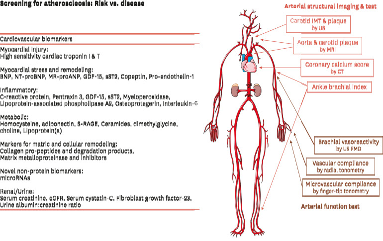
Another approach is to complement fine risk assessment over traditional major risk factors. These non-traditional risk factors help screen subjects at risk for cardiovascular complications and include blood biomarkers, risk factors, and/or markers of subclinical disease (Fig. 1). Each of all has not shown adequate statistical tools to demonstrate the incremental value of an emerging biomarker in addition to global risk scoring.
Fig. 1. Screening for subjects at risk for cardiovascular complications: blood biomarkers/risk factors and/or markers of subclinical disease.
BNP, b-type natriuretic peptide; NT-proBNP, N-terminal pro-B-type natriuretic peptide; MR-proANP, mid-regional pro-A-type natriuretic peptide; GDF-15, growth differentiation factor-15; sST2, soluble suppression of tumorigenicity 2; sRAGE, serum soluble receptor for advanced glycation end products; eGFR, estimated glomerular filtration rate; IMT, intima-media thickness; CT, computed tomography; MRI, magnetic resonance imaging; FMD, flow-mediated dilation.
The 2018 ACC/AHA Guideline on Management of Blood Cholesterol specially mentioned the evidence-based statin benefit group for primary prevention. In non-diabetic adults 40 to 75 years of age, the 10-year ASCVD higher risk group can be used to determine superiority of initiation or intensification of statin therapy. The patients in this category are proposed to initiate statin therapy combined with “risk enhancers” (Table 1). In the general population, risk enhancers may or may not predict ASCVD risk independently using PCE, but they can be useful for identifying specific factors that influence each individual future risk. This review will focus on the potential role of biomarkers such as high-sensitivity C-reactive protein (hsCRP), lipoprotein(a) (LP[a]), apolipoprotein B (apoB), and ankle-brachial index (ABI) for additional risk assessment in patients with borderline or intermediate risk in the view of 2018 ACC/AHA Guideline on Management of Blood Cholesterol. Coronary artery calcium (CAC) measurements have an additive role in the decision to use statin therapy in non-diabetic patients 40–75 years old with intermediate risk in primary prevention. However, this review addressed ASCVD lipid/biomarkers other than CAC.
Table 1. Risk enhancing factors for clinician-patient risk discussion.
| Risk-enhancing factors | |
|---|---|
| Family history of premature ASCVD (male <55 yr, female <65 yr) | |
| Primary hypercholesterolemia (LDL-C, 160–189 mg/dL; non-HDL-C, 190–219 mg/dL)* | |
| Metabolic syndrome (over than 3 marks, diagnosis as metabolic syndrome) | |
| Increased waist circumference | |
| Elevated triglycerides (>175 mg/dL) | |
| Elevated blood pressure | |
| Elevated glucose | |
| Low HDL-C (<40 mg/dL in men; <50 in women mg/dL) | |
| Chronic kidney disease (eGFR 15–59 mL/min/1.73 m2 with or without albuminuria; not treated with dialysis or kidney transplantation) | |
| Chronic inflammatory condition such as psoriasis, RA, or HIV/AIDS | |
| History of premature menopause (before age 40 yr) and history of pregnancy-associated conditions that increase later ASCVD risk such as preeclampsia | |
| High-risk race/ethnicity (e.g., South Asian ancestry) | |
| Lipid/biomarkers: associated with increased ASCVD risk | |
| Persistently* elevated, primary hypertriglyceridemia (≥175 mg/dL) | |
| Elevated high-sensitivity C-reactive protein (≥2.0 mg/L) | |
| Elevated Lp(a): a relative indication for its measurement is family history of premature ASCVD. An Lp(a) ≥50 mg/dL or ≥125 nmol/L constitutes a risk-enhancing factor especially at higher levels of Lp(a) | |
| Elevated apoB ≥130 mg/dL: a relative indication for its measurement is triglycerides ≥200 mg/dL. A level ≥130 mg/dL corresponds to LDL-C >160 mg/dL and constitutes a risk-enhancing factor | |
| ABI <0.9 | |
AIDS, acquired immunodeficiency syndrome; ABI, ankle-brachial index; apoB, apolipoprotein B; ASCVD, atherosclerotic cardiovascular disease; eGFR, estimated glomerular filtration rate; HDL-C, high-density lipoprotein cholesterol; HIV, human immunodeficiency virus; LDL-C, low-density lipoprotein cholesterol; Lp(a), lipoprotein (a); RA, rheumatoid arthritis.
*Optimally, 3 determinations.
EFFECTIVE TARGET GROUP FOR USING BIOMARKERS
1. Status of Korean risk assessment
Many studies have attempted to assess cardiovascular disease (CVD) risk based on a comprehensive review of exposure to various CVD risk factors. Since development of the Framingham risk score (FRS) by the Framingham Heart Study to calculate the 10-year risk of CAD using seven items of information (age, sex, total cholesterol, high-density lipoprotein cholesterol [HDL-C], blood pressure, diabetes, and smoking), various CVD prediction models have been developed, and a CVD prediction recommendation guideline has been formulated.18 In South Korea, several studies have developed a stroke risk model, a CAD risk model, and a CVD risk assessment model using data from health check-up recipients.20,21,22,23 However, Korean treatment guidelines still do not recommend use of these tools in decision-making for drug therapy. There are concerns that the generalizability of these risk assessment tools developed in Korea needs to be further examined as they have not been adequately validated, while other concerns suggest that, even if individual CVD risks are assessed, it is difficult to reflect them in treatment guidelines because evidence supporting the clinical efficacy and cost-effectiveness of drug therapy according to the level of risk is lacking. Additional biomarker studies on CVD risk assessment are not well developed in South Korea. Thus, development of a Korean guideline for management of dyslipidemia is required.
2. Risk group by PCE and biomarker in the 2018 US guideline
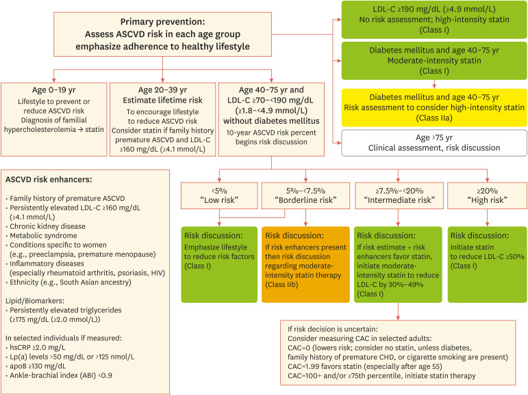
In contrast to the current situation in Korea, in the US, the 2018 ACC/AHA guideline specified recommendations for use of risk-enhancing factors including biomarkers to individualize risk status based on PCE as well as other factors that may inform risk prediction. Adults 40 to 75 years of age with LDL-C level 70 to 189 mg/dL in primary prevention can be classified as 4 groups like this; borderline risk (10-year risk of ASCVD 5% to <7.5%), intermediate-risk (7.5% to <20%), and high-risk (20%). A 10-year risk of ASCVD ≥7.5% was identified as a randomized controlled trial (RCT)-supported threshold for benefit of statin therapy by PCE by the 2013 ACC/AHA guidelines. In intermediate-risk patients, moderate- to high-intensity statin therapy should be considered during risk discussion of treatment option (class Ia); for borderline-risk patients with risk enhancers, moderate-intensity statin therapy initiation should be discussed with patients (class IIb) among adults 40 to 75 years of age (Fig. 2).
Fig. 2. Primary prevention: the role of ASCVD risk enhancer.
ASCVD, atherosclerotic cardiovascular disease; apoB, apolipoprotein B; CAC, coronary artery calcium; HIV, human immunodeficiency virus; hsCRP, high-sensitivity C-reactive protein; LDL-C, low-density lipoprotein cholesterol; Lp(a), lipoprotein (a); CHD, coronary heart disease.
LIPID/BIOMARKERS
1. hsCRP
The high hsCRP level is strongly associated with coronary heart disease (CHD) events. Although a few studies have directly assessed the effect of CRP on risk reclassification in intermediate-risk individuals, adding CRP to risk prediction models among initially intermediate-risk patients moderately improves risk stratification. In patients with intermediate ASCVD risk (7.5–20%), an ASCVD score does not assign statin therapy. An RCT in women ≥60 years of age with a family history of premature ASCVD with elevated hsCRP but without ASCVD showed clinical benefit from high intensity statin therapy.24 In a systemic review, the U.S. Preventive Services Task Force (USPSTF) evaluated the factors' clinical usefulness. The adjusted risk ratio (95% confidence interval [CI]) for major CHD events and comparison was 1.58 (1.37–1.83) for CRP 3.0 mg/L vs. <1.0 mg/L and 1.22 (1.11–1.33) for 1.0–3.0 vs. <1.0 mg/L. Use of CRP level stratified the study population with a FRS of 1% to 20%. A CRP level greater than 3.0 mg/L reclassified 5% of intermediate-risk women in the Women's Health Study.25 In studies in men, high CRP level clearly identified a high-risk subset of persons with a FRS between 15% and 20%.26 As interventions that reduce CRP such as weight loss, exercise, smoking cessation, statins, and fibrates reduce the risk for coronary event, it is unclear whether performing a CRP test to guide treatment goals is more beneficial than intensifying treatment goals in an all intermediate-risk group.
High-intensity statin showed clinical benefit in patients with high hsCRP level and LDL-C less than 130 mg/dL, but there is currently no clinical data in low-risk and intermediate-risk patients. A single primary prevention trial of high-intensity rosuvasatin 20 mg versus placebo in 17,802 patients with a CRP level greater than 2 mg/L and an LDL-C less than 130 mg/dL was terminated early because of overwhelming benefit.27 However, this trial did not clearly classify the number of patients with low- or intermediate-risk on the basis of FRS.
Thus, hsCRP level is regarded as a risk-enhancing factor guiding intensification in intermediate-risk adults and initiation of moderate-intensity statin therapy in borderline-risk adults 75 years of age with LDL-C level 70 to 189 mg/dL in primary prevention.
2. ABI
Role of ABI in primary prevention
An ABI <0.9 is a generally accepted cutoff point used to indicate possible significant PAD. The Framingham cohort study revealed that traditional risk factors were equally applicable as predictors of incidence of PAD, which was classified as a coronary equivalent.28,29,30,31,32 ABI is associated with total CHD risk and leads to significant risk reclassification, but there was no direct evidence that ABI independently predicts the risk for incident CHD events in individuals without symptomatic PAD.
The Ankle-Brachial Index Collaboration reported a meta-analysis of 16 studies in 2008. Overall, the FRS showed relatively poor discrimination, with a C-statistic of 0.646 (95% CI, 0.643–0.657) in men and 0.605 (0.590–0.619) in women. There was an improvement in C-statistic in both men (0.655 [0.643–0.666]) and women (0.658 [0.644–0.672]) when ABI was added to a model with FRS. The improvement in C-statistic was greater and significant in women but was not significant in men. The pattern of reclassification is different by sex. Among men, the effect is to down-classify high-risk individual, while among women, the result is to up-classify low-risk.28 Another ABI-related risk assessment was published by the USPSTF regarding the utility of assessing ABI in 2013. There was no evidence that ABI independently predicts risk for incident CHD events in asymptomatic PAD patients. The USPSTF found no evidence that screening for and treatment of PAD in asymptomatic patients lead to clinically important benefits. It also reviewed the potential benefits of adding the ABI to the FRS and found evidence that this results in some patient risk reclassification. However, how often reclassification is appropriate or whether it results in improved clinical outcomes is not known. The Work Group notes that this review provides some evidence that assessing ABI may improve risk assessment; however, no evidence was found by the USPSTF reviewers as to whether measuring ABI leads to better patient outcomes.33 However, these meta-analyses had some limitations. First, these analyses did not discriminate whether participants with a known history of stroke, transient ischemic attack, or symptomatic PAD were excluded from the analysis. Second, adequacy of Framingham risk factor measurement from the pooled discrimination statistics reported in the studies cannot be judged. For example, adjusted 10-year risk for major CHD events among men in the FRS high-risk range from 20% to 29% was under-adjusted to 15.3%.
ABI in diabetes
Diabetes is one of the 4 beneficial statin groups in the 2013 Guideline on the Management of Blood Cholesterol. However, in adults 20 to 39 years of age with diabetes mellitus that is either of long duration (≥10 years of type 2 diabetes mellitus, ≥20 years of type 1 diabetes mellitus), albuminuria (≥30 mcg of albumin/mg creatinine), estimated glomerular filtration rate (eGFR) less than 60 mL/min/1.73 m2, retinopathy, neuropathy, or ABI <0.9 were not fully recommended for initiation of statin therapy. In these patients, “Clinician-Patient Discussion” is recommended prior to statin therapy. These discussion points include benefits of risk reduction, statin adverse effects, and drug interaction; lifestyle modifications; patient preference; and when decision is unclear, patient clinical factors such as primary LDL-C ≥160 mg/dL, family history of premature ASCVD, lifetime ASCVD risk, abnormal CAC score or ABI, or hs-CRP ≥2 mg/L should be considered to initiate statin therapy.
Two population cohort studies of diabetes with decreased ABI revealed increased risk of ASCVD.34,35 Therefore, ABI has role in initiation primary prevention of statin therapy in adults 40 to 75 years of age with LDL-C level 70 to 189 mg/dL. In adults 20 to 39 years of age with diabetes mellitus who have ABI <0.9, it may be reasonable to initiate statin therapy (class IIb).
3. ApoB
ApoB is the major apolipoprotein embedded in low-density lipoprotein (LDL) and very-low-density lipoprotein, and there is an association between apoB and ASCVD.3,36,37 A previous epidemiologic study showed evidence of the rough equivalence of associations of CVD with non-HDL-C and apoB after multivariable adjustment (including HDL-C).36 However, a more large-scale review showed that apoB was the most potent marker of cardiovascular risk (relative risk reduction [RRR], 1.43; 95% CI, 1.35–1.51), and LDL-C was the least potent marker (RRR, 1.25; 95% CI, 1.18–1.33).37 Therefore, apoB is a stronger indicator of atherogenicity compared with LDL-C alone.
The measurement of apoB may have advantages especially in patients with hypertriglyceridemia.38 However, apoB measurement is costly, and its measurement in some laboratories may not be reliable.39
A relative indication for apoB measurement is triglycerides ≥200 mg/dL. ApoB level ≥130 mg/dL corresponds to LDL-C >160 mg/dL and constitutes a risk-enhancing factor. In intermediate-risk adults, apoB is a risk-enhancing factor that favors initiation or intensification of statin therapy. In patients at borderline risk, presence of risk-enhancing factors may justify initiation of moderate-intensity statin therapy.
4. Lp(a)
Lp(a) consists of an LDL-like particle and the specific apolipoprotein(a) that is bound covalently to the apoB of the LDL-like particle. Lp(a) is a modified form of LDL that appears to possess atherogenic potential.40 Lp(a) in a Framingham offspring cohort setting provided elevated plasma Lp(a) is an independent risk factor for development of premature CHD in men and woman, comparable in magnitude and prevalence (i.e., attributable risk) to a total cholesterol level of 240 mg/dL or more or an high-density lipoprotein level less than 35 mg/dL.41,42
High Lp(a) is considered a risk-enhancing factor,43 especially in patients with higher Lp(a) value and in women, only in the presence of hypercholesterolemia.44 However, its usefulness for stratifying intermediate-risk persons is unclear.
The 2018 ACC/AHA Guideline on the Management of Blood Cholesterol recommends that Lp(a) ≥50 mg/dL or ≥125 nmol/L be considered as an increase. However, no available RCT evidence supports Lp(a) level as a target of therapy.
CONCLUSION
Pooled cohort risk estimation is a powerful tool to predict population risk, but it has limitations when applied to individuals. Although risk-enhancing factors may or may not predict risk independently of PCE in the general population, the evidence of risk-enhancing factors revealed association with ASCVD. Before statin initiation, the purpose of the clinician-patient risk discussion is to individualize risk status based on PCE as well as other factors that may inform risk prediction. In primary prevention, adults 40 to 75 years old with LDL-C level 70 to 189 mg/dL, elevated hs-CRP (≥2.0 mg/L), ABI <0.9, apoB ≥130 mg/dL (accompanied by persistent hyper-triglycerides), and Lp(a) ≥50 mg/dL show a high lifetime risk for ASCVD; statin therapy is favored in the borderline risk group, and intensification of statin therapy should be recommended in the intermediate risk group.
Although Korean dyslipidemia guidelines did not adopt these risk-enhancing biomarkers due to lack of long-term large population cohort data, the prospective cohort-based prognostic studies that can be appropriately applied to Koreans need to establish evidence-based patient care that can be applied to Korean individuals.
Footnotes
Funding: None.
Conflict of Interest: The author has no conflicts of interest to declare.
References
- 1.Stamler J, Wentworth D, Neaton JD. Is relationship between serum cholesterol and risk of premature death from coronary heart disease continuous and graded? Findings in 356,222 primary screenees of the Multiple Risk Factor Intervention Trial (MRFIT) JAMA. 1986;256:2823–2828. [PubMed] [Google Scholar]
- 2.Kannel WB, Castelli WP, Gordon T, McNamara PM. Serum cholesterol, lipoproteins, and the risk of coronary heart disease. The Framingham study. Ann Intern Med. 1971;74:1–12. doi: 10.7326/0003-4819-74-1-1. [DOI] [PubMed] [Google Scholar]
- 3.Boekholdt SM, Hovingh GK, Mora S, Arsenault BJ, Amarenco P, Pedersen TR, et al. Very low levels of atherogenic lipoproteins and the risk for cardiovascular events: a meta-analysis of statin trials. J Am Coll Cardiol. 2014;64:485–494. doi: 10.1016/j.jacc.2014.02.615. [DOI] [PMC free article] [PubMed] [Google Scholar]
- 4.Cannon CP, Blazing MA, Giugliano RP, McCagg A, White JA, Theroux P, et al. Ezetimibe added to statin therapy after acute coronary syndromes. N Engl J Med. 2015;372:2387–2397. doi: 10.1056/NEJMoa1410489. [DOI] [PubMed] [Google Scholar]
- 5.Sabatine MS, Giugliano RP, Keech AC, Honarpour N, Wiviott SD, Murphy SA, et al. Evolocumab and clinical outcomes in patients with cardiovascular disease. N Engl J Med. 2017;376:1713–1722. doi: 10.1056/NEJMoa1615664. [DOI] [PubMed] [Google Scholar]
- 6.Silverman MG, Ference BA, Im K, Wiviott SD, Giugliano RP, Grundy SM, et al. Association between lowering LDL-C and cardiovascular risk reduction among different therapeutic interventions: a systematic review and meta-analysis. JAMA. 2016;316:1289–1297. doi: 10.1001/jama.2016.13985. [DOI] [PubMed] [Google Scholar]
- 7.Stone NJ, Robinson JG, Lichtenstein AH, Bairey Merz CN, Blum CB, Eckel RH, et al. 2013 ACC/AHA guideline on the treatment of blood cholesterol to reduce atherosclerotic cardiovascular risk in adults: a report of the American College of Cardiology/American Heart Association Task Force on Practice Guidelines. J Am Coll Cardiol. 2014;63:2889–2934. doi: 10.1016/j.jacc.2013.11.002. [DOI] [PubMed] [Google Scholar]
- 8.Grundy SM, Stone NJ, Bailey AL, Beam C, Birtcher KK, Blumenthal RS, et al. 2018 AHA/ACC/AACVPR/AAPA/ABC/ACPM/ADA/AGS/APhA/ASPC/NLA/PCNA Guideline on the Management of Blood Cholesterol. Executive summary: a report of the American College of Cardiology/American Heart Association Task Force on Clinical Practice Guidelines. Circulation. 2019;139:e1046–e1081. doi: 10.1161/CIR.0000000000000624. [DOI] [PubMed] [Google Scholar]
- 9.Catapano AL, Graham I, De Backer G, Wiklund O, Chapman MJ, Drexel H, et al. 2016 ESC/EAS Guidelines for the Management of Dyslipidaemias. Eur Heart J. 2016;37:2999–3058. doi: 10.1093/eurheartj/ehw272. [DOI] [PubMed] [Google Scholar]
- 10.Committee of Clinical Practice Guideline of the Korean Society of Lipid and Atherosclerosis (KSoLA) 2018 Korean Guidelines for the Management of Dyslipidemia. 4th ed. Seoul: Korean Society of Lipid and Atherosclerosis; 2018. [Google Scholar]
- 11.Stone NJ, Robinson JG, Lichtenstein AH, Bairey Merz CN, Blum CB, Eckel RH, et al. 2013 ACC/AHA guideline on the treatment of blood cholesterol to reduce atherosclerotic cardiovascular risk in adults: a report of the American College of Cardiology/American Heart Association Task Force on Practice Guidelines. Circulation. 2014;129:S1–S45. doi: 10.1161/01.cir.0000437738.63853.7a. [DOI] [PubMed] [Google Scholar]
- 12.Muntner P, Colantonio LD, Cushman M, Goff DC, Jr, Howard G, Howard VJ, et al. Validation of the atherosclerotic cardiovascular disease Pooled Cohort risk equations. JAMA. 2014;311:1406–1415. doi: 10.1001/jama.2014.2630. [DOI] [PMC free article] [PubMed] [Google Scholar]
- 13.Dawber TR, Kannel WB, Lyell LP. An approach to longitudinal studies in a community: the Framingham Study. Ann N Y Acad Sci. 1963;107:539–556. doi: 10.1111/j.1749-6632.1963.tb13299.x. [DOI] [PubMed] [Google Scholar]
- 14.Fried LP, Borhani NO, Enright P, Furberg CD, Gardin JM, Kronmal RA, et al. The Cardiovascular Health Study: design and rationale. Ann Epidemiol. 1991;1:263–276. doi: 10.1016/1047-2797(91)90005-w. [DOI] [PubMed] [Google Scholar]
- 15.Kannel WB, Feinleib M, McNamara PM, Garrison RJ, Castelli WP. An investigation of coronary heart disease in families. The Framingham offspring study. Am J Epidemiol. 1979;110:281–290. doi: 10.1093/oxfordjournals.aje.a112813. [DOI] [PubMed] [Google Scholar]
- 16.Friedman GD, Cutter GR, Donahue RP, Hughes GH, Hulley SB, Jacobs DR, Jr, et al. CARDIA: study design, recruitment, and some characteristics of the examined subjects. J Clin Epidemiol. 1988;41:1105–1116. doi: 10.1016/0895-4356(88)90080-7. [DOI] [PubMed] [Google Scholar]
- 17.The ARIC investigators. The Atherosclerosis Risk in Communities (ARIC) Study: design and objectives. Am J Epidemiol. 1989;129:687–702. [PubMed] [Google Scholar]
- 18.Wilson PW, D'Agostino RB, Levy D, Belanger AM, Silbershatz H, Kannel WB. Prediction of coronary heart disease using risk factor categories. Circulation. 1998;97:1837–1847. doi: 10.1161/01.cir.97.18.1837. [DOI] [PubMed] [Google Scholar]
- 19.Mora S, Wenger NK, Cook NR, Liu J, Howard BV, Limacher MC, et al. Evaluation of the Pooled Cohort risk equations for cardiovascular risk prediction in a multiethnic cohort from the Women's Health Initiative. JAMA Intern Med. 2018;178:1231–1240. doi: 10.1001/jamainternmed.2018.2875. [DOI] [PMC free article] [PubMed] [Google Scholar]
- 20.Asia Pacific Cohort Studies Collaboration. Barzi F, Patel A, Gu D, Sritara P, Lam TH, et al. Cardiovascular risk prediction tools for populations in Asia. J Epidemiol Community Health. 2007;61:115–121. doi: 10.1136/jech.2005.044842. [DOI] [PMC free article] [PubMed] [Google Scholar]
- 21.Jee SH, Park JW, Lee SY, Nam BH, Ryu HG, Kim SY, et al. Stroke risk prediction model: a risk profile from the Korean study. Atherosclerosis. 2008;197:318–325. doi: 10.1016/j.atherosclerosis.2007.05.014. [DOI] [PubMed] [Google Scholar]
- 22.Jee SH, Jang Y, Oh DJ, Oh BH, Lee SH, Park SW, et al. A coronary heart disease prediction model: the Korean Heart Study. BMJ Open. 2014;4:e005025. doi: 10.1136/bmjopen-2014-005025. [DOI] [PMC free article] [PubMed] [Google Scholar]
- 23.Park GM, Han S, Kim SH, Jo MW, Her SH, Lee JB, et al. Model for assessing cardiovascular risk in a Korean population. Circ Cardiovasc Qual Outcomes. 2014;7:944–951. doi: 10.1161/CIRCOUTCOMES.114.001305. [DOI] [PubMed] [Google Scholar]
- 24.Buckley DI, Fu R, Freeman M, Rogers K, Helfand M. C-reactive protein as a risk factor for coronary heart disease: a systematic review and meta-analyses for the U.S. Preventive Services Task Force. Ann Intern Med. 2009;151:483–495. doi: 10.7326/0003-4819-151-7-200910060-00009. [DOI] [PubMed] [Google Scholar]
- 25.Cushman M, Arnold AM, Psaty BM, Manolio TA, Kuller LH, Burke GL, et al. C-reactive protein and the 10-year incidence of coronary heart disease in older men and women: the cardiovascular health study. Circulation. 2005;112:25–31. doi: 10.1161/CIRCULATIONAHA.104.504159. [DOI] [PubMed] [Google Scholar]
- 26.Koenig W, Löwel H, Baumert J, Meisinger C. C-reactive protein modulates risk prediction based on the Framingham Score: implications for future risk assessment: results from a large cohort study in southern Germany. Circulation. 2004;109:1349–1353. doi: 10.1161/01.CIR.0000120707.98922.E3. [DOI] [PubMed] [Google Scholar]
- 27.Ridker PM, Danielson E, Fonseca FA, Genest J, Gotto AM, Jr, Kastelein JJ, et al. Rosuvastatin to prevent vascular events in men and women with elevated C-reactive protein. N Engl J Med. 2008;359:2195–2207. doi: 10.1056/NEJMoa0807646. [DOI] [PubMed] [Google Scholar]
- 28.Newman AB, Siscovick DS, Manolio TA, Polak J, Fried LP, Borhani NO, et al. Ankle-arm index as a marker of atherosclerosis in the Cardiovascular Health Study. Circulation. 1993;88:837–845. doi: 10.1161/01.cir.88.3.837. [DOI] [PubMed] [Google Scholar]
- 29.Weatherley BD, Nelson JJ, Heiss G, Chambless LE, Sharrett AR, Nieto FJ, et al. The association of the ankle-brachial index with incident coronary heart disease: the Atherosclerosis Risk In Communities (ARIC) study, 1987–2001. BMC Cardiovasc Disord. 2007;7:3. doi: 10.1186/1471-2261-7-3. [DOI] [PMC free article] [PubMed] [Google Scholar]
- 30.Newman AB, Shemanski L, Manolio TA, Cushman M, Mittelmark M, Polak JF, et al. Ankle-arm index as a predictor of cardiovascular disease and mortality in the Cardiovascular Health Study. Arterioscler Thromb Vasc Biol. 1999;19:538–545. doi: 10.1161/01.atv.19.3.538. [DOI] [PubMed] [Google Scholar]
- 31.Abbott RD, Petrovitch H, Rodriguez BL, Yano K, Schatz IJ, Popper JS, et al. Ankle/brachial blood pressure in men >70 years of age and the risk of coronary heart disease. Am J Cardiol. 2000;86:280–284. doi: 10.1016/s0002-9149(00)00914-0. [DOI] [PubMed] [Google Scholar]
- 32.Resnick HE, Lindsay RS, McDermott MM, Devereux RB, Jones KL, Fabsitz RR, et al. Relationship of high and low ankle brachial index to all-cause and cardiovascular disease mortality: the Strong Heart Study. Circulation. 2004;109:733–739. doi: 10.1161/01.CIR.0000112642.63927.54. [DOI] [PubMed] [Google Scholar]
- 33.Moyer VA U.S. Preventive Services Task Force. Screening for peripheral artery disease and cardiovascular disease risk assessment with the ankle-brachial index in adults: U.S. Preventive Services Task Force recommendation statement. Ann Intern Med. 2013;159:342–348. doi: 10.7326/0003-4819-159-5-201309030-00008. [DOI] [PubMed] [Google Scholar]
- 34.Ogren M, Hedblad B, Engström G, Janzon L. Prevalence and prognostic significance of asymptomatic peripheral arterial disease in 68-year-old men with diabetes. Results from the population study ‘Men born in 1914’ from Malmö, Sweden. Eur J Vasc Endovasc Surg. 2005;29:182–189. doi: 10.1016/j.ejvs.2004.11.013. [DOI] [PubMed] [Google Scholar]
- 35.Pang XH, Han J, Ye WL, Sun X, Ding Y, Huang WJ, et al. Lower extremity peripheral arterial disease is an independent predictor of coronary heart disease and stroke risks in patients with type 2 diabetes mellitus in China. Int J Endocrinol. 2017;2017:9620513. doi: 10.1155/2017/9620513. [DOI] [PMC free article] [PubMed] [Google Scholar]
- 36.Emerging Risk Factors Collaboration. Di Angelantonio E, Sarwar N, Perry P, Kaptoge S, Ray KK, et al. Major lipids, apolipoproteins, and risk of vascular disease. JAMA. 2009;302:1993–2000. doi: 10.1001/jama.2009.1619. [DOI] [PMC free article] [PubMed] [Google Scholar]
- 37.Sniderman AD, Williams K, Contois JH, Monroe HM, McQueen MJ, de Graaf J, et al. A meta-analysis of low-density lipoprotein cholesterol, non-high-density lipoprotein cholesterol, and apolipoprotein B as markers of cardiovascular risk. Circ Cardiovasc Qual Outcomes. 2011;4:337–345. doi: 10.1161/CIRCOUTCOMES.110.959247. [DOI] [PubMed] [Google Scholar]
- 38.Sniderman AD, Tremblay A, De Graaf J, Couture P. Phenotypes of hypertriglyceridemia caused by excess very-low-density lipoprotein. J Clin Lipidol. 2012;6:427–433. doi: 10.1016/j.jacl.2012.04.081. [DOI] [PubMed] [Google Scholar]
- 39.Grundy SM, Vega GL, Tomassini JE, Tershakovec AM. Comparisons of apolipoprotein B levels estimated by immunoassay, nuclear magnetic resonance, vertical auto profile, and non-high-density lipoprotein cholesterol in subjects with hypertriglyceridemia (SAFARI Trial) Am J Cardiol. 2011;108:40–46. doi: 10.1016/j.amjcard.2011.03.003. [DOI] [PubMed] [Google Scholar]
- 40.Writing Committee. Lloyd-Jones DM, Morris PB, Ballantyne CM, Birtcher KK, Daly DD, Jr, et al. 2016 ACC expert consensus decision pathway on the role of non-statin therapies for LDL-cholesterol lowering in the management of atherosclerotic cardiovascular disease risk: a report of the American College of Cardiology Task Force on Clinical Expert Consensus Documents. J Am Coll Cardiol. 2016;68:92–125. doi: 10.1016/j.jacc.2016.03.519. [DOI] [PubMed] [Google Scholar]
- 41.Bostom AG, Cupples LA, Jenner JL, Ordovas JM, Seman LJ, Wilson PW, et al. Elevated plasma lipoprotein(a) and coronary heart disease in men aged 55 years and younger. A prospective study. JAMA. 1996;276:544–548. doi: 10.1001/jama.1996.03540070040028. [DOI] [PubMed] [Google Scholar]
- 42.Bostom AG, Gagnon DR, Cupples LA, Wilson PW, Jenner JL, Ordovas JM, et al. A prospective investigation of elevated lipoprotein (a) detected by electrophoresis and cardiovascular disease in women. The Framingham Heart Study. Circulation. 1994;90:1688–1695. doi: 10.1161/01.cir.90.4.1688. [DOI] [PubMed] [Google Scholar]
- 43.Tsimikas S. A test in context: lipoprotein(a): diagnosis, prognosis, controversies, and emerging therapies. J Am Coll Cardiol. 2017;69:692–711. doi: 10.1016/j.jacc.2016.11.042. [DOI] [PubMed] [Google Scholar]
- 44.Cook NR, Mora S, Ridker PM. Lipoprotein(a) and cardiovascular risk prediction among women. J Am Coll Cardiol. 2018;72:287–296. doi: 10.1016/j.jacc.2018.04.060. [DOI] [PMC free article] [PubMed] [Google Scholar]


