In Brazil, the first COVID-19 case was confirmed on February 26, 2020, and by May 26, 2020, the number of confirmed cases was of 391,222, with 24,512 confirmed deaths [1]. CT and [18F]FDG PET/CT findings in COVID-19 have been described in several reports [2, 3], but to our knowledge findings in [68Ga]-labeled radiopharmaceuticals PET/CT remain to be described.
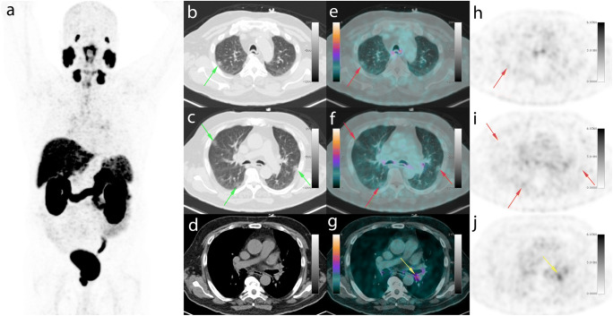
An asymptomatic 66-year-old man from Rio de Janeiro underwent [68Ga]Ga-PSMA-11 PET/CT on March 20, 2020, requesting for primary staging of prostate cancer. The patient denied any history of foreign travel. Prior biopsy showed a Gleason score of 8 (4 + 4) and the PSA level was 21.6 ng/ml. PET/CT identified two foci with [68Ga]Ga-PSMA-11 uptake in an enlarged prostate, one in the right prostate lobe in the transition zone (SUVmax 23.5) and other in the prostate basis (SUVmax 16.9). No other radiotracer uptake related to the primary disease was present (a, MIP image). However, peripheral ground-glass opacities in superior and inferior lobes of both lungs were identified on CT (b–d, axial CT; green arrows), presenting mild radiopharmaceutical uptake with an SUVmax of 2.5 (e, f, axial PET/CT; h, i, axial PET; red arrows). This CT pattern is described as a typical COVID-19 appearance at the RSNA consensus statement [4]. In addition, mild bilateral bronchial uptake was seen with an SUVmax of 4.4 (g, axial PET/CT; j, axial PET; yellow arrows), which could represent an often seen physiological variant. The attending physician and patient were informed of the possibility of COVID-19 infection and the requirement of social isolation. The patient was able to perform a serologic test only on April 25 which confirmed past COVID-19 infection: IgM negative and IgG positive. PSMA expression is not exclusive to prostate cells and it is described that benign processes such as inflammation or infection could have a variable, often mild, [68Ga]Ga-PSMA uptake [5]. Nonetheless, different from what has been described with [18F]FDG, the uptake of [68Ga]Ga-PSMA-11 was low in our patient. We hypothesize that this low uptake of [68Ga]Ga-PSMA-11 could be expected in cases of pulmonary infiltrates associated with COVID-19. It is necessary for nuclear medicine services to be alert to the risk of COVID-19 infection, even in asymptomatic patients, and to consider adequate measures after detecting a suspected case.
Compliance with ethical standards
Conflict of interest
The authors declare that they have no conflict of interest.
Informed consent
Informed consent was obtained from the participant for publication of this case report.
Footnotes
This article is part of the Topical Collection on Image of the month
Publisher’s note
Springer Nature remains neutral with regard to jurisdictional claims in published maps and institutional affiliations.
References
- 1.https://covid.saude.gov.br/ accessed on May 26th, 2020.
- 2.Setti L, Kirienko M, Dalto S, et al. FDG-PET/CT findings highly suspicious for COVID-19 in an Italian case series of asymptomatic patients. Eur J Nucl Med Mol Imaging. 2020;47:1649–1656. doi: 10.1007/s00259-020-04819-6. [DOI] [PMC free article] [PubMed] [Google Scholar]
- 3.Albano D, Bertagna F, Bertolia M, et al. Incidental findings suggestive of Covid-19 in asymptomatic patients undergoing nuclear medicine procedures in a high prevalence region. J Nucl Med. 2020;61:632–636. doi: 10.2967/jnumed.120.246256. [DOI] [PubMed] [Google Scholar]
- 4.Simpson S, Kay FU, Abbara S, et al. Radiological Society of North America Expert Consensus statement on reporting chest CT findings related to COVID-19. Endorsed by the Society of Thoracic Radiology, the American College of Radiology, and RSNA. Radiol Cardiothorac Imaging. 2020. 10.1148/ryct.2020200152. [DOI] [PMC free article] [PubMed]
- 5.Barbosa FG, Queiroz MA, Nunes RF, Costa LB, Zaniboni EC, Marin JFG, Cerri GG, Buchpiguel CA. Nonprostatic diseases on PSMA PET imaging: a spectrum of benign and malignant findings. Cancer Imaging. 2020;20:23. doi: 10.1186/s40644-020-00300-7. [DOI] [PMC free article] [PubMed] [Google Scholar]


