Abstract
A 14-year-old boy who had ocular motility disorder which started 2 weeks following retinal surgery (scleral buckling) secondary to rhegmatogenous retinal detachment, was referred to the strabismus clinic. He had significant ocular movement limitations in adduction and elevation under general anesthesia. The forced duction test (FDT) was positive in both adduction and elevation. After buckle removal, FDT was negative. The eye was orthotropic without ocular movement limitation at final follow-up. In conclusion, FDT at the end of the scleral buckling procedure needs to be performed. It may prevent restrictive strabismus after scleral buckling surgery.
Keywords: Forced duction test, Strabismus, Scleral buckling
Introduction
Strabismus following retinal detachment surgery has been reported in up to 50% of patients. In 5–25% of cases, it usually resolves spontaneously within 3–6 months [1], although it can persist in approximately 3.6–25% of cases [2]. This kind of strabismus may have multiple mechanisms such as sensory causes [3], periocular anesthesia toxicity [4], or mechanical factors related to scleral buckle implantation. These mechanical factors may arise secondary to direct muscle injury [5], adhesions between muscle and sclera, fat prolapse [6], size of scleral buckle or redirection of muscle forces either directly or indirectly by the buckle [7]. This kind of adhesion can manifest as incomitant deviation with horizontal, vertical, or torsional components [8]. The success rate of strabismus surgery after scleral buckle ranges from 47 to 80% compared to 60–80% in congenital strabismus [8]. Here, we present a patient who complained of strabismus immediately after retinal buckle surgery.
Case Report
A 14-year-old boy was referred to the strabismus clinic due to ocular movement disorder after retinal surgery. He had a history of macula-on rhegmatogenous retinal detachment with a large superior break in the left eye 2 weeks before admission. He underwent a scleral buckling procedure consisting of sponge 507/200, cryopexy, and air injection with successful anatomical attachment.
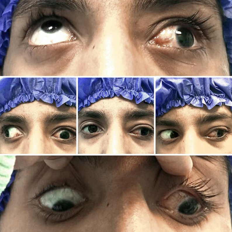
On ophthalmic examination, the best corrected visual acuity of the left eye was 20/40. Relative afferent pupillary defect was negative. Gross and biomicroscopic evaluation showed lid swelling and conjunctival hemorrhage. Funduscopic evaluation showed a good buckle effect, completely attached retina, and a small air bubble in the vitreous cavity. Ocular motility exam revealed large-angle exotropia (90 Prism dpt) in primary position and severe limitation in adduction and elevation and to some extent in depression (Fig. 1).
Fig. 1.
The different gazes of the patient before surgery for buckle removal. Top: up gaze. Left: right gaze. Centre: primary position. Right: left gaze. Down: down gaze.
The globe could not pass the midline; therefore, the patient was considered as a candidate for strabismus surgery under general anesthesia. The forced duction test (FDT) was positive on both adduction and elevation. Conjunctival sutures were removed, and medial, superior, and lateral rectus muscles were hooked. Exposure revealed a large-size sponge beneath the three rectus muscles. Its fixations were released, and sponge was removed. FDT was performed again, and it was negative. Conjunctiva was sutured with vicryl 8-0 (Ethicone), and the eye was patched.
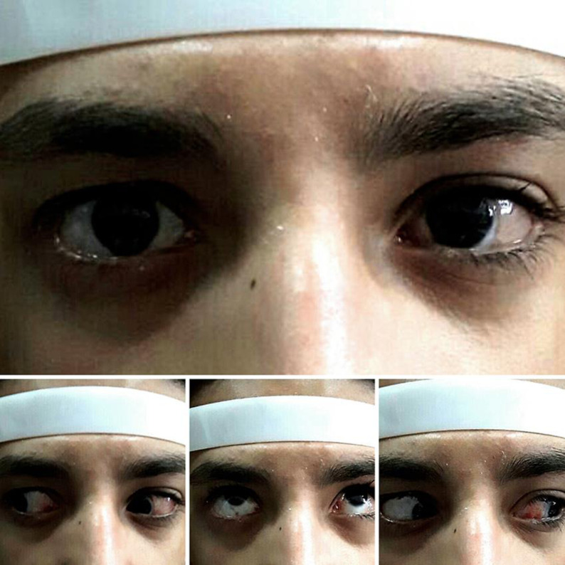
The patient was orthotropic in the primary position and had a nearly full range of ocular movement postoperatively (Fig. 2). His retina was attached completely at 1 month after surgery without any further intervention.
Fig. 2.
The different gazes of the patient after scleral buckle removal. Top: primary position. Left: right gaze. Centre: up gaze. Right: left gaze.
Discussion
Several factors are associated with strabismus after scleral buckling procedures. They are operative trauma, fat adhesion syndrome, and mismatch between size of the buckle and globe. Sometimes, strabismus can present before retinal detachment. Borderline fusion may be disrupted in several ways like blurring of vision due to retinal detachment, ocular occlusion after surgery, or haziness of ocular media [9].
Operative trauma is a cause of ocular motility disorder after scleral buckle procedures, so minimizing operative trauma by careful and gentle surgical technique has been stressed repeatedly [9].
Disinsertion of rectus muscle can cause strabismus after scleral buckle procedures [9]. Also size, type, and location of buckle can influence ocular movement [10, 11]. Sometimes, a mismatch between size of buckle and globe can cause mechanical restriction. In such cases, diplopia and limitation in ocular movements occur soon after surgery. FDT at the time of surgery after buckle insertion may reveal mechanical limitation of extraocular muscles and lead the surgeon to revise the buckle at the time of scleral buckle procedure and avoid such strabismus.
In conclusion, strabismus after scleral buckle procedures is multifactorial. Meticulous surgical techniques and FDT at the end of the scleral buckling procedure may avoid acute-onset diplopia, ocular movement disturbances, and second surgeries for buckle revision.
Statement of Ethics
This manuscript was approved by the Ethics Committee of Mashhad University of Medical Sciences. The subject has given his written informed consent.
Disclosure Statement
No conflict of interest.
Funding Sources
Eye Research Center, Mashhad University of Medical Sciences.
Author Contributions
Data collection, writing, and submission of the manuscript was done by Dr. Sharifi, and Dr. Ansari Astaneh. We approve the final version of the manuscript.
Acknowledgement
We would like to acknowledge everyone who played a role in the preparation of the manuscript, especially our colleagues in Khatam Eye Hospital.
References
- 1.Mets MB, Wendell ME, Gieser RG. Ocular deviation after retinal detachment surgery. Am J Ophthalmol. 1985 Jun;99((6)):667–72. doi: 10.1016/s0002-9394(14)76033-7. [DOI] [PubMed] [Google Scholar]
- 2.Seaber JH, Buckley EG. Strabismus after retinal detachment surgery: etiology, diagnosis, and treatment. Semin Ophthalmol. 1995 Mar;10((1)):61–73. doi: 10.3109/08820539509059981. [DOI] [PubMed] [Google Scholar]
- 3.Maurino V, Kwan A, Khoo BK, Gair E, Lee JP. Ocular motility disturbances after surgery for retinal detachment. J AAPOS. 1998 Oct;2((5)):285–92. doi: 10.1016/s1091-8531(98)90085-4. [DOI] [PubMed] [Google Scholar]
- 4.Salama H, Farr AK, Guyton DL. Anesthetic myotoxicity as a cause of restrictive strabismus after scleral buckling surgery. Retina. 2000;20((5)):478–82. doi: 10.1097/00006982-200009000-00008. [DOI] [PubMed] [Google Scholar]
- 5.Wu TE, Rosenbaum AL, Demer JL. Severe strabismus after scleral buckling: multiple mechanisms revealed by high-resolution magnetic resonance imaging. Ophthalmology. 2005 Feb;112((2)):327–36. doi: 10.1016/j.ophtha.2004.09.015. [DOI] [PubMed] [Google Scholar]
- 6.Wright KW. The fat adherence syndrome and strabismus after retina surgery. Ophthalmology. 1986 Mar;93((3)):411–5. doi: 10.1016/s0161-6420(86)33726-6. [DOI] [PubMed] [Google Scholar]
- 7.Kushner BJ. Superior oblique tendon incarceration syndrome. Arch Ophthalmol. 2007 Aug;125((8)):1070–6. doi: 10.1001/archopht.125.8.1070. [DOI] [PubMed] [Google Scholar]
- 8.Metz HS, Norris A. Cyclotorsional diplopia following retinal detachment surgery. J Pediatr Ophthalmol Strabismus. 1987 Nov-Dec;24((6)):287–90. doi: 10.3928/0191-3913-19871101-05. [DOI] [PubMed] [Google Scholar]
- 9.Kanski JJ, Elkington AR, Davies MS. Diplopia after retinal detachment surgery. Am J Ophthalmol. 1973 Jul;76((1)):38–40. doi: 10.1016/0002-9394(73)90007-x. [DOI] [PubMed] [Google Scholar]
- 10.Kutschera E, Antlanger H. Influence of retinal detachment surgery on eye motility and binocularity. Mod Probl Ophthalmol. 1979;20:354–8. [PubMed] [Google Scholar]
- 11.Sewell JJ, Knobloch WH, Eifrig DE. Extraocular muscle imbalance after surgical treatment for retinal detachment. Am J Ophthalmol. 1974 Aug;78((2)):321–3. doi: 10.1016/0002-9394(74)90096-8. [DOI] [PubMed] [Google Scholar]


