

Highlights
-
•
Information regarding average width and length of goats’ dairy claws is provided.
-
•
Increased width of the front claws was associated with increased likelihood of having deformation.
-
•
There was a significant correlation between lameness score and the number of deformed claws.
Keywords: Dairy goat, Claw overgrowth, Claw size, Lameness, Animal welfare assessment
Abstract
Lameness due to claw overgrowth remains one of the main welfare challenges in dairy goat farms. Although claw trimming is a crucial part of the solution, most times there is a delay in its implementation, with no perceived consequences. The goal of this cross-sectional study was to assess the correlation between the size and deformation of dairy goats claws with lameness score. The width and length of the claws of 38 adult dairy goats were taken and classified as deformed (DEF) or non-deformed (NO_DEF). Lameness was also scored in the majority of the animals assessed for claw deformation. Deformation of at least one claw was present in 34 animals (89% of the total sample). From the 34 goats with deformed claws, 33 presented at least one deformed rear claw and 18 presented at least one deformed front claw. From the 152 claws assessed 58% were deformed (n = 88), of which 19% (n = 29) were front claws and 39% were rear claws (n = 59). Increased width of the front claws was associated with increased likelihood of having deformation with odds of 1.24, and the increased length explained 16% of the variation in lameness scores. A positive relation between lameness score and the number of deformed claws was also shown. Overall, these results suggest that the size of dairy goats’ claws influences the prevalence of deformation and lameness severity and that the number of deformed claws affects goats’ gait. They also help to build the argument in favor of regular trimming in dairy goat farms.
1. Introduction
Claw disorders have been reported along the years as one of the major health and welfare problems in intensive dairy goat farms being claw overgrowth one of the main challenges, with 60.5% to 95.5% of goats having at least one overgrown claw (Anzuino, Bell, Bazeley, & Nicol, 2010; Hill et al., 1997; Muri, Stubsjøen, & Valle, 2013). Claw overgrowth is a common problem in intensive goat farms, mainly due to permanent housing in pens with soft surfaces (e.g. straw bedding) and lack of exercise, leading to reduced horn wear (Smith, & Sherman, 2005). It has been previously demonstrated that the unbalanced wearing of the claws leads to malformations, leading to an increase of the angle of the claw with the floor surface or a thicker horn (Franck, & De Belie, 2006). These malformations, characteristic of overgrown claws, may affect the locomotion and muscular skeletal structure, increasing vulnerability to mechanical injury or penetration of infectious agents (Somers, Schouten, Frankena, Noordhuizen-Stassen, & Metz, 2005). In permanently housed dairy goats, only regular and frequent claw trimming prevents this condition (Anzuino et al., 2010; Muri et al., 2013). Hill et al. (1997) referred that a regular claw trimming is associated with a low lameness prevalence. Anzuino et al. (2010) reported a 0.5 positive correlation between overgrown claws and lameness score in dairy goat farms. A similar relationship between overgrown claws and lameness has been reported in cattle (Gitau, Mbiuki, & McDermott, 1997), sheep (Winter, 2011), horses (Hoffman, Moore, Vanegas, & Wenz, 2014) and pigs (Pluym et al., 2011). Furthermore, the persistent presence of overgrown claws can lead to chronic deformed morphology due to the continuous incorrect placement of bones and joints when walking (Hill et al., 1997; Pinsent, 1989). This incorrect and prolonged misplacement of the claws leads to a rotation of the horn tissue in such a way that the claw starts to curl and the weight bearing is transferred to the heel area (Hill et al., 1997). The link between the length and the wrongfully positioning of the claw leading to deformation, has been studied in other ungulate species, such as horses (Hinterhofer, Stanek & Haider, 2000; Roberts, Ochoa, & Haynes, 1980), bovines (Franck & De Belie, 2006; Somers et al., 2005) and sheep (Winter, 2008). However, to the authors’ knowledge the impact of the actual claw size on lameness, as well as its possible involvement in deformation, has not been described in goats.
The aim of this preliminary study is to assess the relationship between claw size and deformation and lameness score, with the overarching goal of demonstrating the importance of hoof trimming in indoor-housed goats (Ajuda, 2014).
2. Material and methods
A cross-sectional study was conducted in March 2012 in adult dairy goats (Saanen and Alpine breeds) selected from a commercial dairy farm with 1400 lactating animals in Portugal. Thirty eight goats (20 Saanen and 18 Alpine) were randomly selected from a pen with 120 high yield animals. The goats in the sample had between two and five years (mean 3.4 years, SD 0.4) and with an average body condition score of 3.5 (SD 0.6) vy using Hervieu and Morand-Fehr (1999) method. The sampled animals were indoor-housed in a straw bedded pen and were milked twice a day on a milking parlor with concrete floor. Outdoor access was not provided. Walking distance from the pen entrance to the milking parlor was 14 m. The average days in milking was 200 (SD 1.3, ranging from 160 to 300 days. Goats had to support the front claws on a cement step to get to the feed trough.
Each goat was immobilized manually by the trimmer (licensed veterinary). The trimmer followed the appropriate method for goat immobilization, by tipping up the goat and allowing the head to fall backward between the handler's thighs so that the goat's back was resting on the handler's shins (Smith, & Sherman, 2005). Each goat underwent a throughout physical exam to exclude other possible and evident causes of lameness such as CAEV or trauma. The measurements of the medial and lateral claws of each limb (304 claws in total) were taken on the widest and longest points with a digital caliper (Fig. 1). The average of the medial and lateral claws’ measures was considered as the value for the respective limb, being this average treated as the unit claw (hoof) from now on in this paper. A claw was classified as deformed (DEF) whenever a rotation of either or both the medial or the lateral claw wall(s) was present, leading to a change of the weight bearing surface from the sole to the wall, or whenever there was a loss of the normal triangular shape of one or both claws, independently of their size. A claw was classified as non-deformed (NO_DEF), whenever the normal triangular shape of both claws (medial and lateral) was maintained (Fig. 2).
Fig. 1.
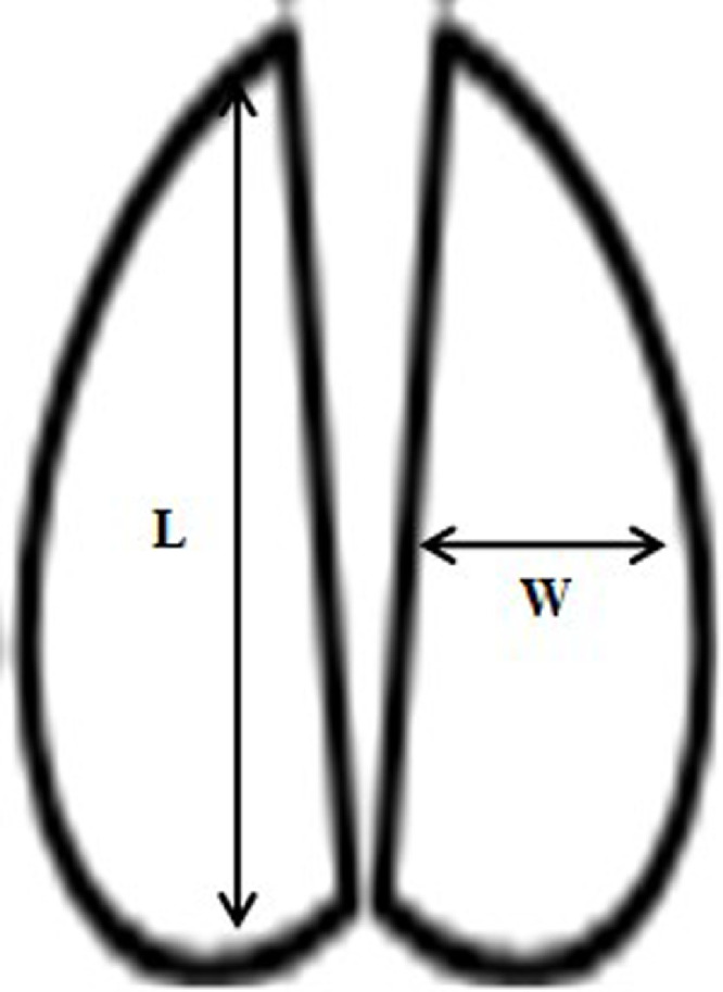
Schematization of the measurement method used to take length (L) and width (W) of the medial and lateral claws of the rear and front limbs, of 38 adult dairy goats.
Fig. 2.

Scoring method for non-deformed and deformed claws. Claws with a rotation of either or both the medial or the lateral claw wall(s), leading to a change of the weight bearing surface from the sole to the wall were also classified as deformed.
Goat's lameness was scored two days after measuring the claws, so this activity did not interfere with lameness classification. Only 34 animals out of the 38 animals were scored, due to culling, selling and removing animals to the hospital pen (in any case this removal was not related to a locomotion problem). The 34 goats were scored by a trained and experienced veterinarian while walking on an even, dry and hard surface, using the 4-point-scale system proposed by Anzuino et al. (2010). Animals classified as 0 were considered non lame, and animals classified as 1, 2 and 3, were considered lame (Table 1).
Table 1.
Lameness score (Anzuino et al., 2010).
| Score | Description |
|---|---|
| Score 0 | Goat places full weight on all four limbs, moves forward freely with an even gait |
| Score 1 | Goat has a definite limp on one or more legs, but bearing weight and moves forward freely |
| Score 2 | Goat has some difficulty moving forward, severe limp, bearing little weight on one or more legs, may be a degree of goose-stepping |
| Score 3 | Goat has some difficulty moving forward, not bearing weight on one or more legs, or may ‘goose-step’ high or walk on the knees |
Data were compiled using Microsoft Excel (Microsoft Office Professional Plus 2010, Microsoft Corp., Redmond, WA). Statistical analysis was performed using IBM SPSS Statistics 21.0. For any of the statistical models used the experimental unit was the claw within each goat, hence the sample was controlled for the unit ‘goat’. Co-variates used were age, BCS, days in milking and breed. Descriptive statistics were calculated. ANOVA was used to analyze the differences between measurements of the different claws, and between measurements of DEF and NO_DEF front and hind claws. Furthermore, a regression model (binary logistic) was used to ascertain the effect of the measures of the claw, which presented a difference on the previous analyses of variance, on the deformation of the claw itself. Spearman correlation was used to evaluate the correlation between the number of deformed claws and lameness score.
3. Results
No difference was found in claws’ size between breeds (Saanen and Alpine), ages or days in lactation (P>0,05), so data was pooled.
From the 152 claws assessed 58% were deformed (n = 88), of which 19% (n = 29) were front claws and 39% were rear claws (n = 59). From the initial 38 goats, deformation of at least one claw was present in 34 animals (89% of the total sample). All of these goats (except one) presented at least one deformed rear claw and 18 (47% of the total sample) presented at least one deformed front claw.
Differences (P<0.001) were found in the width and length of the DEF and NO_DEF front claws (Table 2). It was also shown that DEF front claws were wider than DEF rear claws (P = 0.004; Table 2).
Table 2.
Mean width and length values of the front and rear claws (mm) in 38 Saanen and Alpine dairy goats sampled for this study (76 Rear claws and 76 Front claws, DF = 75).
| Front claws1 |
Rear claws1 |
|||
|---|---|---|---|---|
| NO_DEF | DEF | NO_DEF | DEF | |
| Length (mm) | 72.9α (± 2.3) | 81.9α (± 1.7) | 76.7 (± 3.7) | 80.9 (± 2.3) |
| Width (mm) | 27.2β (± 1.2) | 33.8β,δ (± 2.5) | 26.0 (± 1.4) | 28.6δ (± 1.5) |
α-δMeans within a row with different superscript letters are statistically different (P, 1 <0.005).
Front and rear claws: NO_DEF = claws without deformation; DEF = claws with deformation.
The logistic regression model was statistically significant (χ2=27.203, P<0.001). The model explained 40.1% (Negelkerke R2) of the variance in deformation and correctly classified 77.6% of the deformation cases based on the width and length of the claws. Increased width, both in front and rear claws, was associated with increased likelihood of having deformation with odds of 1.24 (P<0.001).
The length of the front claws was the only measure that significantly explained a variation in the lameness score. The logistic regression model was statistically significant, χ2=4.19, P = 0.041. The model explained 16% (Negelkerke R2) of the variance in lameness and correctly classified goats as lame or non-lame in 69.7% of the cases. Increased length was associated with a significant tendency for increased likelihood of being lame with odds of 1.07 (P = 0.052).
A correlation of 36% (P = 0.041) between lameness score and the number of claws deformed by animal, was also shown. Although there was one goat that was classified as lame but did not have any deformed claws, it was possible to observe a significant tendency for an increase in lameness with the increase in number of deformed claws.
4. Discussion
The present study is the first attempt to collect and interpret the link between claw size and its deformation in goats: DEF claws were wider and longer than NO_DEF claws both in front and rear claws. The prevalence of animals with deformed claws in this study was much higher than the one showed by Hill et al. (1997), who found a maximum of 34% with at least one deformed claw. This can be explained by the fact that in the study conducted by Hill et al. (1997) the definition given to deformed (or slippered) claws was “claw with chronic overgrowth of the horn resulting in a long curling toe”. With this definition only animals with an excess of horn tissue were included, excluding animals that had been trimmed recently but could continue to have a chronic rotation of the claw. The classification adopted in the present study is wider and also includes goats with chronic deformations, giving a more comprehensive idea of what may affect the welfare of goats.
Hill et al. (1997) also referred to rear claws as being the most affected, in agreement with our study in which the prevalence of deformation involving the rear limbs was approximately 20% higher than the prevalence of deformed front ones. Only one goat with deformed front claws did not show deformed rear ones, indicating that front claws are probably subjected to a higher wear. This can be explained by the structure of the pen where the study was conducted.
In our research, the length of the front claws explained the percentage of animals detected as lame, supporting the theory that the location of the overgrowth is an important factor influencing lameness severity. Neveux, Weary, Rushen, von Keyserlingk, and de Passillé (2006) found a shift in weight bearing to the hind limbs when there was a discomfort present in the front claws. However, an opposite shift was not found when hind limbs were exposed to the same discomfort. This weight shift could lead to a more obvious expression of lameness. Flower and Weary (Flower & Weary, 2006) also suggest that the location of the affected claw could influence the evaluation of lameness. In infectious diseases like foot rot, a poor claw conformation was also proven to be associated with a greater risk of lameness (Kaler et al., 2010).
In our study a higher number of deformed claws was correlated with a higher probability for lameness. There are very few published studies correlating the number of affected claws with impaired gait. However, because lameness is generally a sign of pain Shearer, Stock, Van Amstel, and Coetzee. (2013); Winter (2011), it seems correct to assume a correlation between the amount of discomfort and an increase in lameness.
This study presents a strong evidence that trimming should be a priority, for the farmer as well as for the practitioner. Also, it demonstrates the possible influence of the building structure on the wearing of the claws, highlighting the importance of an enriched environment were goats can climb and exercise, as well as wear off their claws (Zobel et al., 2017). Further studies looking at the prolonged effect of claw overgrowth and deformation on internal structures (e.g. joints and bones) are needed. It is also important to highlight that although trimming is definitely important for these cases it has been reported in other species, such as sheep, that overtrimming can also be an issue (Dickins et al., 2016) demonstrating how important it is to perform this management procedure in a right manner.
5. Conclusions
Evaluation of claw overgrowth is currently used in animal welfare assessment in goat farms, being considered a very important animal-based indicator (Anzuino et al., 2010; Muri et al., 2013). With this study it was possible to conclude that the size of dairy goat's claws is highly correlated with the occurrence of claw deformation and that the length of the front claws and the number of deformed claws in the same animal are correlated with the presence of lameness in dairy goats.
Nevertheless, more information about the impact and long term consequences of these conditions is still necessary. In order to do so, further studies are needed to perceive if lameness associated with claw overgrowth and deformation is due to inflammatory pain or mechanical restriction, or both. It would be also important to understand if there are long term consequences to the animal, from being exposed for a long period to long and deformed claws.
Declaration of Competing Interest
The authors declare that they have no competing interests.
Acknowledgements
The Animal Welfare Indicators (AWIN) project (FP7-KBBE-2010–4) was supported by the European Union Seventh Framework Programme for research, technological development and demonstration (grant number 266213). The studies were also possible thanks to the funding by CIISA Project UID/CVT/00276/2013 and Project UID/CVT/00276/2019 by CIISA-FMV-Universidade de Lisboa (Lisbon, Portugal). The authors would also like to thank the farms and working personnel of Barão & Barão farm and all the masters’ students that helped in the data collection.
References
- Anzuino K., Bell N.J., Bazeley K.J., Nicol C.J. Assessment of welfare on 24 commercial UK dairy goat farms based on direct observations. The Veterinary Record. 2010;167:774–780. doi: 10.1136/vr.c5892. [DOI] [PubMed] [Google Scholar]
- Dickins A., Clark C.C.A., Kaler J., Ferguson E., O'Kane H., Green L.E. Factors associated with the presence and prevalence of contagious ovine digital dermatitis: A 2013 study of 1136 random English sheep flocks. Preventive Veterinary Medicine. 2016;130:86–93. doi: 10.1016/j.prevetmed.2016.06.009. [DOI] [PubMed] [Google Scholar]
- Flower F.C., Weary D.M. Effect of hoof pathologies on subjective assessments of dairy cow gait. Journal of Dairy Science. 2006;89:139–146. doi: 10.3168/jds.S0022-0302(06)72077-X. [DOI] [PubMed] [Google Scholar]
- Franck A., De Belie N. Concrete floor-bovine claw contact pressures related to floor roughness and deformation of the claw. Journal of Dairy Science. 2006;89:2952–2964. doi: 10.3168/jds.S0022-0302(06)72567-X. [DOI] [PubMed] [Google Scholar]
- Gitau T., Mbiuki S.M., McDermott J.J. Assessment of bovine hoof conformation and its association with lameness, animal factors and management practices on small-scale dairy farms in Kiambu district, Kenya. The Onderstepoort Journal of Veterinary Research. 1997;64:135–140. [PubMed] [Google Scholar]
- Hervieu J., Morand-Fehr. P. Comment noter l’état corporel des chèvres. Réussir Chèvre. 1999;231:22–34. [Google Scholar]
- Hill N.P., Murphy P.E., Nelson A.J., Mouttotou N., Green L.E., Morgan K.L. Lameness and foot lesions in adult British dairy goats. The Veterinary Record. 1997;141(16):412–416. doi: 10.1136/vr.141.16.412. [DOI] [PubMed] [Google Scholar]
- Hinterhofer C., Stanek C., Haider H. The effect of flat horseshoes, raised heels and lowered heels on the biomechanics of the equine hoof assessed by finite element analysis (FEA) J. Vet. Med. Ser. A. 2000;47:73–82. doi: 10.1046/j.1439-0442.2000.00263.x. [DOI] [PubMed] [Google Scholar]
- Hoffman A.C., Moore D.A., Vanegas J., Wenz J.R. Association of abnormal hind-limb postures and back arch with gait abnormality in dairy cattle. Journal of Dairy Science. 2014;97:2178–2185. doi: 10.3168/jds.2013-7528. [DOI] [PubMed] [Google Scholar]
- Kaler J., Medley G.F., Grogono-Thomas R., Wellington E.M.H., Calvo-Bado L.A., Wassink G.J. Factors associated with changes of state of foot conformation and lameness in a flock of sheep. Preventive Veterinary Medicine. 2010;97:237–244. doi: 10.1016/j.prevetmed.2010.09.019. [DOI] [PubMed] [Google Scholar]
- Muri K., Stubsjøen S., Valle P. Development and testing of an on-farm welfare assessment protocol for dairy goats. Anim. Welf. 2013;22:385–400. doi: 10.7120/09627286.22.3.385. [DOI] [Google Scholar]
- Neveux S., Weary D.M., Rushen J., von Keyserlingk M.A.G., de Passillé A.M. Hoof discomfort changes how dairy cattle distribute their body weight. Journal of Dairy Science. 2006;89:2503–2509. doi: 10.3168/jds.S0022-0302(06)72325-6. [DOI] [PubMed] [Google Scholar]
- Pinsent P. The care and diseases of the goat's foot. Br. Goat Soc. 1989;82:72–73. [Google Scholar]
- Pluym L., Nuffel A.Van, Dewulf J., Cools A., Vangroenweghe F., Hoorebeke S.Van. Prevalence and risk factors of claw lesions and lameness in pregnant sows in two types of group housing. Vet. Med. (Praha). 2011;3(No. 3):101–109. [Google Scholar]
- Roberts E.D., Ochoa R., Haynes P.F. Correlation of dermal-epidermal Laminar Lesions of Equine Hoof with Various Disease Conditions. Veterinary Pathology. 1980;17:656–666. doi: 10.1177/030098588001700601. [DOI] [PubMed] [Google Scholar]
- Shearer J.K., Stock M.L., Van Amstel S.R., Coetzee J.F. Assessment and management of pain associated with lameness in cattle. The Veterinary Clinics of North America. Food Animal Practice. 2013;29:135–156. doi: 10.1016/j.cvfa.2012.11.012. [DOI] [PubMed] [Google Scholar]
- Smith M.C., Sherman D.M. Wiley; Ames, Iowa, USA: 2005. Goat medicine. [Google Scholar]
- Somers J.G.C.J., Schouten W.G.P., Frankena K., Noordhuizen-Stassen E.N., Metz J.H.M. Development of claw traits and claw lesions in dairy cows kept on different floor systems. Journal of Dairy Science. 2005;88:110–120. doi: 10.3168/jds.S0022-0302(05)72668-0. [DOI] [PubMed] [Google Scholar]
- Winter A.C. Treatment and control of hoof disorders in sheep and goats. The Veterinary Clinics of North America. Food Animal Practice. 2011;27:187–192. doi: 10.1016/j.cvfa.2010.10.018. [DOI] [PubMed] [Google Scholar]