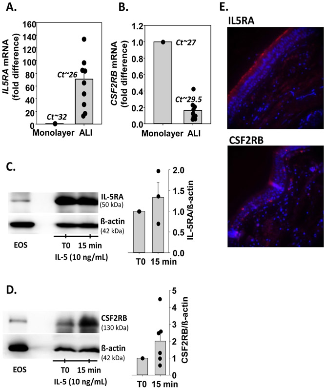
Figure 1. Human bronchial epithelial cells (HBEC) express the two subunits of the IL-5 receptor.
A and B. HBEC were either cultured undifferentiated as a monolayer (1 donor) or fully differentiated at an air liquid interface (ALI; 9 donors). A. IL5RA mRNA levels were determined by RT-qPCR, normalized to GUSB, and expressed as a fold difference compared to the monolayer. Dots identify fold difference for each individual culture. Mean Ct values are shown. B. CSF2RB mRNA levels. Analysis is identical to that for IL5RA mRNA in A. C and D. Differentiated HBEC in ALI cultures were treated with rh-IL-5 (10ng/mL) for 15 minutes. Western blots were performed to visualize IL5RA (C) and CSF2RB (D) proteins. Eosinophils were used as a positive control. Graphs represent ratios of IL5RA (n=3) and CSF2RB (n=6) to β-actin normalized to untreated HBEC (T0). E. In red, protein expression of the IL-5 receptor α-(IL5RA, upper panel) and β- (CSF2RB, lower panel) subunits in bronchial tissue from one lung donor as determined by immunofluorescence. In blue, DAPI (nuclei) staining.

