

Notes
Editorial note
This review has been superseded by Cochrane Review 'Treatment for telangiectasias and reticular veins': www.cochranelibrary.com/cdsr/doi/10.1002/14651858.CD012723.pub2/full
Abstract
Background
Sclerotherapy has been used in clinical practice for centuries, but there is still no consensus about which, if any, sclerosing agent provides the best results.
Objectives
To assess the effectiveness and safety of sclerosing agents in the treatment of telangiectasias of the lower limbs.
Search methods
The Cochrane Peripheral Vascular Diseases (PVD) Group searched their Specialised Register (last searched 26 May 2011) and CENTRAL (2011, Issue 2). We searched references within identified studies and from the Cited References in the Web of Science. We contacted study authors and pharmaceutical companies. There were no language restrictions.
Selection criteria
We included randomised or quasi‐randomised controlled trials on the treatment of telangiectasias comparing sclerotherapy with a normal saline placebo, no treatment or an alternative sclerotherapy regimen.
Data collection and analysis
Both authors determined which studies to include, extracted the data and rated risk of bias. One author (LS) contacted study authors and pharmaceutical companies and analysed the results.
Main results
Ten studies involving 484 patients were included. There was no evidence suggesting superior efficacy of any one sclerosant over another, but there was evidence of superiority of sclerotherapy to placebo.
The evidence did not suggest an increase in patient satisfaction with any one agent versus another, but there was evidence that patients were less satisfied with placebo.
There was some evidence suggesting that polidocanol (POL) was more likely to cause adverse reactions at a concentration of 1% compared with lower concentrations or hypertonic saline, and that sodium tetradecyl sulfate (STS) was more likely to cause adverse reactions at a concentration of 1% compared with POL at 0.5%.
There was some evidence suggesting that STS was more painful than POL, heparsal (20% saline mixed with heparin 100 units/mL) or placebo, and that POL was no more painful than placebo. Evidence from one study suggested that hypertonic saline (HS) was more painful than POL.
The data were not suitable for meta‐analysis.
Authors' conclusions
The evidence did not suggest superior efficacy or patient satisfaction for any one sclerosing agent used in the treatment of telangiectasias of the lower limbs, but the agents studied showed superiority to a normal saline placebo. However, the amount of available evidence in this field is small and the overall methodological quality of the research was poor, as was the quality of reporting. More research is needed to determine the optimal agent(s) and the ideal dosing to achieve the best results and maximize patient satisfaction. Future research efforts should incorporate more demographic data and symptom measures to allow for comparison with findings from observational studies, thereby aiding assessment of how various risk groups respond to treatment.
Plain language summary
Sclerotherapy (injection techniques) for spider veins on the legs
Spider or thread veins (telangiectasias) are small superficial veins that widen and become visible, often on the legs. They are sometimes, but not always, associated with chronic venous disease affecting the deeper veins. Risk factors for developing spider veins include a family history, pregnancy, taking female hormones, topical steroid use, local trauma, and prolonged sitting or standing. Some people experience pain, cramps, burning, throbbing, itching or leg fatigue, and women in particular may be concerned about the cosmetic appearance. People are increasingly seeking treatment.
Sclerotherapy has been used for centuries to treat spider veins. The technique involves the injection of a chemical into the veins. This is sometimes followed by compression with bandages or stockings.
The liquid or foam sclerosing agent is injected into the vein to cause localised damage to the inner lining (endothelium) of the vein. This leads to inflammation, a blood clot, collapse and thickening or scarring of the vessel. The blood stops flowing and the vein loses its red or purple appearance. There is currently no agreement about which sclerosing agent is most effective with the fewest side effects and least discomfort to patients.
We included 10 randomised controlled trials involving 484 people in our review. Sodium tetradecyl sulfate (STS), polidocanol (POL), and heparsal (20% saline mixed with heparin 100 units/mL) cleared the veins more effectively than an injection of normal saline. There was no evidence that one agent was better than any other sclerosant, that patients were more satisfied with one agent than another, and which dose of an agent was best. There was some evidence that POL was less painful than heparsal and STS, and that STS was more painful than heparsal. At higher doses, some of the agents appeared to cause more pain and side effects such as mild brown discoloration, a flare or blush next to the injected vein, or itching; however, we do not have enough evidence to determine the optimal concentration to use. The trials were designed in very different ways and used various agents, which meant we were unable to combine the studies to help form firm conclusions. The amount of available evidence was limited and the overall methodological quality of the research was poor, as was the quality of reporting.
Background
Description of the condition
The term telangiectasia was coined by Von Graf in 1807 from the Greek words for end (telos), vessel (angeion), and dilation (ektasis) (Goldman 2007). He used the term to refer to visible superficial blood vessels in the skin. Colloquially they are known by many names, such as spider veins, thread veins, stellate veins, sunburst veins, hyphen webs, and venus flares (Ruckley 2008). They are clinically divided into four different types that were characterised by Redisch and Pelzer based on their visual appearance: simple linear, arborising (branched like a tree), spider (radiating from a central locus), and papular (discrete round spots) (Goldman 2007). Telangiectasias can form anywhere on the body and appear at any age. They can be derived from either the arterial or venous system. However, when they appear on the lower limbs in association with chronic venous disease, their origin stems from the venous and capillary systems (Vitale‐Lewis 1995).
The CEAP classification for chronic venous disease was developed in 1994 by an ad hoc committee of the American Venous Forum to set a standard for worldwide reporting, investigation and management. These criteria were updated in 2004 by Eklöf et al (Eklöf 2004). CEAP is an acronym based on the relevant criteria (clinical‐etiology‐anatomy‐pathophysiology) and consists of seven main categories (C0 to C7), where C0 is no visible or palpable venous disease. Telangiectasias are classified as C1 along with the slightly larger reticular veins. More specifically, telangiectasias are defined as "a confluence of dilated intradermal venules of less than 1 mm in caliber" (Eklöf 2004, p. 1250).
The precise pathophysiology of telangiectasias is unknown. While many researchers have proposed that they originate from similar mechanisms to varicose veins of larger vessels, whereby turbulent flow leads to venous hypertension and valvular damage causing reflux and dilatation, telangiectasias can occur in the absence of larger vessel pathology. A study by Thibault and Lewis found venous incompetence in only 22.9% of patients with telangiectasias (Thibault 1992). Other potential causes include local cellular inflammatory processes in the endothelium and vascular neogenesis in response to anoxia (lack of oxygen) (Goldman 2007).
Risk factors for the development of telangiectasias include genetic predisposition or family history, hormonal factors including pregnancy and exogenous female hormones, topical steroid use, local trauma, and prolonged sitting or standing (Goldman 2007; Vitale‐Lewis 1995).
Epidemiological studies of the incidence and prevalence of telangiectasias have found that the majority of adults will develop them over the course of a lifetime. A cross‐sectional study of 1566 randomly‐selected members of the adult population of Edinburgh, Scotland found telangiectasias in the right leg of 79% of men and 88% of women. The majority of those affected (92%) had the mildest manifestation of telangiectasias (Ruckley 2008). A non‐random cross‐sectional study of 4288 adults in Italy found that women were four times as likely as men to develop telangiectasias (Chiesa 2005). While women were more at risk, especially as the number of pregnancies increased, men were more likely to have more severe underlying chronic venous disease. The incidence of telangiectasias increases with age.
Not all telangiectasias are associated with symptoms but many people experience pain, cramps, burning, throbbing, itching and leg fatigue (Vitale‐Lewis 1995). One survey of 350 people undergoing sclerotherapy for CEAP 1 venous disease documented symptoms in 53% of respondents. After treatment, 50% of women reported improvement of symptoms (Weiss 1990).
A survey of American women found that the women were more concerned about telangiectasias of the lower limbs than any other cosmetic problem (Goldman 2007). The presence of visible leg veins can affect self‐esteem and social behavior because of emotional distress about the appearance of the affected leg. Many people, especially women, are increasingly seeking sclerotherapy as a treatment for spider veins. Sometimes this treatment is associated with more severe venous disease, but often it is considered cosmetic and patients pay out‐of‐pocket. Some experts in the field do not advocate treatment of mild telangiectasias since they feel the risks outweigh the benefits (Shami 2003). Others disagree citing the potential for improvement in quality of life, symptom relief and the opportunity to address risk factors for more serious chronic venous disease, and they suggest early intervention (Goldman 2007; Vitale‐Lewis 1995).
Description of the intervention
Sclerotherapy is a technique used to treat telangiectasias as well as varicose veins that are amenable to non‐surgical intervention. The term sclerotherapy is derived from the Greek word skleros, meaning hard. A liquid or foam sclerosing agent is injected into the vein with the aim of causing targeted, localised damage to the endothelium that leads to inflammation, thrombus formation, collapse and fibrosis of the vessel. In the absence of blood flow, the vein loses its red or purple appearance.
One of the earliest attempts to treat veins with an injected substance was in 1682, when D. Zollikofer of Switzerland used an acid to achieve thrombus formation (Goldman 2007). After the invention of the hypodermic syringe, in 1845, more people experimented with sclerotherapy, such as Cassaignac and Debout who used a ferric chloride solution as a sclerosing agent (Goldman 2007; Shami 2003). For more than a century thereafter, numerous sclerosing agents have been administered in the quest for the ideal one that is capable of producing efficacy while minimising side effects. The candidates included a mixture of iodine and tannin, sublimant, perchloride of mercury, Luargol solution, quinine, salicylate of soda, grape sugar, and ethanolamine oleate (Goldman 2007). Unfortunately, many of the substances initially used caused high levels of allergic reaction, tissue and kidney damage, pain, and even death (Goldman 2007). The technique lost popularity in the early part of the 20th century but has regained credibility as more effective sclerosing agents and techniques have been developed, making sclerotherapy an increasingly popular option for those suffering from telangiectasias.
How the intervention might work
Sclerosing agents fall into the three major categories of detergents, osmotics, and chemical irritants (Vitale‐Lewis 1995). Agents in all three groups produce endothelial damage leading ultimately to fibrosis of the vessel; they differ by their mechanism of action. The detergents (for example polidocanol, sodium tetradecyl sulfate) have a hydrophilic and a hydrophobic pole and they form micelles that produce injury through alteration of the surface tension around the cells. The osmotic agents (for example hypertonic saline, hypertonic saline‐dextrose) cause injury by dehydrating the endothelial cells. The chemical irritants (for example chromated glycerin) are corrosives that cause damage through cauterisation. Foam sclerosing agents are produced through combining the liquid agents with gas, thereby increasing the surface area of the endothelium that has contact with the sclerosing agent. The strength of sclerosing agents used in the treatment of telangiectasias is diluted relative to the doses required for treatment of larger veins.
Why it is important to do this review
While sclerosing agents have a long history of use for the treatment of telangiectatic leg veins and a number of agents have been approved for use, there is currently no consensus about which ones are superior, or whether the procedure has clinical utility beyond cosmetic indications. Sclerotherapy for varicose veins is the subject of another three Cochrane reviews: sclerotherapy versus compressions stockings or ‘observation’, or comparing different sclerosants (Tisi 2006); surgery versus sclerotherapy (Rigby 2004); and radiofrequency ablation, laser ablation and foam sclerotherapy versus conventional surgery (Nesbitt 2011). Despite potential overlap with one of these reviews (Tisi 2006), the problem of telangiectasias as a clinical entity, separate from the broad categorisation of 'all varicose veins' covered in the review by Tisi et al, was considered sufficiently important to be the subject of a separate review. There is considerable potential for people in the general population to be seeking treatment specifically for spider veins and for healthcare professionals to be treating people with limited superficial disease. With the increasing popularity of this procedure, and with the medical costs incurred that are frequently out‐of‐pocket, we anticipate that many medical providers and healthcare consumers will be interested in the findings of this review.
Objectives
To assess the effectiveness of sclerotherapy with various agents, in comparison to each other or a normal saline placebo, in the treatment of telangiectasias with respect to visual improvement and resolution including timing of effect, adverse events, patient satisfaction and quality of life measures.
Methods
Criteria for considering studies for this review
Types of studies
Randomised or quasi‐randomised controlled trials on the treatment of telangiectasias comparing sclerotherapy with a normal saline placebo, no treatment or an alternative sclerotherapy regimen. We did not include trials of sclerotherapy versus other treatment modalities because we anticipated that such comparisons are more suitable to separate reviews. There were no language restrictions.
Types of participants
Adult women and men with clinically diagnosed telangiectasias of the lower limbs.
Types of interventions
1. Sclerotherapy versus a normal saline placebo
2. Sclerotherapy versus no treatment
3. Sclerotherapy with one agent versus sclerotherapy with one or more other agents
Types of outcome measures
Primary outcomes
1. Clinically or photographically assessed resolution or improvement, or both. We recorded the assessments used by the authors of the individual studies
2. Adverse events, e.g. hyperpigmentation, bruising, anaphylaxis
3. Patient satisfaction
4. Quality of life
5. Alteration in symptoms
6. Recurrence (we recorded follow‐up periods as listed in the individual studies; we intended, if possible, to subgroup different time points but we were unable to do this)
Secondary outcomes
1. Pain at injection site (during the procedure and post‐procedure)
2. Time to resolution
Search methods for identification of studies
Electronic searches
The Cochrane Peripheral Vascular Diseases Group searched their Specialised Register (last searched 26 May 2011) and the Cochrane Central Register of Controlled Trials (CENTRAL) part of The Cochrane Library at www.thecochranelibrary.com (last searched 2011, Issue 2). See Appendix 1 for details of the search strategy used to search CENTRAL. The Specialised Register is maintained by the Trials Search Co‐ordinator and is constructed from weekly electronic searches of MEDLINE, EMBASE, CINAHL and AMED; and through handsearching relevant journals. The full list of the databases, journals and conference proceedings which have been searched, as well as the search strategies used, are described in the Specialised Register section of the Cochrane Peripheral Vascular Diseases (PVD) Group module in The Cochrane Library (www.thecochranelibrary.com).
Searching other resources
We searched references within identified studies and from the Cited References in the Web of Science. We contacted study authors to inquire about ongoing or unpublished studies. We also asked pharmaceutical companies to provide information on both published and unpublished trials. Where necessary, we contacted authors for more clarification and detail. There were no language restrictions.
Data collection and analysis
Selection of studies
Both authors independently assessed for inclusion all studies that we identified through the search strategy. We resolved disagreements through discussion.
Data extraction and management
For all eligible studies, both authors extracted data onto the Cochrane PVD Group Data Extraction Table (Appendix 2). Where there were discrepancies, we resolved them through discussion. We entered data into Review Manager 5 (RevMan 2008) and checked for accuracy. When clarification was required, we contacted the original authors.
Assessment of risk of bias in included studies
Both authors independently assessed included studies for risk of bias using the Cochrane Collaboration's tool in the Cochrane Handbook for Systematic Reviews of Interventions (Higgins 2008) (Appendix 3).
(1) Sequence generation (checking for possible selection bias)
We assessed the method used to generate the allocation sequence in included studies in order to determine whether the allocation sequence was sufficient to produce comparable groups.
(2) Allocation concealment (checking for possible selection bias)
We assessed each included study for the adequacy of the concealment of the allocation and whether investigators and participants could have foreseen their allocation at any stage of the recruitment or intervention.
(3) Blinding (checking for possible performance bias)
We assessed each included study for the adequacy of the methods used, if any, to blind study participants and investigators to intervention allocations. We judged studies to have low risk of bias if they were blinded, if we determined that the lack of blinding could not have influenced the results, or if the blinding was unlikely to have been broken. Blinding was assessed separately for different outcomes or classes of outcomes.
(4) Incomplete outcome data (checking for possible attrition bias through withdrawals, dropouts, protocol deviations)
We assessed each included study for the completeness of data for each outcome, including attrition and exclusions from the analysis. We stated whether attrition and exclusions were reported and if explanations were provided. We assessed the likelihood that missing outcome data had biased the results, and whether the missing data and their causes were balanced across groups.
(5) Selective outcome reporting
We analysed each included study to determine whether all of the pre‐specified primary and secondary outcomes were reported, and whether outcome measures or analyses that were not pre‐specified were reported. We analysed the completeness of reporting and whether data were presented suitably for incorporation in a meta‐analysis.
(6) Other potential threats to validity
We assessed each included study for other possible sources of bias, such as risk of bias introduced by the chosen study design, early termination, baseline study group imbalance, fraud, or other noted contributors to bias.
(7) Overall risk of bias
We made explicit judgements about whether studies were at high risk of bias according to the criteria given in the Cochrane Handbook for Systematic Reviews of Interventions (Higgins 2008). For all relevant areas of bias, we assessed the likely magnitude and direction of the bias and its potential impact on the outcomes.
Measures of treatment effect
Categorical data
We intended to present the results as summary risk ratios with 95% confidence intervals.
Continuous data
We intended to use the mean difference where there was consistency of measurement of outcomes or the standardised mean difference to combine trials that measured the same outcome but used different methods.
Unit of analysis issues
We considered data from trials where the comparison intervention was performed in opposite limbs of the same participant. For these trials, we specifically considered how the participants and investigators were blinded in our risk of bias assessments.
Dealing with missing data
We noted the level of attrition for all included studies and examined the impact of missing data on the assessment of outcome measures. We intended, where possible, to analyse all outcome measures on an intention‐to‐treat basis by including data from all participants. If future studies are found, we will perform an intention‐to‐treat analysis and subtract those with missing data from the denominator in cases where outcome data are unavailable.
Assessment of heterogeneity
Our intention was to measure heterogeneity among the trials in each analysis using the Chi² and I² statistics. If substantial heterogeneity had been identified, we would have explored it further by pre‐specified subgroup analysis.
Assessment of reporting biases
If eligible trials had been available, we intended to explore publication bias through the use of funnel plots and to explore the presence of time‐lag bias in both published and unpublished trials. We also intended to examine the association between funding source and publication of favourable results. We were careful to avoid duplication of data from multiple publications of the same research so as not to perpetuate duplicate publication bias in our systematic review. Our inclusion of studies in all languages further reduced bias.
Data synthesis
We used the Review Manager software provided by The Cochrane Collaboration to prepare our review. We considered the results separately for each sclerosing agent used and examined the effect on outcomes for each sclerotherapy method in comparison to placebo or to other methods.
We were not able to undertake meta‐analysis of the data. If meta‐analysis had been possible, the exact methods used would have depended on the data found in the included trials. In the absence of heterogeneity, we would have used a fixed‐effect method to combine data in trials of the same intervention type. Where clinical or methodological heterogeneity was present, we would have performed a random‐effects meta‐analysis. Analysis by both methods would have been undertaken where there were concerns about small sample bias.
Subgroup analysis and investigation of heterogeneity
If we had undertaken a meta‐analysis and if the data had permitted, we would have performed a subgroup analysis to explore differences in outcomes among those participants with CEAP 1 classification venous disease only and those with concomitant more severe chronic venous disease. Other subanalyses we were interested in undertaking included exploring differences in outcomes based on pregnancy history in women, body mass index (BMI) in men and women, and differences among those treated with and without compression following sclerotherapy.
Sensitivity analysis
We were unable to undertake sensitivity analysis to investigate the effect of allocation concealment on trial quality. Our intention was to exclude studies with inadequate concealment and trials with losses to follow up of more than 35%. If we are able to perform a meta‐analysis in the future, a sensitivity analysis will be carried out that will be restricted to randomised studies.
Results
Description of studies
Results of the search
Our search identified 26 studies for possible inclusion. Please see Figure 1 for a flow chart describing our search. After review and discussion, we selected 10 studies for inclusion. We did not identify any ongoing or unpublished studies through correspondence with study authors in the field and through contacting Omega Laboratories Ltd., STD Pharmaceuticals, and Kreussler Pharma. We were initially going to exclude one of the studies (Rabe 2010) because the research focused on both telangiectasias and reticular veins, with the published results not separating outcome data by vein size. However, we were supplied with unpublished outcome data separated by vein size after contacting the study authors and we were able to include this study.
1.

Quorum (quality of reporting meta‐analyses) flow chart of study selection
Included studies
Please also refer to the table Characteristics of included studies.
There are 10 included studies (Benigni 1999; Carlin 1987; Goldman 2002; Kahle 2006; Leach 2003; McCoy 1999; Norris 1989; Prescott 1992; Rabe 2010; Rao 2005). We included studies that investigated telangiectasias alone or telangiectasias in addition to larger veins as long as outcome data that were specific to veins less than 1 mm in size were available. The studies were published between 1987 and 2010.
Design
All of the included studies were randomised trials. Six of these used a split‐body design whereby the comparison was in an opposite lower limbs or a lower limb quadrant (Benigni 1999; Carlin 1987; Leach 2003; McCoy 1999; Norris 1989; Rao 2005).
Sample sizes
The 10 included studies provided data on telangiectasias for 484 patients. Six of the studies, comprising a total of 242 patients, investigated telangiectasias alone (Carlin 1987; Kahle 2006; Leach 2003; McCoy 1999; Norris 1989; Prescott 1992). The other four studies contributed data on telangiectasias for another 242 patients (Benigni 1999; Goldman 2002; Rabe 2010; Rao 2005). The smallest study included 13 patients and the largest included 157 patients. A total of five studies had fewer than 40 patients.
None of the studies presented a sample size calculation or addressed the impact of sample size on the conclusions from their statistical analysis.
Setting
Five of the studies were conducted in the United States (Carlin 1987; Goldman 2002; Leach 2003; Norris 1989; Rao 2005). Two were done in Germany (Kahle 2006; Rabe 2010). The rest were done in France (Benigni 1999), Australia (McCoy 1999), and Canada (Prescott 1992). All of the participants were seen on an outpatient basis. Three studies specified that they used more than one site (Benigni 1999; Goldman 2002; Rabe 2010); the others were not specific about the precise location where the patients were seen.
Participants
The majority of patients that were included were female. Eight men were included in one study (Rabe 2010) and one man participated in another (Rao 2005). Two studies did not specify the gender of participants (Goldman 2002; Kahle 2006) and together they accounted for 90 patients with telangiectasias. The age of participants ranged from 18 to 76 years. Among the four studies (Benigni 1999; Leach 2003; McCoy 1999; Rabe 2010) that presented a mean age, the mean ranged from 37 to 57 years. Three studies did not present information on age and together they accounted for 109 patients with telangiectasias (Goldman 2002; Kahle 2006; Rao 2005). Only two studies provided demographic data other than sex and age (Benigni 1999; Rabe 2010). Both of these studies provided information on mean BMI and mean height, and one provided additional information on mean weight, history of pregnancy, employment status, athletic status, smoking status, family history of venous disease, and oral contraceptive use (Benigni 1999). While none of the studies provided information on race or ethnicity, one study listed "clear phototype" as an inclusion criterion (Benigni 1999).
Interventions
There was only one study that compared a single sclerosing agent with placebo alone; the comparison was polidocanol (POL) 0.25% versus normal saline (NS) (Kahle 2006). Two studies included NS as a comparator to more than one sclerosant. Rabe 2010 compared both POL 0.5% and sodium tetradecyl sulfate (STS) 1% to NS in a randomised design with unequal ratios (POL:STS:NS: 3:2:1). Carlin 1987 compared POL 0.25%, STS 0.5%, and heparsal (20% saline mixed with heparin 100 units/mL) to NS in a split‐body design using four quadrants of each participant's legs. Using a similar quadrant design, Norris 1989 compared four concentrations of POL (0.25%, 0.5%, 0.75%, and 1%). The remainder of the studies compared one sclerosant to another. Two compared POL 0.5% and STS 0.25% (Goldman 2002; Rao 2005). One compared POL 0.25% in foam and liquid solution (Benigni 1999). The others compared STS 0.25% with chromated glycerin (CG) 72% (Leach 2003), POL 1% with hypertonic saline (HS) (McCoy 1999), and STS 0.15% with hypertonic dextrose (HD) 10% (Prescott 1992).
Outcomes
All included studies presented data on visual improvement or resolution, or both. Outcome data for telangiectasias was assessed visually in each case, and the data was therefore subjective since a gold standard visual scale does not exist. Each study used its own scale or method to categorize and compare the visual appearances. Eight studies used an ordinal scale to measure improvement (Benigni 1999; Carlin 1987; Goldman 2002; Kahle 2006; McCoy 1999; Norris 1989; Rabe 2010; Rao 2005); two assessed the percentage of treatments that achieved vessel clearance (Leach 2003; Prescott 1992), and one study used an ordinal scale and calculated per cent disappearance (Norris 1989). Five studies included assessments made by blinded external physicians who judged improvement by reviewing photographs taken at baseline and at varied, subsequent time points (Goldman 2002; Kahle 2006; McCoy 1999; Rabe 2010; Rao 2005). Two of these used blinded photographic assessment as the only measure of efficacy (Goldman 2002; Rao 2005). Eight studies included assessments made by treating doctors (Benigni 1999; Carlin 1987; Kahle 2006; Leach 2003; McCoy 1999; Norris 1989; Prescott 1992; Rabe 2010); only three of these studies mentioned that the treating physician was blinded (Carlin 1987; Norris 1989; Rabe 2010). Two studies included patient assessment of per cent disappearance (Benigni 1999; Prescott 1992) but only one of them presented these data separately for telangiectasias (Prescott 1992).
Nine studies measured adverse events but only five presented the data separately for patients with telangiectasias (Carlin 1987; Leach 2003; McCoy 1999; Norris 1989; Prescott 1992). Itching was assessed in two of these studies (Carlin 1987; Norris 1989). Pain at the time of the procedure was assessed in three of the studies (Carlin 1987; Leach 2003; Prescott 1992). Four of these studies reported the percentage of hyperpigmentation or haemosiderin staining (Leach 2003; McCoy 1999; Norris 1989; Prescott 1992) and three reported the percentage of telangiectatic matting or neovascularisation (McCoy 1999; Norris 1989; Prescott 1992). Two of these studies assessed bruising (Leach 2003; Prescott 1992) and only one assessed swelling (Leach 2003). Finally, one of the studies reported the number of patients who experienced thromboses requiring intervention (McCoy 1999).
Patient satisfaction was assessed in five studies (Carlin 1987; Goldman 2002; Kahle 2006, McCoy 1999; Rabe 2010). One study did not separate the satisfaction score by treatment received (Carlin 1987) and another mentioned the average of satisfied patients in each category of treatment, in the abstract but not in the main text (Goldman 2002). One of the studies did not report patient satisfaction based on vein size in the published study, but we were able to obtain these data for veins < 1 mm from the study authors (Rabe 2010). The fourth study presented an average summary statistic for patient satisfaction in each treatment group, computed from ratings on an ordinal scale (Kahle 2006).
One study assessed the quantity of sclerosant used and the number of points of injection but did not present separate data for telangiectasias (Benigni 1999). Two studies recorded the number of treatments required for clearance (Carlin 1987; Prescott 1992) and one reported the percentage of clearance after each number of treatments (Norris 1989).
None of the included studies looked specifically at symptom reduction, quality of life or recurrence over time.
Excluded studies
Please also refer to the table Characteristics of excluded studies.
We excluded 16 studies. Two of these, one of which is ongoing (Anon 2010), compared sclerotherapy of lower limb telangiectasias with laser treatment (Lupton 2002). Six studies were excluded because they compared compression after sclerotherapy versus either no compression or compression for varied time periods (Hamel‐Desnos 2010; Isiklar 2003; Kern 2007; Nootheti 2009; Schul 2009; Weiss 1999). Additionally, one of these studies did not address telangiectasias (Hamel‐Desnos 2010). One study was excluded because it compared a topical treatment versus placebo in patients post‐sclerotherapy (Neto 2001). Another excluded study compared a topical anaesthetic versus placebo for pain control during sclerotherapy (Reis 1991). One study compared sclerosing agents in patients with venous disease of the lower limbs but did not include CEAP class 1 veins (Blaise 2010). Two studies compared sclerosing agents in patients with telangiectasias and larger veins but the studies were excluded because they did not present separate outcome data for telangiectasias (Kern 2004; Uncu 2010). One study that investigated the incidence of side effects in patients treated with sclerotherapy for telangiectasias was excluded because it was not randomised and there was no treatment comparison (Weiss 1990a). Two other non‐randomised studies were excluded. One did not address sclerotherapy as an intervention but rather assessed the incidence, classification and relationship of telangiectasias with the deep venous system in patients treated with sclerotherapy (Raymond‐Martimbeau 1995). The other studied the effect of increasing sclerosant concentrations and added heparin in patients receiving sclerotherapy for varicose and telangiectatic leg veins (Sadick 1991).
Risk of bias in included studies
Please see the 'Risk of bias' graph (Figure 2) and the 'Risk of bias' summary (Figure 3).
2.

Risk of bias graph: review authors' judgements about each risk of bias item presented as percentages across all included studies.
3.
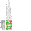
Risk of bias summary: review authors' judgements about each risk of bias item for each included study.
Allocation
We rated four of the studies as unclear in their method of sequence allocation; in each case there was no mention of the method used and we were unsuccessful in our attempts to contact the authors for clarification (Carlin 1987; McCoy 1999; Norris 1989; Prescott 1992). Of the six studies that were rated low risk of bias in this category, only three were explicit about their method in the published text (Benigni 1999; Kahle 2006; Rabe 2010). The other three studies did not mention the method of sequence generation in the text, but through correspondence with Dr Mitchell P Goldman, an author on all three studies, we were informed that a coin toss method was used (Goldman 2002; Leach 2003; Rao 2005).
Regarding concealment of allocation, only one study specifically detailed the method they used (Norris 1989). None of the other studies addressed concealment of allocation in the text; however, we learned through correspondence with Dr Mitchell P Goldman that syringes were prepared by a nurse and handed to the doctor for injection in two studies (Goldman 2002; Rao 2005).
Blinding
We rated three studies as not blinded (Benigni 1999; Carlin 1987; Leach 2003). In two of these studies, blinding was not mentioned but the comparison sclerosing agents had distinct appearances on injection (Benigni 1999; Leach 2003). In the third study, the authors claimed the study was double‐blind but the patients received agents of differing viscosities during the intervention, thereby allowing for unblinding of the treating physician during the intervention (Carlin 1987). We rated the blinding in one study as unclear because there was no mention of blinding and we could not make a definitive judgement as to whether this was related to inadequate reporting or it was an unblinded study (Prescott 1992). Of the remaining studies, two used single blinding for the treatment phase but had blinded assessors rate photographs to assess efficacy (McCoy 1999; Rao 2005). Two studies blinded patients, treating doctors, and photograph assessors (Goldman 2002; Rabe 2010) and in a third study the patients and photograph assessors were blinded but it was unclear whether the treating doctors were blinded (Kahle 2006). The remaining study blinded patients and treating doctors (Norris 1989).
Incomplete outcome data
While seven studies either addressed incomplete outcome data or did not appear to have incomplete data, we rated three studies as unclear in this area. One of these mentioned the omission of four randomised patients from the analysis but the author did not state the reason for the omission (Benigni 1999). Communication with the study author revealed that the patients were "lost of view" but we felt this explanation did not completely clarify the reason for omission or address how this may have affected the study analysis and conclusions. In the second study (Goldman 2002), we could not discern why the number of participating patients mentioned in the text did not correlate with the assignments to treatment groups as presented in the efficacy outcome data. There was no mention of withdrawals or exclusions and our attempt to clarify this discrepancy with the author did not result in a satisfactory explanation. We rated Prescott 1992 as unclear because while the text mentioned that some participants chose to discontinue treatment early, the number who did so and the effect this had on outcome data were not addressed.
Selective reporting
We rated three studies as at low risk of bias in this area (Leach 2003; McCoy 1999; Norris 1989). Seven studies were categorized as at high risk of bias. In one of these studies the data were reported incompletely and inconsistently for some outcome measures, and statistical analysis and descriptive data were not presented (Prescott 1992). The second study did not separate all outcome data for telangiectasias and the lack of statistical analysis and standard deviations prevented inclusion of the data in a meta‐analysis (Benigni 1999). In Rao 2005, some of the outcome measures were not reported separately for telangiectasias; the data were presented without a statistical analysis or standard deviations thereby precluding its incorporation into a meta‐analysis. For the fourth study (Goldman 2002), as addressed in the section on incomplete outcome data, we were unable to clarify the reason for the discrepancy in the number of patients included in the study and the number of outcomes presented. Because of this inconsistency, we couldn't include the data in a meta‐analysis since we couldn't determine the number of analysed participants. The fifth study presented the efficacy outcome from the blinded independent evaluators in a satisfactory manner but the efficacy outcome measure of the treating physician and the patient satisfaction data were not statistically analysed or presented sufficiently for inclusion in a meta‐analysis (Kahle 2006). In Carlin 1987, the patient satisfaction data and some of the adverse reactions were not statistically analysed by the sclerosing agent used. Finally, in Rabe 2010 the outcome data for telangiectasias and reticular veins were not separated in the publication; however, we were able to obtain separate data from the trial authors that we could use in a future meta‐analysis.
Other potential sources of bias
We rated four studies as having low risk of bias in this category (Benigni 1999; Kahle 2006; Leach 2003; McCoy 1999). Four studies had an unclear risk of bias for varied reasons. Three of them received funding from pharmaceutical companies (Goldman 2002; Rabe 2010; Rao 2005). Additionally in one of these studies (Rabe 2010), the choice of dose for the sclerosing agent not represented by the funder was higher than that used in clinical practice or in other studies in this area. The authors addressed this potential risk of bias in their discussion and provided an explanation for their decision to use this dose in accordance with FDA labelling within a trial conducted with the aim of obtaining FDA approval for the solution manufactured by the funder. The fourth study we rated as unclear had baseline imbalance in the comparison groups whereby one treatment group had 20% more telangiectasias (Prescott 1992). The author acknowledged this imbalance but we were unclear about whether or how this might affect the outcome data since no power calculation, statistical analysis or descriptive data were provided. Finally, we rated two studies as having high risk of bias in this category based on their shared design (Carlin 1987; Norris 1989). These studies used a complex split‐body design that separated each participant's legs into four quadrants that were each assigned to receive treatment with a different sclerosing agent. This design risks bias in two major areas. First, it presents the risk of a carry‐across effect (Lesaffre 2009). We could not state with certainty that sclerosing agents injected simultaneously and adjacently would not impact the outcome of the different treatments. Second, we felt that non‐blinded assessments of efficacy on neighbouring quadrants could be biased when viewed together, allowing their comparative appearances to alter subjective impressions.
Effects of interventions
The included studies provided a number of comparisons between sclerosing agents and against placebo. We present the comparisons below along with the relevant outcome measures that we were able to assess for each comparison. We did not undertake a meta‐analysis for the reasons detailed at the end of this section.
1. Polidocanol (POL) foam versus polidocanol (POL) solution
This comparison was addressed for telangiectasias in only one of the included studies (Benigni 1999). This was a pilot study that included 24 patients but which analysed data for only 20. The author reported improvement scores based on a summary score computed from the ordinal ratings of non‐blinded treating physicians who assessed improvement using visual analogue scales ranging from zero to 10. Summary scores ranged from 3.2/10 to 5.4/10 in the group treated with solution and from 3.6/10 to 7.3/10 in the foam‐treated group. The ratings of the patients, adverse events, quantity of sclerosant required, and the number of points of injection were not presented separately for telangiectasias. This was a small study in which no statistical analysis was undertaken and which provided insufficient evidence to determine whether POL foam and solution differ in efficacy. The data as presented were insufficient for inclusion in a future meta‐analysis.
2. Sodium tetradecyl sulfate (STS) versus glycerin
Only one very small study addressed this comparison (Leach 2003). Data from 13 patients were presented and no statistical analysis was performed. The authors reported vessel clearance and side effects in this split‐body design trial, as assessed by non‐blinded treating physicians who made determinations based on visual appearance. Seven of 13 patients achieved vessel clearance in the glycerin‐treated group compared with one of 13 in the STS‐treated group. Twelve patients in the STS group experienced hyperpigmentation and only one patient had this side effect in the glycerin group. The numbers who experienced other side effects in the glycerin versus STS groups were, respectively, 3/13 versus 2/13 for pain, 1/13 versus 7/13 for bruising, and 0/13 versus 3/13 for swelling. Given the small sample size of this single study, insufficient evidence was available to make a conclusion regarding the comparison of these two sclerosants.
3. Sodium tetradecyl sulfate (STS) versus hypertonic dextrose (HD)
One study compared these agents (Prescott 1992). Sixty patients were included, 36 of whom were treated with HD and 24 of whom were treated with STS. The summary statistics for efficacy were the mean values and ranges of the estimated per cent clearance as determined by patients and the treating doctor, and assessment of before and after pictures. The author reported an average of 70% clearance for each group as determined by the treating doctor (range 25% to 90% in the HD group, 25% to 85% in the STS group). For before and after pictures, average clearance in the HD group was 72.5% (range 25% to 90%) and in the STS group it was 70% (range 30% to 85%). For patients' assessments, average clearance in the HD group was 75% (range 25% to 100%) and in the STS group it was 70% (range 25% to 90%). Mild pain was reported by 28% of patients in the HD group and 17% of patients in the STS group. Data on side effects were provided for the following: ecchymosis ('mild to moderate' with HD, 'mild' with STS but no numeric data provided), telangiectatic matting (present in 6% of the HD group, 4% of the STS group), and post‐sclerotic pigmentation (3% with 'light' pigmentation in the HD group, 8% 'light to moderate' pigmentation in the STS group).
The author of this trial noted that STS required fewer treatment sessions (average of 4.2 sessions for HD and 2.6 for STS) and that the group treated with HD had 20% more telangiectasias at the start of the study yet they required 61% more treatments to achieve the same level of disappearance. He also suggested that his results indicated that STS was more painful on injection and caused more postsclerotic pigmentation, based on the fact that 28% in the STS group reported mild pain versus 17% in the HD group. However, no statistical analysis was performed and he did not provide data for categories other than mild with respect to pain. The data as presented are insufficient for inclusion in a future meta‐analysis. Based on this single study, there is currently insufficient evidence to judge the comparative effect of these two sclerosing agents.
4. Polidocanol (POL) at different concentrations
One study compared POL solution at 0.25%, 0.5%, 0.75% and 1% (Norris 1989). Twenty patients were randomised but only 16 were analysed. The legs of each participant were divided into four quadrants that were randomised to receive each of the four doses of POL. Data on efficacy were presented as the percentage of patients who achieved vessel clearance according to the treating physicians' visual assessment, as well as the per cent improvement as rated in each of three ordinal categories (moderate to poor, good, excellent) based on the investigators' review of photographs taken at baseline and before each treatment. The authors also presented the percentage of patients whose vessels cleared after each treatment. The adverse events of itching, neovascularisation and hyperpigmentation were recorded as absent or present.
The statistical significance of observed differences among pairwise dose comparisons was assessed using the McNemar test for the data on improvement, itching, neovascularisation, hyperpigmentation, and percentage clearing. Only one comparison yielded a P value at the specified 0.05 significance level, and this pertained to the difference in hyperpigmentation occurring between the 0.25% and 1% quadrants. For this same comparison in the 1% versus 0.5% quadrants, the P value was 0.07.
The authors concluded that patients did not have a dose preference and noted that, overall, 80% of patients were satisfied or very satisfied with the results of their treatment. Because these results come from a small study using a complex split ‐body study at high risk of bias, we concluded that there is currently insufficient evidence to determine the optimal dose of POL to achieve clearance, reduce adverse events and maximize patient satisfaction.
5. Heparsal versus polidocanol (POL), sodium tetradecyl sulfate (STS) and normal saline (NS)
Only one study (Carlin 1987) assessed heparsal (20% saline mixed with heparin 100 units/mL) as a sclerosing agent and all comparisons included in this trial are discussed together in the present section. Carlin (Carlin 1987) compared heparsal, STS 0.5%, POL 0.25% and a normal saline placebo in a split‐body four‐quadrant design involving 20 patients (the legs of 20 participants were divided into four quadrants where telangiectasias were treated with one of the four agents). Statistical analyses for outcomes of improvement, itching and pain were undertaken across all four agents using ANOVA, but not between individual treatments. Results of the ANOVA revealed that summary scores for improvement (presented for each agent as a mean of the treating physician's scores rating the level of improvement on a five point ordinal scale) differed significantly according to the treatment (heparsal 3.95, STS 3.90, POL 3.80, NS 1.05; P = 0.0001, ANOVA). Scrutiny of mean improvement scores suggested that these were higher in body quadrants treated with heparsal compared with placebo (3.95 and 1.05 respectively) but similar in quadrants treated with heparsal compared with other sclerosants (heparsal 3.95, STS 3.90, POL 3.80). However, no post hoc tests were performed to assess the statistical significance of any of these specific comparisons.
Pain and itching were rated similarly to improvement but on three‐point scales. Itching scores did not differ significantly according to treatment (heparsal 2.95, STS 1.10, POL 1.15, NS 1.00; P = 0.56, ANOVA). Pain scores differed significantly according to treatment (heparsal 2.95, STS 3.85, POL 1.15, NS 1.00; P = 0.007) although whether pain in the heparsal related quadrants differed significantly from that in the other quadrants was unclear due to lack of post hoc testing. Pain in the quadrant treated with heparsal was rated as moderate or severe in 16/20 patients and as none or mild in 4/20 patients, 2/20 patients experienced pain with POL, no patients experienced pain with NS, and 18/20 patients experienced severe pain with STS.
Given the small size of this study and the use of a complex split‐body design, we concluded that there is currently insufficient evidence regarding this comparison.
6. Polidocanol (POL) versus hypertonic saline (HS) (20% saline and 2% lignocaine)
Only one study addressed this comparison (McCoy 1999).
This split‐body trial included 81 patients who were randomised to treatment with POL on one leg and HS on the other. Efficacy was measured by the unblinded treating doctor using a scale of 0 to 10 for improvement. Efficacy was also assessed by a blinded non‐treating doctor who rated improvement from before and after photographs on the scale of 0 to 10. Analysis of the mean scores given by the unblinded treating doctor for POL and HS showed no statistically significant difference (POL 7.26 (SD 0.21), HS 7.56 (SD 0.14); P = 0.5, Wilcoxon signed ranks test). The scores for POL and HS as rated by the blinded assessments of photographs showed evidence of a difference (POL 6.93 (SD 0.20), HS 7.30 (SD 0.19); P = 0.04, Wilcoxon signed ranks test). Based on this one study that demonstrated conflicting evidence on efficacy depending on the method of assessment used, we were unable to form a conclusion about the superiority of POL versus HS. The P value of 0.04 for the analysis comparing blinded assessments of photographs indicated that the observed difference, or more extreme difference, was only 4% likely to have occurred by chance alone. The blinded assessments eliminated allocation bias and performance bias; however, we can't be certain that the photographic technique didn't introduce measurement bias. The data from this study are suitable for inclusion in a future meta‐analysis, so further research into this comparison could assist in addressing this question.
Patient satisfaction was assessed using a 0 to 10 scale, and there was no statistically significant difference between POL and HS (POL 7.20 (SD 0.19), HS 7.23 (SD 0.14); P = 0.4, paired t‐test).
Pain of injection rated on a scale of 0 to 10 by patients was greater for HS than for POL (mean scores POL 2.79 (SD 0.15), HS 3.84 (SD 0.18); P = 0.00001, paired t‐test).
Telangiectatic matting and haemosiderin staining were rated by the unblinded treating physician on a scale of 0 to 3, and for each adverse event there was evidence of a higher incidence in the POL group than the HS group, although in both cases these effects were relatively mild. Mean scores and P value obtained with the Wilcoxon signed ranks test were: POL 0.54 (SD 0.84), HS 0.33 (SD 0.52) (P = 0.04). The results for haemosiderin staining were: POL 1.15 (SD 0.91), HS 0.77 (SD 0.83) (P = 0.003).
7. Polidocanol (POL) versus sodium tetradecyl sulfate (STS)
Four studies included a comparison of POL versus STS (Carlin 1987; Goldman 2002; Rabe 2010; Rao 2005). Two of these studies looked solely at POL versus STS (Goldman 2002; Rao 2005) and the other two included these agents in trials comparing multiple treatments (Carlin 1987; Rabe 2010).
In Goldman 2002, 129 patients were included and 42 were reported to have telangiectasias. Each patient received STS on one leg and POL on the other. Only efficacy data were reported specifically for veins < 1 mm in size. In this category, there were 32 legs treated with STS and 26 treated with POL. Given that 42 patients were reported to have veins < 1 mm, we could not ascertain whether the missing data in each treatment category were related to patient withdrawal, exclusion, refusal of treatment, leg vein asymmetry or another cause. The data presented for each vein category also did not add up to the reported number of participants with each vein size. We considered that perhaps some patients were included in more than one group but, because these concerns were not clarified in the methods section, we were unable to explain the contradiction between the numbers presented in the data table and the reported methodological approach. We were unsuccessful in our attempt to clarify this discrepancy through correspondence with the author. Because we could not determine the number of participants represented by these data, we could not include them in a meta‐analysis. The efficacy data were presented as an average rating computed from combining the scores given by three blinded vascular surgeons rating overall disappearance on a 5‐point scale. Statistical analysis revealed no significant difference in disappearance between POL and STS at the 5% level. Patients were reported to be 70% to 72% satisfied with treatment in each vein category. Data on adverse events were not presented separately for telangiectasias.
In Rao 2005, 20 patients were included and their veins were treated with STS on one leg and POL on the other. Veins < 1 mm were treated with either STS in a 0.25% solution or POL in a 0.5% solution. Foam was used only for larger veins. Data for efficacy were presented as the average rating on a 5‐point ordinal scale provided by four evaluators, based on reviewing high‐quality digital photographs taken at baseline and 12 weeks post‐treatment. Standard deviations were not provided and no statistical analysis was performed, so we could not include the data in a meta‐analysis. Data on adverse events, patient tolerability and satisfaction were not presented separately for telangiectasias. This small study provided insufficient evidence for us to make conclusions about the comparison between STS and POL.
Results of the trial by Carlin et al (Carlin 1987) have been described in section 5. In addition to assessing STS and POL, this trial included heparsal and a normal saline placebo. Results of ANOVA undertaken across all four treatment groups indicated statistically significant differences in improvement scores according to treatment (STS 3.90, POL 3.80, heparsal 3.95, normal saline 1.05; P = 0.0001, ANOVA) and pain (heparsal 2.95, STS 3.85, POL 1.15, NS 1.00; P = 0.007, ANOVA) but not itching (heparsal 2.95, STS 1.10, POL 1.15, NS 1.00; P = 0.56, ANOVA). Mean improvement scores were similar for STS and POL (3.90 and 3.80 respectively) as were mean itching scores (1.10 and 1.15 respectively). However, the statistical significance of differences in mean scores for these specific comparisons was not determined in post hoc testing.
Data for hyperpigmentation and neovascularisation were not statistically analysed or presented sufficiently as descriptive data that could be incorporated into a future meta‐analysis. Patient satisfaction was also not statistically analysed and results were not presented by sclerosant. This small study did not provide evidence of a difference in the level of improvement achieved with POL versus STS but suggests that there may be increased pain with STS relative to POL. However, this study is small and prone to bias because of the complex split‐body design.
In the Rabe 2010 study, 160 of the total 316 randomised patients had spider veins. These patients were randomised to receive either POL 0.5%, STS 1%, or normal saline placebo in a 3:2:1 ratio. A total of 157 patients were included in the full analysis. While the data were not presented separately for telangiectasias in the published study, we were able to obtain these data from the study authors. Efficacy scores were presented as a summary score, along with the standard deviation (SD), derived from the median ratings on a 5‐point ordinal scale as provided by the investigator, and two blinded independent observers who reviewed photographs taken using a custom‐built digital imaging system. Twelve weeks after the last injection, this score was 4.42 (SD 0.69) for POL, and 4.38 (SD 0.83) for STS. At 26 weeks post‐treatment, the score was 4.44 (SD 0.75) for POL and 4.31 (SD 0.81) for STS. These data were not statistically analysed but were presented in a manner suitable for future meta‐analysis. The authors also provided descriptive data on patient satisfaction with the treatment at 12 and 26 weeks after treatment, as rated on a 5‐point Likert scale. These data were not statistically analysed but were suitable for inclusion in a meta‐analysis. While more patients were very or somewhat unsatisfied with STS than with POL (20% versus 4% at 12 weeks, 19% versus 7% at 26 weeks, respectively) and more were satisfied or very satisfied with POL (68% for POL versus 25% for STS at 12 weeks, 64% versus 27% at 26 weeks), we noted, as acknowledged by the authors, that the 1% dose of STS used in this trial is not used in clinical practice for veins smaller than 1 mm.
We decided not to perform a meta‐analysis for this comparison. As stated above, two of the studies did not present data in a way that was suitable for meta‐analysis (Goldman 2002; Rao 2005), leaving us with two studies for consideration (Carlin 1987; Rabe 2010). Further discussion of our decision not to combine these studies is provided at the end of this section.
8. Polidocanol (POL) versus normal saline (NS) placebo
Three studies were included this comparison. Two of them, as previously mentioned, included these agents in a multiple treatment design (Carlin 1987; Rabe 2010). The third study compared POL to normal saline in a randomised, double‐blind placebo‐controlled trial (Kahle 2006).
The Kahle 2006 study included 48 patients who were randomised to receive one treatment with either POL or normal saline placebo. Efficacy was assessed through evaluation of before and after photographs by two blinded consultants who rated improvement on a scale of 0 to 100, as well as by assessment of the treating doctor who rated improvement on a scale of 0 to 3. The mean scores for POL and placebo as rated by one consultant were 31 and 15.3 respectively (P = 0.0004, Mann‐Whitney U Test). The mean scores for the other consultant were 30 for POL and 16.3 for placebo (P = 0.0004, Mann‐Whitney U Test). A Wilcoxon matched pairs signed rank test performed using the ratings of the two consultants yielded a median of 17.25 for the POL group (P = 0.0013) and a median of 0 for the placebo group (P = 0.292). The treating doctor judged the efficacy of POL on a scale of 0 to 3 with a mean 1.41, and rated the placebo group a mean value of 0.22. The mean of patient satisfaction scores was 2.09 with POL and 0.91 with placebo. Treating doctor and patient satisfaction scores were not statistically analysed or presented with standard deviations so they cannot be included in a future meta‐analysis. The efficacy data obtained from the analysis of the blinded independent raters contributed evidence supporting the superiority of POL versus placebo in treating telangiectasias of the lower limbs.
Results of the trial by Carlin et al (Carlin 1987) have been described in section 5. In addition to assessing POL and normal saline, this trial included heparsal and STS. Results of the ANOVA undertaken across all four treatment groups indicated statistically significant differences according to treatment for improvement scores (POL 3.80, normal saline 1.05, heparsal 3.95, STS 3.90; P = 0.0001, ANOVA) and pain (POL 1.15, NS 1.00, heparsal 2.95, STS 3.85; P = 0.007, ANOVA) but not itching (POL 1.15, NS 1.00, heparsal 2.95, STS 1.10; P = 0.56, ANOVA). The mean improvement score for POL (3.80) was higher than that for normal saline (1.05) and itching scores were similar for the two agents (POL 1.15, normal saline 1.00), as were mean pain scores (POL 1.15, normal saline 1.00). However, the statistical significance of differences in mean scores for these specific comparisons was not determined by post hoc testing.
In Rabe 2010, the mean improvement ratings for POL and NS were 4.42 (SD 0.69) and 2.11 (SD 0.58) respectively at 12 week follow up. At 26 week follow up, the ratings were 4.44 (SD 0.75) for POL and 2.19 (SD 0.62) for NS. At 12 weeks and 26 weeks, the scores for patient satisfaction were much higher in the POL group than in the NS group but we do not know the statistical significance of this finding. In the POL group, 87.2% of patients were either satisfied or very satisfied at 12 weeks, and the figure was 81% at 26 weeks. In the NS group, only 7.4% of patients were satisfied or very satisfied at 12 weeks, and 3.7% were in these categories at 26 weeks. While these results were not statistically analysed, the data were presented in a way suitable for inclusion in a future meta‐analysis.
As discussed previously, we decided not to combine Carlin 1987 and Rabe 2010 in a meta‐analysis. We also decided not to combine Kahle 2006 with either of these studies. Our reasoning was that Kahle 2006 differed greatly from the other studies in the length of follow up. In both Carlin 1987 and Rabe 2010, patients received multiple treatments with the aim of achieving resolution. In Carlin 1987, patients were treated every four weeks up to a maximum of six treatments. In Rabe 2010, patients were treated every three weeks up to a maximum of three treatments and efficacy was analysed at 12 and 26 weeks post‐treatment. This contrasts sharply from the follow up in Kahle 2006 where patients were treated once only and efficacy was assessed at one week and four weeks. The data from Kahle 2006, if converted to a scale similar to that used in the other studies, would have contributed lower efficacy scores based on this difference in study design. These data would be more effectively combined if we had outcome measures of efficacy from all three studies after only one treatment was administered. Including the Kahle 2006 data in a meta‐analysis with either of these studies at this point would needlessly bias the summary effect statistic in the direction of no effect.
9. Sodium tetradecyl sulfate (STS) versus normal saline (NS) placebo
Two studies were included in this comparison (Carlin 1987; Rabe 2010).
Results of the trial by Carlin et al (Carlin 1987) have been described previously. In addition to assessing STS and normal saline, this trial included heparsal and POL. Results of ANOVA undertaken across all four treatment groups indicated statistically significant differences according to treatment for improvement scores (POL 3.80, normal saline 1.05, heparsal 3.95, STS 3.90; P = 0.0001, ANOVA) and pain (POL 1.15, NS 1.00, heparsal 2.95, STS 3.85; P = 0.007, ANOVA) but not itching (POL 1.15, NS 1.00, heparsal 2.95, STS 1.10; P = 0.56, ANOVA). The mean improvement score for STS (3.90) was higher than that for normal saline (1.05) but itching scores were similar for the two agents (STS 1.10, normal saline 1.00). Mean pain scores were higher for STS (STS 3.85, normal saline 1.00) with 18 of the 20 patients experiencing severe pain in the STS quadrant and none of the patients experiencing pain in the placebo quadrant. However, the statistical significance of differences in mean scores for the specific comparison of STS versus normal saline was not determined by post hoc testing.
In Rabe 2010, the mean score at 12 weeks follow up was 4.38 (SD 0.83) for STS and 2.11 (SD 0.58) for NS. At 26 weeks, the mean score was 4.31 (SD 0.81) for STS and 2.19 (SD 0.62) for NS. Patient satisfaction scores at both 12 and 26 weeks revealed that only 7.4% of patients were satisfied or very satisfied with NS. At 12 weeks 49% were satisfied or very satisfied with STS, and 53% were satisfied or very satisfied at 26 weeks. These data were not statistically analysed but they were presented in a manner suitable for a future meta‐analysis.
Why we decided not to perform a meta‐analysis
At the conclusion of our review, we found that only two studies lent themselves to consideration of meta‐analysis (Carlin 1987; Rabe 2010). Both studies included multiple treatment groups, three in Rabe 2010 and four in Carlin 1987, and in theory we could have analysed STS versus POL, STS versus placebo, and POL versus placebo. Combining these two studies in a meta‐analysis introduced complexity not only with respect to the need for a multiple treatments meta‐analysis but also with respect to the challenges posed by the merging of a complex split‐body design with a parallel design. When analysing data from a study with multiple treatment groups, care must be taken to avoid double‐counting groups in the shared intervention, thereby creating a unit of analysis error in the meta‐analysis (Higgins 2008). We would have risked error in this regard were we to conduct pair‐wise comparisons using data from these two studies to meta‐analyse STS versus POL and then, additionally, POL versus placebo and STS versus placebo. Approaches used to avoid the unit of analysis error include combining groups, for example all interventions versus placebo, or selecting only one comparison, for example POL versus STS. Another option was to divide the number of participants according to the number of comparisons made and treat them as independent comparisons (Higgins 2008). Finally, we could have carried out a multiple‐treatments meta‐analysis (Higgins 2008). While these options presented statistical solutions to address the combination of data from Rabe 2010 with another study, they did not satisfactorily address the challenges of combining the data with Carlin 1987 because of the complex split‐body design.
The complex split‐body design posed numerous issues with respect to the introduction of bias. At the analysis stage, the investigators risked biased judgments through the unavoidable proximity of comparison groups, especially in this case where blinding was jeopardized by the differing viscosities of the injected substances. At the recruitment stage, participants without symmetrical diagnostic findings were excluded, and this affected the external validity of the results (Lesaffre 2009). More troublesome was the risk of carry‐across effect. In the case of the Carlin 1987 trial, there was no physical barrier and no statistical test (Lesaffre 2009) to ensure that the solutions tested did not alter the outcome in the adjacent quadrants. The statistical analysis of this study design was necessarily complex and, without adequate detail concerning the approach taken, we could not be certain that all relevant aspects were considered. Adding to this concern was the presentation of the Carlin 1987 data, analysed by ANOVA, which included a P value and a summary statistic for each agent tested. Without standard deviations we had the option to impute the F statistic and calculate the within‐group variance mathematically using the numerator and denominator degrees of freedom. Imputation risked making assumptions and, in this case, we would have needed to assume that all four agents had the same within‐group standard deviation. The Cochrane Handbook for Systematic Reviews of Interventions recommends that imputed data be used for only a small proportion of data in a meta‐analysis (Higgins 2008), a rational recommendation considering the bias such assumptions could introduce. In the case of the Carlin 1987 data the within‐group standard deviation that we calculated was 0.22. This was much smaller than standard deviations seen in other studies and it had the potential to increase the weight given to these data in the analysis.
Meta‐regression can be used to combine split‐body and parallel designs; however, care must be taken with this approach given that data from the two designs were analysed through different techniques. Their combination risked biasing the summary confidence interval and distorting the impact of clinical heterogeneity (Lesaffre 2009). A preferred approach was to analyse split‐body designs and parallel deigns separately as subgroups (Lesaffre 2009), rather than combining them.
In summary, we determined that the bias inherent in this poorly blinded, complex split‐body design, combined with the bias introduced through imputing within‐group standard deviations from an ANOVA, would have contributed unacceptable bias to a meta‐analysis that included only one other trial that had an unequal ratio parallel design. The effect estimate in such a meta‐analysis would be highly biased, particularly in a random‐effects analysis where small studies contribute greater weight to the estimate (Higgins 2008). A common criticism of meta‐analysis is that it risks perpetuating bias. If we knowingly combined studies in a meta‐analysis that was at high risk of bias, we violated not only the principles of good meta‐analysis but also risked perpetrating the ethically dubious act of injecting knowingly biased data into the published research literature.
Discussion
Summary of main results
This review included studies which evaluated a number of sclerosing agents used in the treatment of telangiectasias of the lower limbs. The available research does not provide evidence which allows us to make conclusions regarding the comparisons of the following: polidocanol (POL) foam versus POL solution (Benigni 1999), sodium tetradecyl sulfate (STS) versus glycerin (Leach 2003), and sodium tetradecyl sulfate (STS) versus hypertonic dextrose (HD) (Prescott 1992). There is also insufficient evidence regarding the optimal dose of POL solution to use in treating telangiectasias, based on the one trial which addressed this question (Norris 1989).
Statistically significant evidence from the included trials suggests that STS, POL and heparsal have higher efficacy than a normal saline placebo. There is currently no evidence to support the superiority of any one sclerosing agent over any other agent in terms of efficacy.
There is no evidence to show an increase in patient satisfaction with any one agent versus another, but there is evidence that patients are less satisfied with placebo. While the descriptive data from the Rabe 2010 study suggests increased patient satisfaction with POL 0.5% versus STS 1%, this difference must be considered with caution in any future meta‐analysis since the dose of STS used in this study is higher than that used in practice for telangiectasias. Goldman reports equivalent patient satisfaction with POL 0.5% versus STS 0.25%, although these data were not statistically analysed (Goldman 2002).
There is some evidence that POL is less painful than heparsal and STS, and that STS is more painful than heparsal. There is also evidence that POL, unlike STS and heparsal, is no more painful than placebo. These data were statistically significant, although they were obtained using the complex split‐body design that we rated as highly biased. While the finding of increased pain may be statistically significant, it is unclear whether this is clinically significant in terms of overall patient satisfaction. One study revealed more pain with HS than with POL. The authors also noted an increase in mild haemosiderin staining and telangiectatic matting with POL, but the increase in side effects may be attributable to the 1% dose used in this study that is higher than that typically used in clinical practice for veins < 1 mm in diameter.
Overall completeness and applicability of evidence
There is currently no evidence favouring the use of one sclerosing agent over another for the treatment of telangiectasias of the lower limbs. The 10 studies included in our systematic review included comparisons of seven sclerosing agents, as well as normal saline placebo. The data on three of these sclerosing agents were presented in a manner insufficient for us to base conclusions or incorporate in a future meta‐analysis (Benigni 1999; Leach 2003; Prescott 1992). The remaining studies address STS, POL, heparsal, and HS in comparison to one another or to placebo. Heparsal is addressed in only one study that uses a four quadrant split‐body design that we rated as high risk of bias (Carlin 1987). Hypertonic saline is addressed in one study only in comparison to POL at a concentration of 1%. This sclerosant may benefit from further investigation in comparison to agents administered in the doses commonly used in clinical practice (McCoy 1999). Polidocanol and STS are investigated in most of the studies either together with other agents, in comparison to each other, or individually against a different agent. One of the studies that includes both POL and STS used the highly‐biased four quadrant design (Carlin 1987). The two studies that compare these agents to each other both consisted of bias that prevents their inclusion in a future meta‐analysis (Goldman 2002; Rao 2005). The final study comparing these agents met the highest methodological standards (Rabe 2010), but the concentration of STS used is much higher than that commonly used for veins < 1mm in practice. Additionally, while we received separate outcome data for telangiectasias from the authors, they did not present and analyse these data in their published findings.
The studies present very limited demographic data. We specified in our protocol that we were interested in exploring differences in outcomes based on pregnancy history in women, and based on body mass index in men and women. The epidemiological literature on telangiectasias is rich with data on symptoms, quality of life, and the co‐existence of telangiectasias with more severe venous disease. Without detail about the characteristics of the patients studied, we are unable to determine which patients are more likely to benefit from treatment or how patient characteristics impact outcome measures.
Quality of the evidence
The overall quality of the evidence available to investigate sclerotherapy for the treatment of telangiectasias of the lower limbs is poor. Three of the 10 relevant trials did not include statistical analysis or present data in a manner suitable for future inclusion in a meta‐analysis (Benigni 1999; Leach 2003; Prescott 1992). Two trials were conducted in a highly‐biased four‐quadrant split‐body design (Carlin 1987; Norris 1989). Two trials that were funded by pharmaceutical stakeholders presented data at high risk of bias for selective reporting and which was insufficiently presented for inclusion in a meta‐analysis (Goldman 2002; Rao 2005). Of the three studies of the highest quality, one provided limited data but did show evidence of effectiveness of POL versus placebo (Kahle 2006). Another provided evidence of comparability between hypertonic saline and POL at the dose of 1%, which is higher than that used in current clinical practice, and suggested fewer side effects but more pain with hypertonic saline (McCoy 1999). The third (Rabe 2010) was of the highest methodological quality and presented data that could be included in a future meta‐analysis. However, this study was funded by a pharmaceutical stakeholder and was conducted specifically for the purpose of obtaining FDA approval of POL. This aim appears to have biased the choice of dose for the comparison sclerosant STS and risks biasing a future meta‐analysis that includes these data. No studies reported a power calculation.
Potential biases in the review process
While we conducted a thorough search according to the Cochrane review protocols, we may have missed some unpublished studies or relevant grey literature. We attempted to contact trial authors and, where we were successful, we gained information that altered our risk of bias scores in the direction of low bias in the allocation category (Goldman 2002; Leach 2003; Rao 2005). Without this communication we would have rated the risk of allocation bias as unclear for these studies. We would also have excluded the Leach et al trial for unclear explanation in the text that it was a randomised trial. Conversely, we did not have the opportunity to clarify information with some study authors. Given that we cannot be certain that our corrections represent all those that were possible had we obtained clarification, we may have created new bias about the overall evidence base. However, the alternative would have been to base our decisions on the published texts alone and, given discrepancies in journal standards and authors' knowledge and compliance with the CONSORT guidelines (Schulz 2010), we would risk biasing our judgments in the opposite direction.
Agreements and disagreements with other studies or reviews
There are no other reviews addressing this specific question for comparison.
Authors' conclusions
Implications for practice.
There is currently no evidence suggesting superior efficacy or increased patient satisfaction with any one sclerosant. There is evidence that STS, POL and heparsal are superior to placebo. Patient preference seemed to be unaffected, even in studies that showed some evidence of increased pain or adverse events with one agent versus another. Perhaps these differences, while statistically significant, are not clinically significant. There is evidence to suggest that POL causes less pain than STS, heparsal and hypertonic saline; however, some of this research was conducted using a study design that we considered biased.
The available evidence on the optimal dose of POL for the treatment of telangiectasias is obtained from one study with a highly biased design. This study shows no statistically significant evidence for improved efficacy with any particular dose, but does suggest that the 1% strength contributes to more hyperpigmentation than the lower doses. Another study compared POL to hypertonic saline and found a statistically significant increase in mild telangiectatic matting and haemosiderin staining with POL. However, the dose of POL used in this study was 1%, the same dose found to be associated with increased side effects in the comparative dose trial.
The unpublished descriptive data we have available from one trial comparing POL, STS and placebo demonstrate decreased patient satisfaction with STS; however, the STS was studied at a concentration of 1%. This is not the dose commonly used in practice for veins < 1 mm.
Until further evidence is available regarding the optimal sclerosing agent and dose for the treatment of lower limb telangiectasias, it may be advisable to use doses of POL and STS in clinical practice that are lower than 1% to reduce side effects.
Implications for research.
More research is needed to contribute to evidence‐based decision‐making concerning the optimal sclerosing agent(s) and dosing required for achieving maximum efficacy while reducing adverse reactions and enhancing patient satisfaction. We recommend randomised double‐blind trials that are sufficiently powered for statistical analysis. We advise caution in using split‐body designs as this jeopardizes blinding, risks carry‐across effects, and may contribute to performance bias. More emphasis needs to be placed on publishing trial findings in accordance with the CONSORT guidelines (Schulz 2010). Demographic data on participants would enhance our ability to define the impact of treatment on different risk groups and would allow the recent advances in our epidemiological understanding about venous disease to contribute to treatment advances in different populations. We also recommend that researchers present data in a manner that allows for future incorporation in a meta‐analysis, and that they consider this contribution in their choice of study design.
What's new
| Date | Event | Description |
|---|---|---|
| 15 October 2021 | Review declared as stable | This review has been superseded by Cochrane Review 'Treatment for telangiectasias and reticular veins': www.cochranelibrary.com/cdsr/doi/10.1002/14651858.CD012723.pub2/full and will no longer be updated. |
History
Protocol first published: Issue 11, 2010 Review first published: Issue 12, 2011
Notes
Acknowledgements
We would like to acknowledge Ashma Krishan for her statistical expertise on meta‐analysis and Bianca Jacubowski for her assistance with German translation.
Appendices
Appendix 1. CENTRAL search strategy
#1 MeSH descriptor Sclerotherapy explode all trees (426) #2 MeSH descriptor Sclerosing Solutions explode all trees (344) #3 (sclero*):ti,ab,kw (5286) #4 (tetradecyl near2 (sulfate or sulphate)):ti,ab,kw (53) #5 MeSH descriptor Sodium Tetradecyl Sulfate explode all trees (35) #6 MeSH descriptor Saline Solution, Hypertonic explode all trees (315) #7 MeSH descriptor Ethanolamines, this term only (1221) #8 (polydocanol or polidocanol or lauromacrogol):ti,ab,kw (145) #9 (saline):ti,ab,kw (12610) #10 (ethanolamine near2 oleate):ti,ab,kw (56) #11 (sodium near2 morrhuate):ti,ab,kw (17) #12 (sotradecol):ti,ab,kw (6) #13 (aetoxisclerol or aethoxysclerol):ti,ab,kw (14) #14 (aetoxiskerol or aethoxyskerol):ti,ab,kw (1) #15 (Turbofoam):ti,ab,kw (2) #16 (foam* or microfoam*):ti,ab,kw (824) #17 (varisolve):ti,ab,kw (2) #18 (#1 OR #2 OR #3 OR #4 OR #5 OR #6 OR #7 OR #8 OR #9 OR #10 OR #11 OR #12 OR #13 OR #14 OR #15 OR #16 OR #17) (19880) #19 MeSH descriptor Telangiectasis explode all trees (82) #20 ((reticular or thread or spider) near3 (vein* or venous)):ti,ab,kw (21) #21 (microvaric* or telangiectas*):ti,ab,kw (214) #22 (#19 OR #20 OR #21) (221) #23 (#18 AND #22) (31)
Appendix 2. Data extraction form
| RevMan Headings |
Information to be Recorded: |
|
Methods |
Study design: Method of randomisation: Concealment of allocation Blinded: No o Single o Double o Triple o Unclear o Power calculation: Number of patients randomised: Number of patients analysed: Number of exclusions post randomisation: Number of withdrawals and reasons: ÒIntention to treatÓ analysis: Source of funding: Not stated o Pharmaceutical o Other o Description: |
|
Participants |
Country: Setting/Location: Number of centres: No: Age: Sex: Inclusion criteria: Exclusion criteria: |
| Interventions |
Treatment(s): Control /Placebo Duration: |
| Outcomes |
Primary outcomes: Secondary outcomes: |
| Notes |
Add any additional relevant information Invitation to authors to add and audit review data: not necessary o no reply o declined o extra data provided o review data confirmed o |
Appendix 3. Risk of bias form
| Item |
Information to be Recorded: |
|
Item: Adequate sequence generation |
Q: Was the allocation sequence adequately generated? Judgement: Yes o Unclear o No o Description: |
|
Item: Allocation concealment |
Q: Was allocation adequately concealed? Judgement: Yes o Unclear o No o Description: |
| Item: Blinding |
Q: Was knowledge of the allocated interventions adequately prevented during the study? Judgement: Yes o Unclear o No o Description: |
| Item: Incomplete outcome data addressed |
Q: Were incomplete outcome data adequately addressed? Judgement: Yes o Unclear o No o Description: |
| Item: Free of selective reporting |
Q: Are reports of the study free of suggestion of selective outcome reporting? Judgement: Yes o Unclear o No o Description: |
| Item: Free of other bias |
Q: Was the study apparently free of other problems that could put it at a high risk of bias? Judgement: Yes o Unclear o No o Description: |
Characteristics of studies
Characteristics of included studies [ordered by study ID]
Benigni 1999.
| Study characteristics | ||
| Methods | Study design: multicentre, split‐body, randomised comparative trial (a pilot study) comparing aetoxisclerol in foam and liquid solution Method of randomisation: drawing of lots Blinding: patient ‐ no, treating doctor ‐ no Power calculation: no Number of patients randomised: 24 Number of patients analysed: 20 Number of exclusions post‐randomisation: 4 (information obtained through personal correspondence with Jean‐Patrick Benigni, MD) Number of withdrawals and reasons: none Source of funding: not stated |
|
| Participants | Setting: 4 outpatient centres (information obtained through personal correspondence with Jean‐Patrick Benigni, MD) Country: France Total number: 20 Age: mean age 37.7 (range of ages 18 to 65) Sex: female Weight: mean weight 58.1kg Height: mean height 162.7cm Body mass index (BMI): mean 22.0 History of pregnancy: mean 0.75 Employed: 17/20 Sports: 7/20 Smoking: 2/20 Family history of venous disease: 17/20 Oral contraceptive use: 5/20 Inclusion criteria: "clear phototype" (classifications I to III on the Fitzpatrick classification scale), "roughly symmetrical reticular veins and telangiectases of the lateral thigh surface", reflux of reticular varices of at least 1 point on continuous Doppler Exclusion criteria: incompetence of the large saphenous veins or their collaterals, incompetence of the deep veins, perineal varices, skin condition of the lateral face of the thigh, obesity (BMI > 30), pregnancy, psychiatric trouble, a contraindication to sclerotherapy, hormonal treatment within 6 months of enrolment |
|
| Interventions | After randomisation, one thigh was treated with polidocanol foam 0.25% by the Monfreux method, and the other was treated with polidocanol solution 0.25%. Duration of follow up: 5 sessions every 15 ± 2 days, and a control visit 15 days after the last session |
|
| Outcomes | Efficacy: appearance of telangiectasias on visual analogue scales (0 to 10) as assessed by the treating doctors (days 0, 15, 30, 45, 60 & 75) and by patients (days 0 and 75). Four criteria were assessed: colour, density, relief and surface Adverse effects: as assessed by the treating physician: ecchymosis, thrombi, microthrombi, matting, pigmentation, necrosis Number of points of injection The quantity of sclerosant injected |
|
| Notes | Outcomes were not presented separately for telangiectasias for: patient assessment of efficacy on visual analogue scale, adverse reactions, number of injection points, and quantity of sclerosant injected. | |
| Risk of bias | ||
| Bias | Authors' judgement | Support for judgement |
| Random sequence generation (selection bias) | Low risk | Drawing of lots |
| Allocation concealment (selection bias) | Unclear risk | Not stated |
| Blinding (performance bias and detection bias) All outcomes | High risk | Blinding is not mentioned for patients or treating doctors. Foam and liquid sclerosants have distinct appearances on injection. |
| Incomplete outcome data (attrition bias) All outcomes | High risk | 4 people were "lost of view" as per personal communication with Dr. Benigni. No explanation was provided in the published article. |
| Selective reporting (reporting bias) | High risk | The only outcome data separated for telangiectasias is appearance on visual analogue scales as rated by the treating doctors. No statistical analysis was performed, and no standard deviations were presented, thereby preventing inclusion of the data in a meta‐analysis. |
| Other bias | Low risk | No additional contributors to bias. |
Carlin 1987.
| Study characteristics | ||
| Methods | Study design: split‐body, randomised, double‐blinded, double‐paired, placebo‐controlled
study comparing polidocanol (aetoxisclerol), sodium tetradecyl sulfate, heparsal, and normal saline. Method of randomisation: not mentioned. Author contacted, but no reply received. Blinding: patient ‐ yes, treating doctor ‐ yes Power calculation: No Number of patients randomised: 20 (4 quadrants of legs on each patient) Number of patients analysed: 20 Number of exclusions post‐randomisation: 0 Number of withdrawals and reasons: 0 Source of funding: not stated |
|
| Participants | Setting: outpatient site Country: USA Total number: 20 Age: range from 29 to 55 Sex: female Inclusion criteria: "bilaterally symmetrical telangiectasias of the lower extremities" Exclusion criteria: patients taking hormonal therapy, anticoagulants or disulfiram |
|
| Interventions | Each patient's legs were divided into 4 quadrants: 1) treated with sodium tetradecyl sulfate (STS) 0.5% 2) treated with polidocanol (POL) 0.5% 3) heparsal (saline 20% with heparin 100 units/mL) 4) placebo ‐ normal saline 0.9% Patients wore gradient support hose for 48 hours after each sclerotherapy procedure Duration: treated every 4 weeks until resolution or up to a maximum of 6 visits (maximum of 24 weeks) |
|
| Outcomes | 1) efficacy: clinical assessment of improvement: scale of 1 to 5 (poor, fair, moderate, good, excellent) 2) adverse events: a) itching: scale of 1 to 4 (none, mild, moderate, severe) b) hyperpigmentation: scale of 1 to 3 (mild, moderate, severe) c) neovascularisation: scale of 1 to 3 (mild, moderate, severe) 3) patient satisfaction (dissatisfied, neither, satisfied, very satisfied) 4) pain: scale of 1 to 4 (none, mild, moderate, severe) 5) number of treatments required for clearance (from 1 to >6) |
|
| Notes | ||
| Risk of bias | ||
| Bias | Authors' judgement | Support for judgement |
| Random sequence generation (selection bias) | Unclear risk | Not mentioned. Unsuccessful in attempt to contact authors. |
| Allocation concealment (selection bias) | Unclear risk | Not mentioned. Unsuccessful in attempt to contact authors. |
| Blinding (performance bias and detection bias) All outcomes | Unclear risk | States double‐blind, but no further details provided. Sclerosants used are identifiable by differing viscosities. |
| Incomplete outcome data (attrition bias) All outcomes | Low risk | No missing outcome data |
| Selective reporting (reporting bias) | High risk | Some of the adverse reactions were not statistically analysed by sclerosing agent used. Patient satisfaction data was not separated based on sclerosing agent used. |
| Other bias | High risk | The study design divides each person's legs into four quadrants to receive separate simultaneous treatments and analysis, and thereby risks bias by carry‐across effect. |
Goldman 2002.
| Study characteristics | ||
| Methods | Study design: multicentre, randomised, double‐blinded study comparing polidocanol and sodium tetradecyl sulfate Method of randomisation: coin toss (information obtained through personal correspondence with study author Mitchell P Goldman, MD) Blinding: patient ‐ yes, treating doctor ‐ yes, photographic outcome assessors ‐ yes Power calculation: not mentioned Number of patients randomised: 129 (42 with veins < 1 mm) Number of patients analysed: 129 (42 with veins < 1 mm) Number of exclusions post‐randomisation: unclear Number of withdrawals post‐randomisation: unclear Source of funding: Kreussler & Co GmbH (manufacturer of sclerosant), Beisdorf‐Jobst (manufacturer of compression hosiery) |
|
| Participants | Setting: multiple outpatient centres Country: USA Total number: 129 (42 with veins < 1mm) Age: Not mentioned Sex: Not mentioned Inclusion criteria: presence of varicose, reticular, and/or telangiectatic leg veins Exclusion criteria: incompetence at the saphenofemoral or saphenopopliteal junctions |
|
| Interventions | Treatment 1: polidocanol (veins < 1 mm 0.5%; 1 to 3 mm 1.0%; 3 to 6 mm 3%).
Treatment 2: sodium tetradecyl sulfate (veins < 1 mm 0.25%; 1 to 3 mm 0.5%; 3 to 6 mm 1.5%) Duration: 16 weeks |
|
| Outcomes | 1) efficacy: three vascular surgeons blinded to intervention agent assessed photographs from baseline, week 1, week 4, and week 16 on a scale of 1 to 5 2) adverse sequelae ‐ data was not separated for veins < 1mm 3) patient satisfaction ‐ data was not separated for veins < 1mm |
|
| Notes | Data was not separated for veins < 1mm for adverse sequelae or patient satisfaction. | |
| Risk of bias | ||
| Bias | Authors' judgement | Support for judgement |
| Random sequence generation (selection bias) | Low risk | Coin toss as per personal correspondence with Mitchell P Goldman, MD. |
| Allocation concealment (selection bias) | Low risk | Physician was given the solution to inject by nurse (personal communication with Mitchell P Goldman, MD). |
| Blinding (performance bias and detection bias) All outcomes | Low risk | Treating physician and patients were blinded to the agent being injected. Three vascular surgeons blinded to treatment and study centre evaluated photographs. |
| Incomplete outcome data (attrition bias) All outcomes | Unclear risk | 42 patients with veins < 1mm were randomised to receive STS 0.25% or POL 0.5%. Data presented was N = 32 for the STS group and N = 26 of the POL group. No explanation was provided. |
| Selective reporting (reporting bias) | High risk | Not all outcome data was separated based on vein size. Patient satisfaction data was mentioned only in the abstract, and not described in the methods or results section. As addressed in assessment of incomplete outcome data, it is unclear if the low number of veins treated in each group are related to missing data or to baseline asymmetry of vein presence on each patient's legs. Unsuccessful in attempt to clarify this discrepancy via communication with study author. |
| Other bias | Unclear risk | The study was partly funded by Kreussler & Co GmbH (manufacturer of sclerosant) and Beisdorf‐Jobst (manufacturer of compression hosiery). |
Kahle 2006.
| Study characteristics | ||
| Methods | Study design: randomised, double‐blinded, placebo‐controlled study comparing polidocanol and normal saline Method of randomisation: randomised into 4 blocks with the randomisation program of Microsoft Excel Blinding: patient ‐ yes, treating doctor ‐ unclear, photo evaluator ‐ yes Power calculation: not mentioned Number of patients randomised: 48 Number of patients analysed: 48 Number of exclusions post‐randomisation: 0 Number of withdrawals post‐randomisation: 0 Source of funding: not stated |
|
| Participants | Setting: outpatient site Country: Germany Total number: 48 Age: Not mentioned Sex: Not mentioned Inclusion criteria: presence of spider veins Exclusion criteria: not stated |
|
| Interventions | Treatment: injection with polidocanol 0.25% or normal saline placebo followed by compression bandages for 24 hours and then compression stockings for 1 week Duration: 4 weeks |
|
| Outcomes | 1) evaluation of photos by blinded consultants (scale of 0 to 100) 2) evaluation of veins by treating doctor (scale of 0 to 3) 3) Patient satisfaction: scale of 0 to 3 |
|
| Notes | ||
| Risk of bias | ||
| Bias | Authors' judgement | Support for judgement |
| Random sequence generation (selection bias) | Low risk | Computer‐generated program using Microsoft Excel in 4 random blocks. |
| Allocation concealment (selection bias) | Unclear risk | Method not stated |
| Blinding (performance bias and detection bias) All outcomes | Low risk | Examining doctor and patients were blinded to allocation. Photos from visits one and two were sent twice to blinded external consultants using different numbering. |
| Incomplete outcome data (attrition bias) All outcomes | Low risk | No incomplete data |
| Selective reporting (reporting bias) | High risk | The efficacy data on ratings of the treating physician, and the patient satisfaction data were not statistically analysed or presented in a manner that allows for inclusion in a meta‐analysis. |
| Other bias | Low risk | The study appears to be free of other sources of bias. |
Leach 2003.
| Study characteristics | ||
| Methods | Study design: split‐body, randomised study comparing sodium tetradecyl sulfate and glycerin Method of randomisation: coin toss to determine which leg was assigned each treatment (confirmed through correspondence with trial co‐author Mitchell P Goldman, MD) Blinding: patient ‐ no, doctor ‐ no Power calculation: no Number randomised: 13 patients had each lower limb randomised to receive one of two sclerosing agents Exclusions post‐randomisation: none Losses to follow up: none Source of funding: "No significant interest with commercial supporters." No funding source stated |
|
| Participants | Setting: outpatient location Country: USA Total number: 13 patients Age: mean age 57 years (range of 41 to 70) Sex: female Inclusion criteria: presence of leg veins 0.2 to 0.4 mm in diameter without evidence of feeding reticular veins Exclusion criteria: incompetence at the saphenofemoral or saphenopopliteal junctions |
|
| Interventions | Treatment: one leg of each patient was randomised to receive either sodium tetradecyl sulfate (STS) 0.25% or chromated glycerin 72%, and the other leg received the other agent Duration of follow up: ranged from 2 to 6 months |
|
| Outcomes | Efficacy: vessel clearance (yes,no) assessed by treating physician at follow up Adverse events: bruising, swelling, hyperpigmentation (yes, no) assessed by treating physician at follow up Pain: assessed by patient at time of injection (yes, no) |
|
| Notes | ||
| Risk of bias | ||
| Bias | Authors' judgement | Support for judgement |
| Random sequence generation (selection bias) | Low risk | A coin toss (method confirmed through correspondence with study co‐author Mitchell P Goldman, MD). |
| Allocation concealment (selection bias) | Unclear risk | Not mentioned |
| Blinding (performance bias and detection bias) All outcomes | High risk | No blinding mentioned. STS and CG have different viscosities that are visible at time of injection. |
| Incomplete outcome data (attrition bias) All outcomes | Low risk | There were no incomplete data. |
| Selective reporting (reporting bias) | Low risk | All specified outcomes were reported. |
| Other bias | Low risk | The study appears to be free of other sources of bias. |
McCoy 1999.
| Study characteristics | ||
| Methods | Study design: split‐body, randomised, blinded, controlled study comparing polidocanol and hypertonic saline Method of randomisation: unclear Blinding: patient ‐ yes, treating doctor ‐ no, photo assessor ‐ yes Power calculation: no Number of patients randomised: 81 Number of patients analysed: 81 Number of exclusions post randomisation: 0 Number of withdrawals and reasons: 0 Source of funding: not stated |
|
| Participants | Setting: outpatient setting Country: Australia Total number: 81 Age: average 44.3 years (range from 21 to 76) Sex: female Inclusion criteria: patients with primary idiopathic telangiectasia with "symmetric areas of vessels on both legs and identifiable reticular feeding veins" Exclusion criteria: "telangiectasia around the ankles or clusters of microvessels arising secondary to surgical scars", previous sclerotherapy, clinical or duplex doppler evidence of incompetence of major saphenous or large perforator, history of Ischaemic heart disease, vasculitis of any aetiology, diabetes mellitus, current pregnancy, regular us of anticoagulants |
|
| Interventions | Treatment: one leg treated with Hypertonic saline (HS) 20% (2 mL of 30% saline with 1 mL of 2% lignocaine hydrochloride). The other leg treated with polidocanol (POL) 1% Duration: 2 months |
|
| Outcomes | 1) Patient satisfaction: scale of 0 to 10 2) Clinically assessed efficacy: physician rated improvement on scale of 0 to 10 comparing baseline photographs to clinical assessment at 2 month follow‐up visit 3) Blinded photo assessment: non‐treating physician rated improvement on scale of 0 to 10 based on before and after photos 4) Adverse events: 1) telangiectatic matting: treating physician rated on scale of 0 to 3 (not present, mild, moderate, severe) at 2 month follow‐up visit. 2) haemosiderin staining: rated by same method as telangiectatic matting 5) Pain of injection: patients rated pain of each leg on scale of 0 to 10 |
|
| Notes | ||
| Risk of bias | ||
| Bias | Authors' judgement | Support for judgement |
| Random sequence generation (selection bias) | Unclear risk | Not mentioned. Unsuccessful attempt at contacting authors. |
| Allocation concealment (selection bias) | Unclear risk | Not mentioned. Unsuccessful attempt at contacting authors. |
| Blinding (performance bias and detection bias) All outcomes | Low risk | Patient was blinded to treatment although treating physician was not. External assessor of photographs was blinded. |
| Incomplete outcome data (attrition bias) All outcomes | Low risk | No incomplete outcome data. |
| Selective reporting (reporting bias) | Low risk | All specified outcomes were reported. |
| Other bias | Low risk | The study appears to be free of other sources of bias. |
Norris 1989.
| Study characteristics | ||
| Methods | Study design: split‐body, randomised, double‐blinded, double‐paired, controlled trial to evaluate increasing concentrations of polidocanol Method of randomisation: not mentioned Blinding: patient ‐ yes, treating doctor ‐ yes Power calculation: not stated Number of patients randomised: 20 (4 quadrants of each patients's lower limbs) Number of patients analysed: 16 Number of exclusions post randomisation: 0 Number of withdrawals and reasons: 4 Source of funding: not stated |
|
| Participants | Setting: outpatient centre Country: USA Total number: 20 Age: range from 30 to 57 years old Sex: female Inclusion criteria: "bilaterally symmetric telangiectasias of the legs and thighs" Exclusion criteria: history of phlebitis, current hormone, disulfiram or anticoagulant use |
|
| Interventions | Each patient's legs were divided into 4 quadrants: 1) treated with polidocanol (POL) 0.25% 2) treated with POL 0.5% 3) treated with POL 0.75% 4) treated with POL 1% All patients wore anti‐embolism elastic stockings with gradient pressure for at least 48 hours after treatment, and for as much as possible 2 weeks thereafter Duration: patients were treated every 4 weeks until vessel disappearance or up to a maximum of 6 visits (24 weeks) |
|
| Outcomes | 1) Efficacy: treating physician (2 study authors) rating of colour photos taken at baseline and prior to each treatment, rated as poor‐to‐moderate, good, excellent 2) Adverse events: a) itching: rated by patient as none or present b) hyperpigmentation: rated by investigators as none or present c) neovascularisation: rated by investigators as none or present 3) patient satisfaction: dissatisfied, neither satisfied or dissatisfied, satisfied, very satisfied 4) number of treatments to clear each quadrant from 1 to >6 |
|
| Notes | ||
| Risk of bias | ||
| Bias | Authors' judgement | Support for judgement |
| Random sequence generation (selection bias) | Unclear risk | Not mentioned. Unsuccessful in attempt to contact authors. |
| Allocation concealment (selection bias) | Low risk | "Injections were labelled and coded as an unknown and randomly assigned." |
| Blinding (performance bias and detection bias) All outcomes | Low risk | States double‐blinded but no further details given. |
| Incomplete outcome data (attrition bias) All outcomes | Low risk | Explanations were provided for the 4 patients who dropped out, and the reasons provided indicate this was unlikely to impact effect size. |
| Selective reporting (reporting bias) | Low risk | All specified outcomes were reported. |
| Other bias | High risk | The study design divides each person's legs into four quadrants to receive separate simultaneous treatments and analysis, and thereby risks bias by carry‐across effect. |
Prescott 1992.
| Study characteristics | ||
| Methods | Study design: randomised trial comparing hypertonic dextrose solution and sodium tetradradecyl sulfate Method of randomisation: not mentioned Blinding: not mentioned Power calculation: no Number of patients randomised: 60 Number of patients analysed: not stated Number of exclusions post randomisation: none stated Intention to treat analysis: no Source of funding: not stated |
|
| Participants | Setting: outpatient centre Country: Canada Total number: 60 Age: < 60 years old Sex: female Inclusion criteria: patients with telangiectasias who had already been treated for underlying varices Exclusion criteria: none mentioned |
|
| Interventions | Treatment: either hypertonic dextrose solution 10% or sodium tetradecyl sulfate 0.15% Duration: until patient discontinued or resolution. Timeframe not provided |
|
| Outcomes | Efficacy: 1) subjective evaluation by patient, 2) subjective evaluation by operator, 3) evaluation of before and after pictures Complications: pain, ecchymosis, matting, postsclerotic pigmentation |
|
| Notes | ||
| Risk of bias | ||
| Bias | Authors' judgement | Support for judgement |
| Random sequence generation (selection bias) | Unclear risk | Not mentioned. Unsuccessful attempt at contacting author. |
| Allocation concealment (selection bias) | Unclear risk | Not mentioned. Unsuccessful attempt at contacting author. |
| Blinding (performance bias and detection bias) All outcomes | Unclear risk | Blinding is not mentioned. |
| Incomplete outcome data (attrition bias) All outcomes | Unclear risk | While the text mentions some patients chose to withdraw before disappearance was achieved, there is no mention of how many patients discontinued early or how this impacted outcome data and analysis. |
| Selective reporting (reporting bias) | High risk | Complete outcome data for some items, e.g. ecchymosis, and descriptive data are not provided. Statistical analysis was not reported, and standard deviations were not provided, thereby preventing data from being included in a meta‐analysis. |
| Other bias | Unclear risk | "Quantity of telangiectasias: patients in group A (hypertonic solution) appeared to have 20% more telangiectasias at the beginning of treatment than patients in group B (sodium tetradecyl sulfate)." |
Rabe 2010.
| Study characteristics | ||
| Methods | Study design: randomised, double‐blinded, placebo‐controlled, multicentre study comparing polidocanol, sodium tetradecyl sulfate and isotonic saline (placebo) Method of randomisation: computerised randomisation procedure to allocate patients to 3 treatment groups in a ratio of 3:2:1 (polidocanol: sodium tetradecyl sulfate: isotonic saline placebo) Blinding: patient ‐ yes, treating doctor ‐ yes, independent photo assessors ‐ yes Power calculation: not stated Number of patients randomised: 360 (160 with spider veins) Number of patients analysed: for spider veins: 157 patients in full analysis and 127 patients in per protocol analysis Number of exclusions post randomisation: for spider veins: 30 excluded from full analysis due to protocol deviations Number of withdrawals and reasons: for spider veins: 1 withdrawn due to noncompliance with protocol; 2 lost to follow up Source of funding: stated in the abstract from Phlebology 2009 only and not the Phlebology 2010 publication of this research: study funded by Kreussler (manufacturer of polidocanol) |
|
| Participants | Setting: 19 general practice and hospital locations Country: Germany Total number: 316 (157 with telangiectasias) Age: mean age 43.5 SD 11.5 years (range from 18 to 70) Sex: 308 female and 8 male Mean height: 168 SD 6.5 cm Mean BMI: 23.8 SD 3.7 kg/m² Inclusion criteria: patients aged between 18 to 70 years with C1 (CEAP 1) veins Exclusion criteria: patients with C2 to C6 venous insufficiency, CEAP classifications Es and Ec, AD and PO, those who had sclerotherapy in ipsilateral leg in last 12 weeks or contralateral leg in last 4 weeks, laser treatment, surgery, history of acute or major superficial or deep vein thrombosis, coagulopathies, major leg edema, arterial occlusive disease, clinically relevant ECG abnormalities, acute severe systemic disease or poor general health, severe generalized infection, acute febrile states, symptoms of microangiopathy or neuropathy, inflammatory disease in the treatment area |
|
| Interventions | Patients were randomised in 3:2:1 ratio to treatment with (for spider veins): 1) 0.5% POL 2) 1% STS 3) isotonic saline placebo Patients were asked to wear a compression stocking on the treated leg during waking hours and to remove it 24 hours prior to the next treatment Duration: 26 weeks after last of 3 possible injection visits each 3 weeks apart (so up to maximum of 32 weeks) |
|
| Outcomes | Primary outcome: 1) Efficacy: evaluation of digital photos taken pre‐treatment, 12 weeks post and 26 weeks post with a specially designed digital imaging system for the study were evaluated by the investigator and two independent blinded medical experts. They were rated on 5‐point scale (worse than before, same as before, moderate improvement, good improvement, complete treatment success) Secondary outcomes: 1) Patient satisfaction: patient assessed satisfied based on comparing the baseline photo to results at weeks 12 and 26 on a 5 point scale (very unsatisfied, somewhat unsatisfied, slightly satisfied, satisfied, very satisfied) 2) Adverse events: injection site irritation, discolouration, warmth, necrosis, ulcer, erythema, inflammation, hematoma, neovascularisation, itching, scar, pain |
|
| Notes | ||
| Risk of bias | ||
| Bias | Authors' judgement | Support for judgement |
| Random sequence generation (selection bias) | Low risk | Computerised randomisation procedure |
| Allocation concealment (selection bias) | Unclear risk | Not mentioned |
| Blinding (performance bias and detection bias) All outcomes | Low risk | "Syringes for injection were prepared on site by a dedicated non‐blinded person." "Photo images were investigated by the investigator and two blinded independent observers." |
| Incomplete outcome data (attrition bias) All outcomes | Low risk | Explanations provided for withdrawals and exclusions. A full analysis and per protocol analysis were performed. |
| Selective reporting (reporting bias) | High risk | Data for telangiectasias and reticular veins were not separated in the publication; however, we were able to obtain separate data from the trial authors. |
| Other bias | Unclear risk | The study was funded by Kruessler, and 2 of the study authors are employees of Kruessler, as per the study abstract published in Phlebology in 2009. These affiliations were not mentioned in the Phlebology 2010 publication. The dose of STS 1% was used "as requested by the FDA and authorized by the US‐labelling." The authors acknowledge in their discussion that "this may not be completely in line with current use in the USA where it seems to be common practice to dilute this drug." They attribute the high STS concentration to the higher side‐effects and lower patient satisfaction seen in this study. |
Rao 2005.
| Study characteristics | ||
| Methods | Study design: split‐body, randomised, double‐blinded study comparing foam and liquid polidocanol and sodium tetradecyl sulfate Method of randomisation: coin toss (information obtained through personal correspondence with Mitchell P Goldman, MD, one of the study authors) Blinding: patients ‐ yes, treating doctors ‐ no, assessing doctors ‐ yes Power calculation: no Number of patients randomised: 20 (19 with telangiectasias) Number of patients analysed: For telangiectasias: 19 Number of exclusions post randomisation: 0 Number of withdrawals and reasons: 0 Source of funding: Dr Goldman, one of the authors, received a stipend and the Fibro‐Vein from STD Pharmaceuticals, Ltd |
|
| Participants | Setting: outpatient location Country: USA Total Number: 20 (19 for telangiectasias) Age: not specified Sex: 19 female, 1 male Inclusion criteria: "healthy subjects...with varicose, reticular, and/or telangiectatic leg veins" Exclusion criteria: Incompetence at the saphenofemoral or saphenopopliteal junctions |
|
| Interventions | For veins < 1 mm: one leg received STS 0.25%, and the other received POL 0.5% solution. Treatment was followed by 7 days of class III graduated compression stockings Duration: 12 weeks |
|
| Outcomes | 1) Efficacy: four independent, blinded physicians, assessed high‐quality digital photographs taken prior to treatment and at 12 weeks post‐treatment for "overall clearance of treatment based on vessel size." Scale of 1 to 5: (worse, no change, minimal disappearance, moderate disappearance, complete disappearance) 2) Adverse events: ecchymosis, hyperpigmentation, coagulum formation, local urticaria, telangiectatic matting, skin necrosis, allergic reaction. 3) Patient‐assessed tolerability at 12 weeks post‐treatment: scale of 0 to 3: no discomfort, mild discomfort, moderate discomfort, severe discomfort 4) Patient satisfaction at 12 weeks post‐treatment: scale of 1 to 5: (worse, no change, minimal disappearance, moderate disappearance, complete disappearance) |
|
| Notes | ||
| Risk of bias | ||
| Bias | Authors' judgement | Support for judgement |
| Random sequence generation (selection bias) | Low risk | A coin toss (method confirmed through correspondence with study co‐author Mitchell P Goldman, MD). |
| Allocation concealment (selection bias) | Low risk | Physician was given the solution to inject by nurse (personal communication with Mitchell P Goldman, MD) |
| Blinding (performance bias and detection bias) All outcomes | Low risk | "Treating physician was blinded to the agent being injected. An independent panel of four physicians, blinded to treatment performed randomised photographic evaluations." |
| Incomplete outcome data (attrition bias) All outcomes | Low risk | There were no incomplete outcome data. |
| Selective reporting (reporting bias) | High risk | Data for adverse events, subject tolerability, and subject satisfaction were not presented separately by vein size. No statistical analysis was performed, and the data was presented without standard deviations, thereby preventing future inclusion of study data in a meta‐analysis. |
| Other bias | Unclear risk | Dr Goldman, one of the authors, received a stipend and the Fibro‐Vein from STD Pharmaceuticals, Ltd. |
Characteristics of excluded studies [ordered by study ID]
| Study | Reason for exclusion |
|---|---|
| Anon 2010 | Ongoing randomised controlled trial comparing treatment of leg telangiectasias with sclerotherapy versus laser with Nd:YAG 1064nm. |
| Blaise 2010 | A randomised trial comparing ultrasound‐guided sclerotherapy with 1% versus 3% polidocanol foam in patients with incompetence of the great saphenous vein in CEAP clinical classes C2 to 5. Telangiectasias were not included. |
| Hamel‐Desnos 2010 | Randomised, open, prospective study. After undergoing foam sclerotherapy for symptomatic varicose veins of the great or small saphenous, patients were randomised either to wear compression stockings for 3 weeks or to no compression stockings. |
| Isiklar 2003 | Split‐body non‐randomised controlled trial comparing hypercompression versus no hypercompression following microsclerotherapy for venules and telangiectasias. |
| Kern 2004 | Single‐blind, randomised study that compared chromated glycerin, polidocanol solution, and polidocanol foam used in sclerotherapy treatment of telangiectatic leg veins. Reticular veins of 1 mm to 3 mm were included as well as telangiectasias of 0.2 mm to 1 mm. Outcome data for telangiectasias was not presented separately. |
| Kern 2007 | A randomised controlled trial comparing sclerotherapy for telangiectasias and reticular leg veins with compression for 3 weeks afterwards versus no compression afterwards. |
| Lupton 2002 | Split‐body randomised comparison trial of sclerotherapy of telangiectasias with STS versus long‐pulsed 1064 nm Nd:YAG laser irradiation. |
| Neto 2001 | Split‐body randomised controlled trial comparing hirudoid post‐sclerotherapy of microvarices and telangiectasias of the lower limbs to placebo post‐sclerotherapy. |
| Nootheti 2009 | Split‐body, randomised, single‐blind trial comparing an additional 3 weeks of class I compression stockings to no additional stockings in patients whose reticular and telangiectatic leg veins were treated with sclerotherapy followed by one week of class II compression stockings. |
| Raymond‐Martimbeau 1995 | This was a non‐randomised prospective, double‐blind study investigating the incidence, classification, and relationship with the deep venous system of telangiectasias in 525 consecutive patients treated with sclerotherapy. |
| Reis 1991 | Placebo‐controlled trial assessing pain during sclerotherapy for telangiectasias that compares use of a topical anaesthetic cream to a neutral cream. |
| Sadick 1991 | Non‐randomised, double‐blind paired‐comparison study to investigate the effects of increasing concentrations of hypertonic saline with regard to vessel diameter of varicose and telangiectatic leg veins. A second phase involved the non‐randomised addition of heparin to the sclerosant solution. |
| Schul 2009 | Randomised trial comparing compression therapy to sclerotherapy for isolated refluxing reticular veins and telangiectasias. |
| Uncu 2010 | Patients with telangiectasias and larger varicose veins up to 4mm were treated with compression sclerotherapy using polidocanol in foam or liquid form. Outcome data for telangiectasias (vessels <1mm) was not presented separately. |
| Weiss 1990a | Non‐randomised study investigating the incidence of side effects in patients whose telangiectasias were treated with compression sclerotherapy using hypertonic saline (HS), polidocanol (POL) 1%, both HS and POL 1%, or both POL 1% and POL 0.5%. |
| Weiss 1999 | Comparison trial of compression stockings worn for 3 days, 1 week, or 3 weeks versus no compression following sclerotherapy for telangiectases, venulectases and reticular veins of the lower limbs. |
Differences between protocol and review
In our protocol, we specified that we would compare "sclerotherapy with one agent versus sclerotherapy with one or more other agents". We did not specify whether we would consider a single agent compared at different doses, or a single agent in different formulations, as analogous to comparisons between different sclerosants. We made the judgment to include the study by Norris et al (Norris 1989), which investigated POL at different doses, because we felt this comparison was relevant to our objective of determining the optimal sclerosing agent; and the population, intervention and outcome measures met our inclusion criteria. Likewise, we also included a study (Benigni 1999) that compared POL in foam versus liquid form. Because we did not pre‐specify that we would consider trials looking at dosing or different formulations in this review, we could have made an argument to exclude these trials. Given the limited research addressing sclerotherapy for treatment of lower limb telangiectasias, we feel that the inclusion of these studies is relevant and warranted at this time. Should more data become available, these subjects might someday be suitable for separate reviews.
Contributions of authors
LS proposed the title and wrote the protocol and review. HM contributed to the text of the protocol and assisted with editing of the main text of the review. Both authors selected trials, assessed the risk of bias in selected studies and extracted data.
Sources of support
Internal sources
No sources of support provided
External sources
-
Chief Scientist Office, Scottish Government Health Directorates, Scottish Government, UK
The PVD Group editorial base is supported by the Chief Scientist Office.
Declarations of interest
None known
Stable (no update expected for reasons given in 'What's new')
References
References to studies included in this review
Benigni 1999 {published data only}
- Benigni JP, Sadoun S, Thirion V, Sica M, Demagny A, Chahim M. Telangiectases and reticular veins treatment with a 0.25% aetoxisclerol foam. Presentation of a pilot study. Phlébologie 1999;52(3):283-90. [Google Scholar]
Carlin 1987 {published data only}
- Carlin MC, Ratz JL. Treatment of telangiectasia: comparison of sclerosing agents. Journal of Dermatologic Surgery and Oncology 1987;13(11):1181-4. [DOI] [PubMed] [Google Scholar]
Goldman 2002 {published data only}
- Goldman MP. Treatment of varicose and telangiectatic leg veins: double-blind prospective comparative trial between aethoxyskerol and sotradecol. Dermatologic Surgery 2002;28(1):52-5. [DOI] [PubMed] [Google Scholar]
Kahle 2006 {published data only}
- Kahle B, Leng K. Efficiency of sclerotherapy of spider leg veins: A prospective, randomized, double-blind, placebo-controlled study. Vasomed 2006;18(4):146. [Google Scholar]
Leach 2003 {published data only}
- Leach BC, Goldman MP. Comparative trial between sodium tetradecyl sulfate and glycerin in the treatment of telangiectatic leg veins. Dermatologic Surgery 2003;29(6):612-4. [DOI] [PubMed] [Google Scholar]
McCoy 1999 {published data only}
- McCoy S, Evans A, Spurrier N. Sclerotherapy for leg telangiectasia - A blinded comparative trial of polidocanol and hypertonic saline. Dermatologic Surgery 1999;25(5):381-6. [DOI] [PubMed] [Google Scholar]
Norris 1989 {published data only}
- Norris MJ, Carlin MC, Ratz JL. Treatment of essential telangiectasia: effects of increasing concentrations of polidocanol. Journal of the American Academy of Dermatology 1989;20(4):643-9. [DOI] [PubMed] [Google Scholar]
Prescott 1992 {published data only}
- Prescott RJ. A comparative study of two sclerosing agents in the treatment of telangiectasias. In: Phlebology '92. Vol. 2. Paris: John Libbey Eurotext, 1992:803-4.
Rabe 2010 {published data only}
- Rabe E, Schliephake D, Otto J, Breu FX, Pannier F. Sclerotherapy of telangiectases and reticular veins: a double-blind, randomized, comparative clinical trial of polidocanol, sodium tetradecyl sulphate and isotonic saline (EASI study). Phlebology 2010;25(3):124-31. [DOI] [PubMed] [Google Scholar]
- Rabe E, Schliephake D, Otto J. Polidocanol, sodium tetradecyl sulphate and placebo for sclerotherapy of C1- varicose veins: a double-blind, randomized, controlled clinical trial (EASI-study). Phlebology 2009;24:86. [DOI] [PubMed]
Rao 2005 {published data only}
- Duffy D. Double-blind prospective comparative trial between foamed and liquid polidocanol and sodium tetradecyl sulfate in the treatment of varicose and telangiectatic leg veins: Commentary. Dermatologic Surgery 2005;31(6):635. [DOI] [PubMed] [Google Scholar]
- Rao J, Wildemore JK, Goldman MP. Double-blind prospective comparative trial between foamed and liquid polidocanol and sodium tetradecyl sulfate in the treatment of varicose and telangiectatic leg veins. Dermatologic Surgery 2005;31(6):631-5. [DOI] [PubMed] [Google Scholar]
References to studies excluded from this review
Anon 2010 {published data only}
- Anon. Treatment of Leg Telangiectasias With Nd:YAG 1064nm. http://clinicaltrials.gov/ct2/show/NCT01118390.
Blaise 2010 {published data only}
- Blaise S, Bosson JL, Diamand JM. Ultrasound-guided therapy of the great saphenous vein with 1% vs. 3% polidocanol foam: A multi-centre double-blind randomised trial with 3-year follow-up. European Journal of Vascular and Endovascular Surgery 2010;39(6):779-86. [DOI] [PubMed] [Google Scholar]
Hamel‐Desnos 2010 {published data only}
- Hamel-Desnos CM, Guias BJ, Desnos PR, Mesgard A. Foam sclerotherapy of the saphenous veins: randomised controlled trial with or without compression. European Journal of Vascular and Endovascular Surgery 2010;39(4):500-7. [DOI] [PubMed] [Google Scholar]
Isiklar 2003 {published data only}
- Isiklar MH. Hypercompression after microsclerotherapy for venules and telangiectasias. Abstracts of the UIP World Congress Chapter Meeting 2003.
Kern 2004 {published data only}
- Kern P, Ramelet AA, Wutschert R, Bounameaux H, Hayoz D. Single-blind, randomized study comparing chromated glycerin, polidocanol solution, and polidocanol foam for treatment of telangiectatic leg veins. Dermatologic Surgery 2004;30(3):367-72, discussion 372. [DOI] [PubMed] [Google Scholar]
- Kern P. Foam sclerotherapy of telangiectases: a controlled study. Phlébologie 2005;58(2):183-7. [Google Scholar]
Kern 2007 {published data only}
- Kern P, Blattler W, Ramelet A-A, Wutschert R, Hayoz D. Compression stockings improve the result of sclerotherapy of telangiectasias and reticular veins: evidence from a randomized controlled study. Phlebology 2008;23 Suppl 1:8.
- Kern P, Ramelet AA, Wutschert D, Hayoz D. Compression after sclerotherapy for telangiectasias and reticular leg veins: a randomized controlled study. Phlébologie 2008;61(1):111-4. [DOI] [PubMed] [Google Scholar]
- Kern P, Ramelet A-A, Wutschert R, Hayoz D. Compression after sclerotherapy for telangiectasias and reticular leg veins: A randomized controlled study. Journal of Vascular Surgery 2007;45(6):1212-6. [DOI] [PubMed] [Google Scholar]
Lupton 2002 {published data only}
- Lupton JR, Alster TS, Romero P. Clinical comparison of sclerotherapy versus long-pulsed Nd:YAG laser treatment for lower extremity telangiectases. Dermatologic Surgery 2002;28(8):694-7. [DOI] [PubMed] [Google Scholar]
Neto 2001 {published data only}
- Neto HJG, Hueb WC. Clinical efficacy and tolerability evaluation of post-sclerotherapy of Hirudoid microvarices and telangiectasis of lower limbs. Revista Brasileira de Medicina 2001;58(9):665-70. [Google Scholar]
Nootheti 2009 {published data only}
- Nootheti PK, Cadag KM, Magpantay A, Goldman MP. Efficacy of graduated compression stockings for an additional 3 weeks after sclerotherapy treatment of reticular and telangiectatic leg veins. Dermatologic Surgery 2009;35(1):53-7. [DOI] [PubMed] [Google Scholar]
Raymond‐Martimbeau 1995 {published data only}
- Raymond-Martimbeau P, Dupuis JL. Telangiectasias: Incidence, classification, and relationship with the superficial and deep venous systems: a double-blind study. In: Phlebology '95. Vol. 1. Springer, 1995:169-71.
Reis 1991 {published data only}
- Reis JMSM, Guillaumon AT, Rebello A, Monastero F, Ribera JM. [Use of superficial anaesthesia for venous sclerotherapy]. Cir.Vasc Angiol. 1991;7(4):10-2. [Google Scholar]
Sadick 1991 {published data only}
- Sadick NS. Sclerotherapy of varicose and telangiectatic leg veins. Minimal sclerosant concentration of hypertonic saline and its relationship to vessel diameter. The Journal of Dermatologic Surgery and Oncology 1991;17(1):65-70. [DOI] [PubMed] [Google Scholar]
Schul 2009 {published data only}
Uncu 2010 {published data only}
- Uncu H. Sclerotherapy: a study comparing polidocanol in foam and liquid form. Phlebology 2010;25(1):44-9. [DOI] [PubMed] [Google Scholar]
Weiss 1990a {published data only}
- Weiss RA, Weiss MA. Incidence of side effects in the treatment of telangiectasias by compression sclerotherapy: hypertonic saline vs. polidocanol. The Journal of Dermatologic Surgery and Oncology 1990;16(9):800-4. [DOI] [PubMed] [Google Scholar]
Weiss 1999 {published data only}
- Weiss RA, Sadick NS, Goldman MP, Weiss MA. Post-sclerotherapy compression: controlled comparative study of duration of compression and its effects on clinical outcome. Dermatologic Surgery 1999;25(2):105-8. [DOI] [PubMed] [Google Scholar]
Additional references
Chiesa 2005
- Chiesa R, Marone EM, Limoni C, Volonte M, Schaefer E, Petrini O. Demographic factors and their relationship with the presence of CVI signs in Italy: the 24-cities cohort study. European Journal of Vascular and Endovascular Surgery 2005;30(6):674-80. [DOI] [PubMed] [Google Scholar]
Eklöf 2004
- Eklöf B, Rutherford RB, Bergan JJ, Carpentier PH, Gloviczki P, Kistner RL, et al. Revision of the CEAP classification for chronic venous disorders: Consensus statement. Journal of Vascular Surgery 2004;40(6):1248-52. [DOI] [PubMed] [Google Scholar]
Goldman 2007
- Goldman MP, Bergan JJ, Guex JJ. Sclerotherapy Treatment of Varicose and Telangiectatic Leg Veins. Fourth Edition. Mosby Elsevier Inc., 2007. [Google Scholar]
Higgins 2008
- Higgins, JPT, Green, S. Cochrane Handbook for Systematic Reviews of Interventions. Chichester, West Sussex: The Cochrane Collaboration and John Wiley & Sons, Ltd., 2008. [Google Scholar]
Lesaffre 2009
- Lesaffre E, Philstrom B, Needleman I, Worthington H. The design and analysis of split-mouth studies: What statisticians and clinicians should know. Statistics in Medicine 2009;28(28):3470-3482. [DOI] [PubMed] [Google Scholar]
Nesbitt 2011
- Nesbitt C, Eifell RKG, Coyne P, Badri H, Bhattacharya V, Stansby G. Endovenous ablation (radiofrequency and laser) and foam sclerotherapy versus conventional surgery for great saphenous vein varices. Cochrane Database of Systematic Reviews 2011, Issue 10. Art. No: CD005624. [DOI: 10.1002/14651858.CD005624.pub2] [DOI] [PubMed] [Google Scholar]
RevMan 2008 [Computer program]
- The Nordic Cochrane Centre, The Cochrane Collaboration Review Manager (Revman). Version 5.0. Copenhagen: The Nordic Cochrane Centre, The Cochrane Collaboration, 2008.
Rigby 2004
- Rigby KA, Palfreyman SJ, Beverley C, Michaels JA. Surgery versus sclerotherapy for the treatment of varicose veins. Cochrane Database of Systematic Reviews 2004, Issue 4. Art. No: CD004980. [DOI: 10.1002/14651858.CD004980] [DOI] [PMC free article] [PubMed] [Google Scholar]
Ruckley 2008
- Ruckley CV, Evans CJ, Allan PL, Lee AJ, Fowkes FG. Telangiectasia in the Edinburgh Vein Study: epidemiology and association with trunk varices and symptoms. European Journal of Vascular and Endovascular Surgery 2008;36(6):719-24. [DOI] [PubMed] [Google Scholar]
Schulz 2010
- Schulz KF, Altman DG, Moher D, for the CONSORT Group. CONSORT 2010 Statement: updated guidelines for reporting parallel group randomised trials. BMJ 2010;340:c332. [DOI] [PMC free article] [PubMed] [Google Scholar]
Shami 2003
- Shami SK, Cheatle TR. Fegan's Compression Sclerotherapy for Varicose Veins. Second edition. Springer, 2003. [Google Scholar]
Thibault 1992
- Thibault PK, Lewis WA. Recurrent varicose veins part 1: evaluation utilizing duplex venous imaging. The Journal of Dermatologic Surgery and Oncology 1992;18(7):618-24. [DOI] [PubMed] [Google Scholar]
Tisi 2006
- Tisi PV, Beverley C, Rees A. Injection sclerotherapy for varicose veins. Cochrane Database of Systematic Reviews 2006, Issue 4. Art. No: CD001732. [DOI: 10.1002/14651858.CD001732.pub2] [DOI] [PubMed] [Google Scholar]
Vitale‐Lewis 1995
- Vitale-Lewis, V. Sclerotherapy of Spider Veins. Butterworth-Heinemann, 1995. [Google Scholar]
Weiss 1990
- Weiss RA, Weiss MA. Resolution of pain associated with varicose and telangiectatic leg veins after compression sclerotherapy. The Journal of Dermatologic Surgery and Oncology 1990;16(4):333-6. [DOI] [PubMed] [Google Scholar]