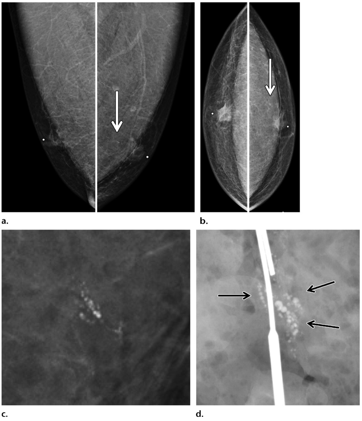
Figure 12.

Calcifications depicted on screening mammogram of a 53-year-old Ashkenazi Jewish man with extensive family history of breast cancer (diagnosed in his father, two sisters, and paternal grandmother), who recently tested positive for BARD1 gene mutation (a BRCA1-associated RING domain 1 gene mutation). (a, b) Screening MLO (a) and CC (b) mammographic views show round calcifications (arrow) in a linear branching distribution in the retroareolar region of the left breast. (c) Magnification mammographic view obtained at a subsequent diagnostic examination best shows the calcifications. Needle localization and excisional biopsy of the calcifications were performed. (d) Surgical specimen radiograph shows the targeted calcifications (arrows). Histopathologic analysis of the biopsy specimen revealed invasive ductal carcinoma and ductal carcinoma in situ (ER positive, PR positive, HER2 negative), with positive surgical margins. A left mastectomy was performed, and tamoxifen therapy was initiated.
