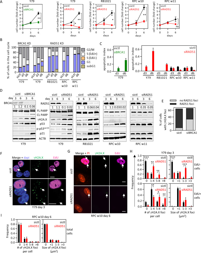
Fig. 2. RAD51 loss kills retinoblastoma but not human fetal retinal cells.
a–d The indicated RB tumor cells or RPC were treated with the indicated siRNAs for 6 days, and growth (a), cell cycle phase (b), apoptosis (c) and protein levels (d) determined. Representative flow cytometry plots used for (b) are shown in Fig. S9A–C. Graph in (c) is quantification of PARP cleavage in (d) (n = 2, mean ± range). e Quantification of nuclear RAD51 and γH2A.X foci in Y79 cells treated with siCtl or siBRCA1, detected by immunostaining at day 6 and analyzed by confocal microscopy. f–i Y79 cells (f) or RPC (g) were treated with siCtl or siRAD51, labeled with EdU (magenta) and γH2A.X (green) at the indicated timepoints, and confocal images obtained. Arrows indicate γH2A.X foci. The number and size of γH2A.X foci were quantified in EdU+ (S-phase) EdU− (non-S-phase) Y79 cells (h) or all RPC (i). In all cases n = 3 (unless specified otherwise). Data in (a), (h), (i) indicate mean ± SD. In (a), *p < 0.05, **p < 0.01, ***p < 0.001, ns nonsignificant, two-way ANOVA, Sidak’s multiple comparisons test. In (h) and (i), *p < 0.05, **p < 0.01, ***p < 0.001 Student t test. Scale bars are 10 μm.

