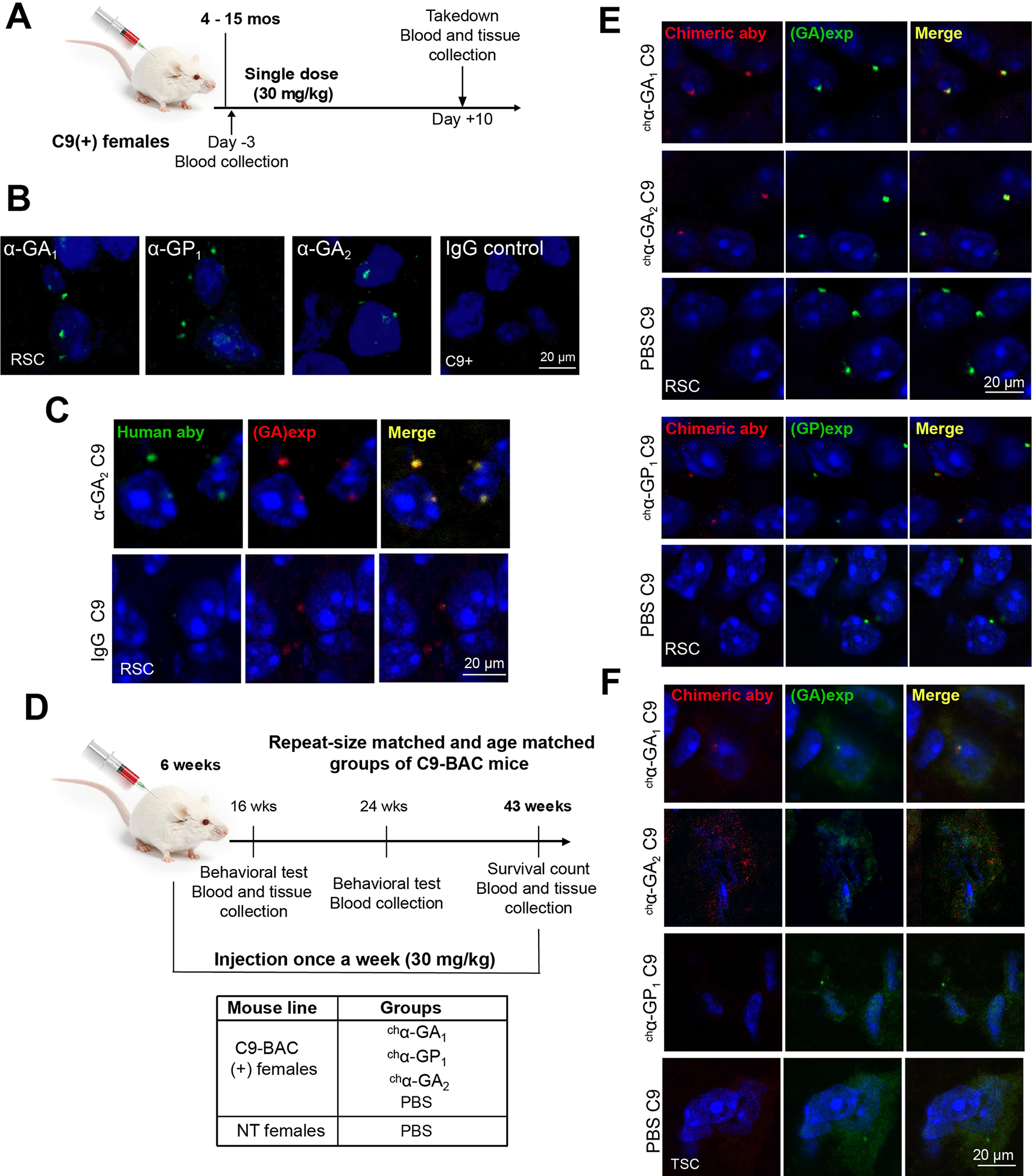
Figure 4. Human antibodies engage in vivo targets in C9 BAC mice.

(A) Experimental design of target engagement study. (B) IF of human antibodies in the RSC 10 days after a single intraperitoneal injection. (C) Double IF staining of α-GA2 and GA aggregates 10 days after single injection. (D) Experimental design of a chronic treatment of antibodies in C9 BAC mice. (E, F) Double IF staining of the retrosplenial cortex (RSC) (E) or thoracic spinal cord (TSC) (F) from mice after chronic treatment with chimeric human antibodies chα-GA1, chα-GP1 or chα-GA2 (detected with α-human IgG, red) and GA or GP aggregates detected with human α-GA1 or α-GP1 antibodies (green). See also Figure S10.
