PLOS ONE logoLink to PLOS ONE
. 2020 Jul 30;15(7):e0236827. doi: 10.1371/journal.pone.0236827

Extraction of radiographic findings from unstructured thoracoabdominal computed tomography reports using convolutional neural network based natural language processing

Mohit Pandey 1,¤a, Zhuoran Xu 1, Evan Sholle 2, Gabriel Maliakal 1,¤b, Gurpreet Singh 1,¤c, Zahra Fatima 1, Daria Larine 3, Benjamin C Lee 1, Jing Wang 1, Alexander R van Rosendael 1,¤d, Lohendran Baskaran 1,¤e, Leslee J Shaw 1, James K Min 1,¤b, Subhi J Al’Aref 4,*
Editor: Yifan Peng5
PMCID: PMC7392233  PMID: 32730362

Abstract

Background

Heart failure (HF) is a major cause of morbidity and mortality. However, much of the clinical data is unstructured in the form of radiology reports, while the process of data collection and curation is arduous and time-consuming.

Purpose

We utilized a machine learning (ML)-based natural language processing (NLP) approach to extract clinical terms from unstructured radiology reports. Additionally, we investigate the prognostic value of the extracted data in predicting all-cause mortality (ACM) in HF patients.

Materials and methods

This observational cohort study utilized 122,025 thoracoabdominal computed tomography (CT) reports from 11,808 HF patients obtained between 2008 and 2018. 1,560 CT reports were manually annotated for the presence or absence of 14 radiographic findings, in addition to age and gender. Thereafter, a Convolutional Neural Network (CNN) was trained, validated and tested to determine the presence or absence of these features. Further, the ability of CNN to predict ACM was evaluated using Cox regression analysis on the extracted features.

Results

11,808 CT reports were analyzed from 11,808 patients (mean age 72.8 ± 14.8 years; 52.7% (6,217/11,808) male) from whom 3,107 died during the 10.6-year follow-up. The CNN demonstrated excellent accuracy for retrieval of the 14 radiographic findings with area-under-the-curve (AUC) ranging between 0.83–1.00 (F1 score 0.84–0.97). Cox model showed the time-dependent AUC for predicting ACM was 0.747 (95% confidence interval [CI] of 0.704–0.790) at 30 days.

Conclusion

An ML-based NLP approach to unstructured CT reports demonstrates excellent accuracy for the extraction of predetermined radiographic findings, and provides prognostic value in HF patients.

Introduction

Heart failure (HF) is a complex chronic condition that is associated with significant morbidity and mortality [1]. Such increased utilization of healthcare resources through frequent medical encounters (either inpatient or outpatient) and performance of diagnostic radiologic studies typically results in the generation of a significant amount of data in the form of unstructured reports. Recently, clinical research in this field has focused on developing predictive models for the occurrence of adverse events within contemporary HF cohorts [24]. Unfortunately, most existent models have limited discriminatory performance as they rely on limited clinical or socio-economic variables that are structured and readily available, and as a result, fail to capture the complexity of such a disease process through incorporation of data that is available in unstructured radiology reports.

The process of data collection and extraction from electronic medical records (EMRs) for such a comprehensive approach can be arduous and time-consuming. Imaging techniques, in particular, typically do not yield structured reports; rather, radiologists dictate or enter free text reports detailing their findings into the EMR. Such a workflow necessitates advanced computational techniques to render the non-structured data computable and amenable to statistical analyses. Natural Language Processing (NLP) is a branch of computer science that focuses on developing computational models for understanding natural (human) language [5,6]. NLP has been increasingly used to automate information extraction from EMRs to streamline data collection for clinical and research purposes [79]. Recently, deep learning architectures, especially the ones based on Recurrent Neural Network (RNN) have been shown to exhibit good performance for clinical NLP tasks. For instance, Long Short-Term Memory (LSTM) and Convolutional Neural Network (CNN) have been used for sentiment analysis and sentence classification, and in the clinical realm an Artificial Neural Network (ANN), Context-LSTM-CNN, that combines the strength of LSTM and CNN with a context encoding algorithm was used to determine the presence of a drug-related adverse event from medical reports [1012]. Further, LSTM was successfully used for the classification of relations within clinical notes [13].

In the present manuscript, we investigate a CNN based NLP pipeline to convert unstructured thoracoabdominal computed tomography (CT) reports into a machine-readable structured format for utilization in outcomes research. We demonstrate that with reasonably limited manual efforts, our NLP pipeline can generate structured dataset of the magnitude of order of big data to facilitate clinical research, which is essential in cohorts of patients with complex and multifactorial disease pathologies. Additionally, we show that such data extracted provides incremental prognostic information for the prediction of all-cause mortality (ACM) in a cohort of HF patients.

Methods

Patient population

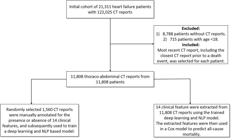
Thoracoabdominal CT reports were obtained from the Clever-Heart registry, which is a single-center registry created to predict outcomes in HF patients. The Weill Cornell Medicine Institutional Review Board (WCM IRB) approved this study and the written informed consent for participants was waived since the registry is retrospective and did not require patient contact or disclosure of identifying information. The registry includes both structured clinical data and unstructured free-text reports. The registry consists of 21,311 patients who were admitted to the New York-Presbyterian Hospital/Weill Cornell Medicine (NYPH/WCM) and discharged with a billing diagnosis of HF (defined by an ICD-9 code of 428. * or an ICD-10 code of I50*) between January of 2008 and July of 2018. The cohort was extracted from the EMR using the Architecture for Research Computing (ARCH) groups Secondary Use of Patients’ Electronic Records (SUPER) landing zone. As detailed in previous work (10), SUPER aggregates data from multiple electronic resources that are in use at NYPH/WCM. For this analysis, exclusion criteria included patients without thoracoabdominal CT reports (n = 8,788) and age <18 at the time of CT acquisition (n = 715). As a result, 11,808 patients were included in the final analysis (Fig 1). The occurrence of death was determined using data extracted from SUPER and was defined as one of the following: (1) death as determined by in-hospital mortality, (2) death as recorded in EMR, (3) an instance of an autopsy report in EMR, or (4) death as recorded in the Social Security Death Master File. Each patient’s most recent follow-up was also calculated using data from SUPER and the various sources from which it draws, including both inpatient and outpatient EMRs. The most recent CT report, including the closest CT report prior to a death event, was selected for each individual.

Fig 1. Overall study design and workflow.

Fig 1

11,808 thoracoabdominal CT reports from 11,808 patients were included in the final analysis.

Choice of radiographic findings

The investigation included a combination of 14 common findings in HF patients as well as commonly reported findings on thoracoabdominal CT reports. These features were: (1) aortic aneurysm, (2) ascites, (3) atelectasis, (4) atherosclerosis, (5) cardiomegaly, (6) enlarged liver, (7) gall bladder wall thickening, (8) hernia, (9) hydronephrosis, (10) lymphadenopathy, (11) pleural effusion, (12) pneumonia, (13) previous surgery, and (14) pulmonary edema. 1,560 CT randomly selected reports were manually annotated by an experienced cardiologist (S.J.A.) for the presence or absence of these 14 radiographic findings, in addition to age and gender. Thereafter, a randomly chosen set representing 15% of the total annotations (225/1500) by physician 1 were independently validated by physician 2 with 3 years of C.T. experience (A.V.R.). The inter-observer variability was less than 2%.

Rule-based NLP for determination of ground truth

Rule-based methods for tasks involving unstructured data perform well for many tasks, especially when there is syntactical uniformity in text and low level of lexical variation [11]. To develop a rule-based approach to extract our target features, a simple ruleset was developed using the manually annotated features from the 1,560 CT reports. This ruleset was based on "phrase-matching" against the corpus of reports: for example, the simple presence of the phrase "pleural effusion" was considered evidence for the presence of the feature "pleural effusion." This approach was based on the assumption that the free-text report would mention a particular finding only if it was present in some measure. However, there are several problems with this approach, including both the relatively high prevalence of negated mentions (i.e. Pleural effusion is not present) as well as lexical variations, including typographical errors (i.e. Pleural effusion present in). Though not very accurate, this provided a good starting place for the generation of ground truths. In addition to the 14 radiographic findings, simple regular expression rules were also identified to extract age (date of birth) and gender. Age and gender extraction were fairly accurate owing to the uniformity of age and gender syntax on CT reports.

Development of Word2vec

Word2vec is an effective way to obtain distributed vector representations of words given a specific vocabulary [12]. It has shown to be very powerful in learning precise syntactic and semantic word relationships. Skip-gram models are used to find word representations by predicting the surrounding words, given a central word [13]. It is imperative to train word-vector representations on a corpus consisting of medical literature, to avoid a significant number of out-of-vocabulary medical words that may appear in the data. Training Word2Vec typically requires medical corpora that are both large and diverse. Since our corpus comprised thoracoabdominal CT reports, we utilized a pre-trained word2vec as detailed by Pyysalo et al [14]. This model was trained using articles from PubMed and PMC texts. Word2Vec was trained using a skip-gram model with a window size of 5. Hierarchical softmax training was employed, with a frequent word-subsampling threshold of 0.001 to create 200 dimensional vectors [15].

CNN model pipeline

The task of retrieving clinical terms from CT reports was treated as a multi-class, multi-label sequence classification task [16]. On average, 67% of each report contained clinical information while the remaining 33% contained patient identifiers. Accordingly, a redaction module was designed to ensure the utilization of only the relevant clinical information. The redaction module assumes that the header and footer of the reports are dedicated principally to patient identifiers. This module trims down the reports by removing the first and last n lines, defining n by scanning through a small vocabulary of start words of radiologic reports to determine the number of lines to be redacted. In a similar fashion, the footers were also removed. The output of the redaction module was then passed to the filtering module for the removal of special characters while retaining alphanumeric characters (S1 Fig).

The annotated 1,560 CT reports were split into training and testing sets at a ratio of 80%-20%. The training set was further split into training and validation sets, also at a ratio of 80%-20%. As proof of the generalizability of the deployed CNN architecture (S2 Fig), the test set was held to ensure the validity of the performance metrics. The validation set was used for a grid search to tune the hyper-parameters of the CNN model. We chose filter sizes of 3, 4 and 5 with 128 filters for each size. To reduce overfitting, we used dropout with 0.5 keep probability. In addition, we also used l2-regularization on the weights, with a value of lambda as 0.01. We trained each model for 50 epochs and chose the one with the highest categorical accuracy on the validation set. We optimized the categorical cross-entropy loss utilizing Adam optimizer with learning rate set to 0.001.

To benchmark the performance of the proposed approach, a classifier was trained by first converting the output of the filter module into word embedding using Term frequency-inverse document frequency (TF-IDF), then training a Naive Bayes and a Support Vector Machine (SVM) classifiers (considered as traditional machine learning algorithms). Python’s scikit-learn library was used to train the benchmark model.

Performance metrics and outcomes investigated

Binary classifiers for each clinical finding were evaluated using the Receiver Operator Characteristic (ROC) curve approach. A ROC curve is a plot of false-positive rates and true positive rates charted over a range of decision thresholds. A purely random guessing classifier would be near the y = x line on the ROC plot, while a perfect classifier would look like a unit-step function for 0<x<1 and will have Area Under Curve (AUC) of 1. We also report F1-score, precision and recall as additional robust metrics given the imbalanced dataset that was available.

Prediction explanation using layer-wise relevance propagation

Bach et al. [17] introduced a layer-wise relevance propagation (LRP) in a feed-forward network to explain pixel-level classification decisions in CNNs. Starting with the output layer, LRP proceeds layer by layer and assigns the relevance of the target neuron to other units of the layers. They proposed two relevance propagation rules for the layer, assuming that the lower layer neurons that mostly contribute to the activation of the higher layer neuron receive a higher relevance. While the convolutional network shows promising performance, the connection between the CNN learned features and the clinical findings might be difficult to see. By visualizing the contribution of specific words toward the classification task by the network proposed by Bach et al and Ancona et al, [17,18] one can identify more clearly the relationships derived by the CNN.

Statistical analysis

Continuous variables were calculated and reported as mean ± standard deviation, whereas categorical variables were calculated and reported as counts and percentages. A Cox proportional hazards model was used for constructing survival models [19]. Backward selection based on the Akaike information criterion (AIC) was utilized for variable selection. A random survival forest model was also used for predicting ACM, defined as death from any cause [17,20]. The inverse probability of censoring weights was used to deal with right-censored survival data [2123]. In addition, the outcome of the survival analysis was time-to-event for both the Cox model and the random survival forest model. Time-dependent AUCs were used to evaluate model performance. Further, feature importance was assessed using VIMP (variable importance) [24]. In the present scenario, VIMP in random forests for a feature, xa, is the difference between prediction error when xa is noised up by permuting its value randomly, compared to prediction error under the original predictor. A p-value of less than 0.05 was considered statistically significant. All analyses were performed using R software (RStudio, Boston, MA) [25].

Results

Dataset

The mean word-length of CT reports was 3,085 words, with an average length of clinical information of 1,330 words (43%). Table 1 shows the prevalence of a positive finding for each of the 14 radiographic findings. While some features had a balanced prevalence of positive and negative occurrences (for example, previous surgery and atelectasis), other features exhibited a significant imbalance between positive and negative findings. Gall bladder thickening, enlarged liver, pulmonary edema, and aortic aneurysm were positive in less than 10% of the reports.

Table 1. Prevalence of radiographic findings between the ground truth cohort (manually annotated reports) and the Clever-Heart cohort.

For the ground truth corpus, the prevalence percentage is calculated based on known, manually annotated ground truths. For Clever-Heart, values are based on predictions from the CNN model.

Ground Truth CT reports Clever-Heart (As Predicted)
No. Radiographic finding Radiographic finding Count Prevalence (%) Radiographic finding Count Prevalence (%)
1 Aortic Aneurysm 120 7.69 754 6.38
2 Ascites 299 19.16 1516 12.83
3 Atelectasis 691 44.29 5625 47.63
4 Atherosclerosis 620 39.74 5639 47.75
5 Cardiomegaly 390 25.00 3374 28.57
6 Enlarged Liver 86 5.50 162 1.37
7 GB Thickening 25 1.60 9 .0007
8 Hernia 366 23.46 2594 21.96
9 Hydronephrosis 65 4.16 136 1.15
10 Lymphadenopathy 484 31.02 2593 21.95
11 Pleural Effusion 673 43.14 4823 40.84
12 Pneumonia 278 17.82 1678 39.96
13 Previous Surgery 778 49.87 4719 39.96
14 Pulmonary Edema 94 6.02 516 4.36

Performance metric for radiographic finding extraction

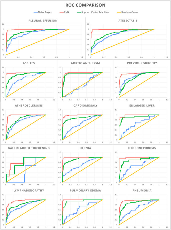
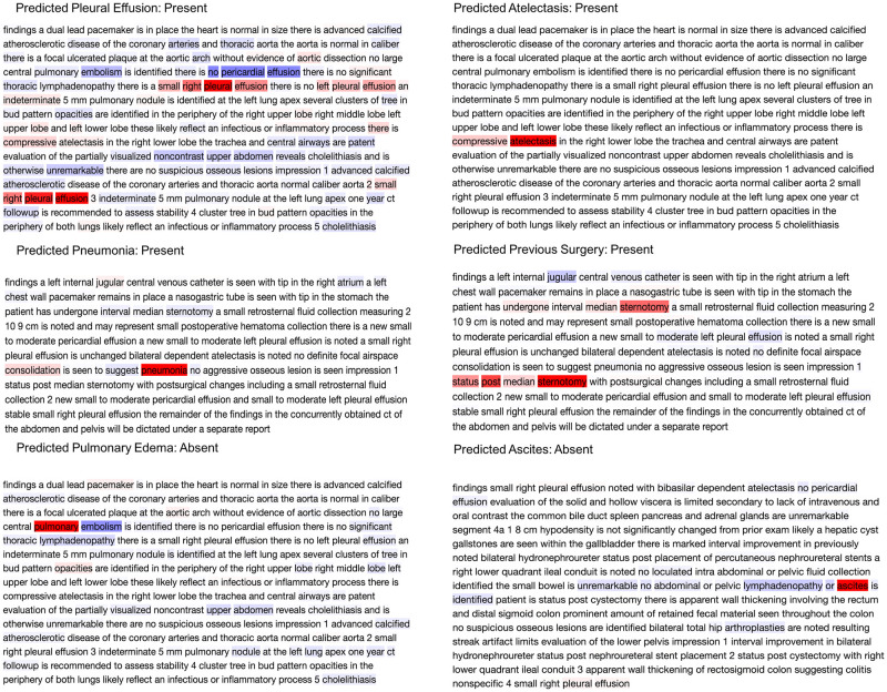
For the held-out test set of 312 CT reports, the average AUC was 0.97 across the 14 radiographic findings using the CNN architecture (average F1 score of 0.90). By comparison, Naive Bayes and SVM had an average AUC of 0.69 and 0.87 respectively (average F1 score of 0.71 and 0.67, respectively) (Table 2 and Fig 2). With regards to the individual 14 radiographic findings, the CNN model outperformed the other models on each radiographic finding. Furthermore, the redaction module for de-identification improves the AUC for 13 of the 14 radiographic findings. For the radiographic finding “aortic aneurysm”, disabling the redaction module produced a slightly better AUC. Fig 3 shows examples of CT reports highlighting words that guided the CNN model towards or away from predicting the presence of a certain radiographic finding.

Table 2. Performance of the Convolutional Neural Network (CNN) model benchmarked against machine learning algorithms for the extraction of 14 pre-selected radiographic findings.

Feature Naive Bayes SVM CNN
Precision Recall F1 Score ROC AUC Precision Recall F1 Score ROC AUC Precision Recall F1 Score ROC AUC
Aortic Aneurysm 0.82 0.91 0.86 0.49 0.82 0.91 0.86 0.94 0.94 0.94 0.91 0.98
Ascites 0.62 0.78 0.69 0.73 0.64 0.8 0.7 0.9 0.87 0.87 0.84 0.98
Atelectasis 0.71 0.57 0.44 0.8 0.31 0.56 0.4 0.85 0.97 0.96 0.96 0.98
Atherosclerosis 0.55 0.58 0.45 0.77 0.34 0.58 0.43 0.83 0.9 0.88 0.89 0.98
Cardiomegaly 0.56 0.75 0.64 0.65 0.52 0.72 0.6 0.88 0.86 0.86 0.84 0.98
Enlarged Liver 0.89 0.95 0.92 0.65 0.89 0.95 0.92 0.92 0.94 0.97 0.95 0.96
Gall Bladder Wall Thickening 0.97 0.99 0.98 0.43 0.98 0.99 0.99 0.77 0.97 0.99 0.98 0.83
Hernia 0.55 0.74 0.63 0.69 0.58 0.76 0.66 0.92 0.97 0.97 0.97 0.99
Hydronephrosis 0.91 0.95 0.93 0.68 0.88 0.94 0.91 0.88 0.89 0.95 0.92 0.99
Lymphadenopathy 0.46 0.68 0.55 0.75 0.41 0.64 0.5 0.82 0.86 0.85 0.84 0.95
Pleural Effusion 0.76 0.57 0.43 0.84 0.35 0.59 0.44 0.9 0.96 0.96 0.96 0.98
Pneumonia 0.69 0.83 0.75 0.67 0.69 0.83 0.75 0.88 0.87 0.88 0.86 0.96
Previous Surgery 0.73 0.73 0.73 0.84 0.23 0.48 0.31 0.78 0.86 0.85 0.85 0.95
Pulmonary Edema 0.88 0.94 0.91 0.65 0.88 0.94 0.9 0.95 0.93 0.92 0.89 1

Fig 2. Receiver Operator Characteristic (ROC) curves for the 14 pre-selected radiographic findings.

Fig 2

The Convolutional Neural Network (CNN) is compared to Naive Bayes, Support Vector Machine (SVM), and random guessing.

Fig 3. Explaining the output of a trained CNN model using layer-wise relevance propagation.

Fig 3

The predicted label is considered as the true class label. The color intensities are normalized to the absolute value of maximum relevance score per report such that the deepest red denotes the word with the highest positive relevance in the class label prediction, while the deepest blue denotes the most negative relevance score in the prediction of the same label.

NLP pipeline for prognostication in HF

The proposed CNN based NLP pipeline was subsequently used to extract these 14 radiographic findings from thoracoabdominal CT reports from 11,808 HF patients in the Clever-Heart registry (mean age of the study cohort 72.8 ± 14.8 years; 52.7% were male). The inter-quartile range of follow-up was between 129 and 1,521 days, with a median follow-up of 606 days. 3,107 death events were observed during the follow-up period. In total, 11,808 CT reports were analyzed, with 9,378 used as the training set and 2,430 used as the test set for ACM prediction. The 14 radiographic findings in addition to age and gender were included in Cox models and random survival forest. The prevalence of the 14 extracted features in this cohort is shown in Table 1. Using Cox modeling, there were significant associations between the 14 extracted radiographic findings and ACM (Fig 4 and S1 Table). For instance, the presence of pleural effusion (hazard ratio [HR] 1.63, 95% confidence interval [CI] 1.48–1.78, p<0.001) and ascites (HR 1.68, 95% CI 1.51–1.88, p<0.001) showed the strongest association with ACM, thus indicating that the presence of such a clinical finding led to higher mortality. Similarly, random survival forest also demonstrated that the presence of pleural effusion and ascites were most highly correlated with mortality.

Fig 4. Correlation between radiographic findings and all-cause mortality.

Fig 4

(A-B): Forest plot for the Cox model with all 14 variables and selected 9 variables. Numbers represent hazard ratios. The range of lines indicates 95% confidence intervals. Color blue implies significant variables with p<0.05. (C) Variable importance plot of the Random Survival Forest model. Large positive values indicate the dependency of the outcome to get high predictive power. Values closer to zero represent a lower contribution to improved predictive accuracy. Negative numbers indicate the predictive accuracy would improve when the variables were unspecified.

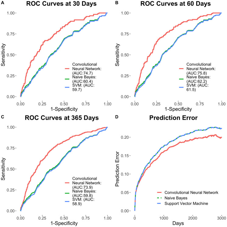
Cox survival models were constructed using features extracted from the developed CNN-based pipeline and compared to that of non-deep learning-based featurization using Naïve Bayes modeling (Fig 5). The CNN-based NLP pipeline for unstructured text featurization yielded enhanced clinical risk prediction, compared to Naïve Bayes modeling for outcomes at 30 days (AUC of 0.747 vs. 0.604, respectively; p<0.01), 60 days (AUC of 0.758 vs. 0.625, respectively; p<0.01) and 365 days (AUC of 0.739 vs. 0.598, respectively; p<0.01). Further, the use of radiographic findings extracted using the CNN-based NLP pipeline resulted in the enhanced prediction of outcomes across multiple survival models. The full Cox model showed the time-dependent AUC for predicting ACM is 0.747 (95% CI of 0.704–0.790) at 30 days (C-statistic of 0.695 ± 0.012), 0.758 (95% CI of 0.720–0.796) at 60 days and 0.739 (95% CI of 0.708–0.770) at 1 year. The Cox model with select features (pneumonia, pleural effusion, lymphadenopathy, hernia, cardiomegaly, atelectasis, ascites, gender, and age) according to AIC showed similar time-dependent AUC results: 0.745 (95% CI of 0.701–0.788) at 30 days, 0.756 (95% CI of 0.719–0.795) at 60 days and 0.738 (95% CI of 0.706–0.769) at 1 year. On the other hand, the random survival forest model performed worse than the Cox models, with time-dependent AUC of 0.701 (95% CI of 0.659–0.744) at 30 days, 0.687 (95% CI of 0.649–0.725) at 60 days and 0.670 (95% CI of 0.638–0.702) at 1 year. Similar results can be observed in the Brier score plot, in which random survival forest exhibited the highest prediction error and the two Cox models performed in a similar fashion, with lower error (S3 Fig).

Fig 5. Prognostication of outcomes using the CNN model.

Fig 5

(A-C) Time-dependent ROC curves at 30, 60 and 365 days. (D) Time-dependent Brier scores.

Discussion

The present investigation highlights the potential of an ML framework that applies NLP to unstructured thoracoabdominal CT reports in the accurate extraction of pre-specified radiographic findings as well as a prognostication of outcomes using the extracted clinical data. While the lack of a standard syntactical structure for free text reports across different sites makes it difficult to retrieve clinical information using traditional rule-based approaches and regular expressions, our analysis shows that the use of a CNN resulted in the accurate extraction of specific clinical terms. The AUC for radiographic finding extraction ranged from 0.88 to 1.0, with an average AUC of 0.96. The study also showed that the CNN model had significantly improved performance when trained against carefully preprocessed input text: removal of headers and footers from the CT reports boosted performance metrics for 13 out of the 14 radiographic findings. In addition, the extracted clinical information was found to be useful for prognostication of outcomes, specifically with the occurrence of ACM, within an HF cohort. Furthermore, such an approach could be curtailed for future clinical applications such as stratification of HF individuals at risk for hospitalizations and readmissions, which could reduce the significant healthcare expenditure associated with HF management.

Free-form language in EMR is unrestricted and is subject to endless interpreter-to-interpreter and site-to-site variations. The theoretical potential for lexical variation in the expression of a given concept is infinite—hence, interpretation and computational modeling of context is crucial for the development of tools that go beyond simple string matching. Our work here demonstrates that a CNN based NLP approach can provide enhanced performance on these tasks by incorporating novel computational models. Illustrative examples are as in Fig 4. The CNN model predicts the presence of atelectasis by assigning the highest relevance to "compressive atelectasis". Similarly, it learns that the clinical term “pneumonia” is present by assessing the highest relevance to "pneumonia" in the report. However, while classifying pulmonary edema, the model learns "pulmonary" with the highest positive relevance but at the same time assigns "embolism" with the highest negative relevance score, demonstrating that the model "understands" that pulmonary edema is different from a pulmonary embolism. In the case of the term “ascites”, even though the model learns that the presence of this term is most important for prediction of ascites, the filters of the CNN learn negation from neighboring words. As a result, even though the report says, "ascites is identified the model aggregates this with the earlier negation in the report and hence was able to classify this report as ascites-absent “no ascites is identified”. The model’s ability to comprehend free-form medical text is further demonstrated in the analysis of “previous surgery” and “pleural effusion” classification. For previous surgery, "median sternotomy" was scored with the most positive relevance. The CNN model coupled with word2vec trained on medical corpora understands that median sternotomy is a surgical procedure even without explicit training. Words like "post" and "undergone" help the model to learn the context and deduce that the procedure was performed in the past and hence flags the report as positive for the target concept "prior surgery." Similarly, the “pleural effusion” prediction model learns with the highest positive relevance that "a small right pleural effusion" was present, while simultaneously taking into consideration the presence of the term "no left pleural effusion", assigning high relevance to that phrase as well. The model also assigns the highest negative relevance score to "no pericardial effusion" to prevent it from falsely flagging the feature of pericardial effusion as an instance of a pleural effusion. Overall, the model was able to average these different inferences about pleural effusion in this report and ultimately correctly deduce the presence of a pleural effusion.

While the inference on “atelectasis” and “pneumonia” could be achieved using string matching and simple rule-sets, our model understands more complex sentences and structures as prevalent in imaging reports. An additional benefit of this technique is the extent to which it minimizes the need for labor-intensive human effort. In recent investigations, recurrent neural networks (RNNs) have shown excellent results for text comprehension tasks—however, their need for copious amounts of annotated examples makes them unsuitable for NLP applications in the clinical field, where human annotation often requires clinical expertise, which can become a burden within a busy clinical setting. Our CNN-based method proved to effectively balance the need for both high performance and minimized requirement for manual effort (as evident by the high extraction accuracy using 1,560 annotated CT reports).

NLP has been increasingly used for clinical applications over the past few years. The Linguistic String Project—Medical Language Processor (LSP-MLP), conducted at New York University in 1987, was among the first large-scale projects using NLP within the context of clinical research [26]. The LSP-MLP sought to help physicians extract and summarize sign/symptom information, drug dosage, and response data, and to identify possible medication side effects. Those results revealed the utility of various computational linguistic experiments in the medical field. In addition, substantial efforts have been directed towards NLP pipelines to supplement or classify conditions based on ground truths provided by the International Classification of Diseases (ICD) codes. For example, Pakhomov et al. found that NLP algorithms consistently retrieve more clinical information from free-text reports, and specifically regarding the presence of angina pectoris, than what is provided with ICD codes [27]. A naïve yet powerful NLP approach in evaluating clinical texts is by studying sentence modifiers. State of the art tools, such as NegEx or NegBio look at finding possible negation to clinical entities in discharge summaries and radiology reports, respectively [2829]. However, the application of NLP to clinical data and radiologic reports is an active area of research, since the field is in the process of establishing a framework for methodological evaluation. Contemporary practices utilize human manual annotations as the ground truth and generate performance metrics based on comparisons with the prediction models. From a clinical perspective, NLP frameworks could prove to be a valuable resource in the care of patients with complex chronic conditions such as HF. As a disease process, HF has intricate pathophysiology as well as disease manifestations, leading to a heterogeneous expression of symptoms and subsequent outcomes. HF patients undergo numerous imaging modalities as part of the initial diagnostic workup, monitoring of therapy, assessment of underlying structural progression, as well as evaluation of concomitant non-cardiovascular pathologies. Extraction of imaging findings from radiologic reports could thus be complementary to patient-level clinical information and could further help prognosticate and better risk-stratify individuals, especially in the setting of HF and similar multifaceted diseases.

While our investigation has clear advantages in terms of clinical applications and novelty of this approach, there are several limitations worthy of mentioning. Firstly, this was a single-center study of thoracoabdominal CT reports obtained from HF patients. While the CNN model had impressive accuracy for the extraction of 14 pre-selected radiographic findings, the performance of this model on free-text CT reports from other sites is unknown. Secondly, several radiographic findings, such as gall bladder wall thickening, had a high rate of false-negative results, which is attributable to severe class imbalance since very few reports had a positive label for this class (i.e. most reports did not mention the presence of gall bladder wall thickening). A better sampling approach and the inclusion of gastrointestinal disease cases could be used in the subsequent analysis to further improve the diagnostic performance of our model. Finally, the study used the latest CT reports in the survival analysis, instead of including all available CT reports. This was done since complex modeling is required in order to utilize time-dependent covariates. The scope of the present investigation was to demonstrate the utility of CNN for clinical finding extraction from unstructured CT reports, while subsequent investigations will aim at developing sophisticated models that include time-dependent variables for prognostication of outcomes.

In summary, we show that a CNN based NLP pipeline applied to unstructured CT reports accurately extracts 14 pre-specified radiographic findings in a cohort of HF patients undergoing thoracoabdominal CT imaging, which in turn provides prognostic value for prediction of ACM in such a cohort. The approach detailed herein offers the potential to supplement the extraction of clinical data, beyond that of existing structured data in EMR systems, for outcomes research and clinical care especially for individuals with chronic and complex medical conditions such as HF.

Supporting information

S1 Fig. Stacked Convolutional Neural Network (CNN) architecture.

The input in the training phase consisted of 1,560 free-text thoracoabdominal computed tomography reports. These reports passed through redaction module to remove headers consisting of non-clinically relevant free text. The filtering module tokenized and removed special characters. After converting these reports into 200 dimensional vectors for each word, they were then used to train the CNN model. The output of the training phase was a trained CNN model. A separate CNN model for each of the 14 clinical findings constituted the model zoo. In the Go Live phase, input consisting of 11,808 free-text reports was passed to the trained CNN models after passing through the redaction module, the filtering module, and conversion to word vectors. The output from this phase generated the database of clinical findings which, along with age and gender, were used for prediction of all-cause mortality.

(TIF)

S2 Fig. Structure of the Convolutional Neural Network (CNN).

(a) Filter bank with kernels of size 2, 3 and 4. Each kernel is a 200-dimensional vector which is the same as the dimensionality of the word embedding used. (b) Each word of the sentence is converted to a 200-dimensional vector using word2vec. For uniformity in length, shorter sentences are zero padded. Filters of size 2, 3 and 4 are individually convolved with the sentence matrix. (c) Feature maps generated for each filter size. (d) Max pooling over time to create single feature vector. (e) Fully connected layer with SoftMax to classify presence or absence of the specific clinical finding.

(TIF)

S3 Fig. Prognostication of outcomes using the deep learning-based method for feature extraction.

(A-C) Time-dependent ROC curves at 30, 60 and 365 days for 3 models created using the deep learning-based method for feature extraction: (1) a full COX model, (2) a COX model with select variables and (3) a random survival forest. Panel D shows the time-dependent Brier scores.

(TIFF)

S1 Table

(A) Full Cox model for correlation between the radiographic finding and all-cause mortality. (B) Select Cox model and correlation between radiographic findings and all-cause mortality.

(DOCX)

Data Availability

All relevant data are within the manuscript and its Supporting Information files.

Funding Statement

This study received support from the New York-Presbyterian Hospital (NYP) and Weill Cornell Medicine (WCM), including the Clinical and Translational Science Center (CTSC) (UL1 TR000457) and Joint Clinical Trials Office (JCTO). The funders had no role in study design, data collection and analysis, decision to publish, or preparation of the manuscript. The funder provided support in the form of salaries for author [E.S.] but did not have any additional role in the study design, data collection and analysis, decision to publish, or preparation of the manuscript. The specific roles of these authors are articulated in the ‘author contributions’ section.

References

  • 1.Benjamin EJ, Muntner P, Alonso A, Bittencourt MS, Callaway CW, Carson AP, et al. Heart Disease and Stroke Statistics-2019 Update: A Report From the American Heart Association. Circulation. 2019. March 5;139(10):e56–528. 10.1161/CIR.0000000000000659 [DOI] [PubMed] [Google Scholar]
  • 2.Donze J, Aujesky D, Williams D, Schnipper JL. Potentially avoidable 30-day hospital readmissions in medical patients: Derivation and validation of a prediction model. JAMA Intern Med. 2013. April 22;173(8):632–8. 10.1001/jamainternmed.2013.3023 [DOI] [PubMed] [Google Scholar]
  • 3.Keenan PS, Normand S-LT, Lin Z, Drye EE, Bhat KR, Ross JS, et al. An administrative claims measure suitable for profiling hospital performance on the basis of 30-day all-cause readmission rates among patients with heart failure. Circ Cardiovasc Qual Outcomes [Internet]. 2008. September [cited 2020 Feb 19];1(1):29–37. Available from: http://www.ncbi.nlm.nih.gov/pubmed/20031785 [DOI] [PubMed] [Google Scholar]
  • 4.Hammill BG, Curtis LH, Fonarow GC, Heidenreich PA, Yancy CW, Peterson ED, et al. Incremental value of clinical data beyond claims data in predicting 30-day outcomes after heart failure hospitalization. Circ Cardiovasc Qual Outcomes [Internet]. 2011. January 1 [cited 2020 Feb 19];4(1):60–7. Available from: http://www.ncbi.nlm.nih.gov/pubmed/21139093 [DOI] [PubMed] [Google Scholar]
  • 5.Wang Y, Wang L, Rastegar-Mojarad M, Moon S, Shen F, Afzal N, et al. Clinical information extraction applications: A literature review. Vol. 77, Journal of Biomedical Informatics. Academic Press Inc.; 2018. p. 34–49. [DOI] [PMC free article] [PubMed] [Google Scholar]
  • 6.Meystre SM, Savova GK, Kipper-Schuler KC, Hurdle JF. Extracting Information from Textual Documents in the Electronic Health Record: A Review of Recent Research. Yearb Med Inform [Internet]. 2008. August 7 [cited 2020 Feb 19];17(01):128–44. Available from: http://www.thieme-connect.de/DOI/DOI?10.1055/s-0038-1638592 [PubMed] [Google Scholar]
  • 7.Chen X, Xie H, Wang FL, Liu Z, Xu J, Hao T. A bibliometric analysis of natural language processing in medical research. BMC Med Inform Decis Mak [Internet]. 2018. March 22 [cited 2020 Feb 19];18(S1):14 Available from: https://bmcmedinformdecismak.biomedcentral.com/articles/10.1186/s12911-018-0594-x [DOI] [PMC free article] [PubMed] [Google Scholar]
  • 8.Velupillai S, Mowery D, South BR, Kvist M, Dalianis H. Recent Advances in Clinical Natural Language Processing in Support of Semantic Analysis. Yearb Med Inform [Internet]. 2015. August 10 [cited 2020 Feb 19];24(01):183–93. Available from: http://www.thieme-connect.de/DOI/DOI?10.15265/IY-2015-009 [DOI] [PMC free article] [PubMed] [Google Scholar]
  • 9.Kreimeyer K, Foster M, Pandey A, Arya N, Halford G, Jones SF, et al. Natural language processing systems for capturing and standardizing unstructured clinical information: A systematic review. J Biomed Inform. 2017. September 1;73:14–29. 10.1016/j.jbi.2017.07.012 [DOI] [PMC free article] [PubMed] [Google Scholar]
  • 10.Hochreiter S, Schmidhuber J. Long Short-Term Memory. Neural Comput. 1997. November 15;9(8):1735–80. 10.1162/neco.1997.9.8.1735 [DOI] [PubMed] [Google Scholar]
  • 11.Kim Y. Convolutional Neural Networks for Sentence Classification. EMNLP 2014–2014 Conf Empir Methods Nat Lang Process Proc Conf [Internet]. 2014 Aug 25 [cited 2020 Feb 19];1746–51. Available from: http://arxiv.org/abs/1408.5882
  • 12.Song X, Petrak J, Roberts A. A Deep Neural Network Sentence Level Classification Method with Context Information. 2018. August 31 [cited 2020 Feb 19];900–4. Available from: http://arxiv.org/abs/1809.00934 [Google Scholar]
  • 13.Luo Y. Recurrent neural networks for classifying relations in clinical notes. J Biomed Inform. 2017. August 1;72:85–95. 10.1016/j.jbi.2017.07.006 [DOI] [PMC free article] [PubMed] [Google Scholar]
  • 14.Pyysalo S, Ginter F, Moen H, Salakoski T, Ananiadou S. Distributional Semantics Resources for Biomedical Text Processing. Proc LBM [Internet]. 2013. [cited 2020 Feb 19];39–44. Available from: https://github.com/spyysalo/nxml2txt [Google Scholar]
  • 15.Chiticariu L, Li Y, Reiss FR. Rule-based Information Extraction is Dead! Long Live Rule-based Information Extraction Systems! Proc 2013 Conf Empir Methods Nat Lang Process. 2013;18–21.
  • 16.Mikolov T, Chen K, Corrado G, Dean J. Distributed Representations of Words and Phrases and their Compositionality. 2013.
  • 17.Bach S, Binder A, Montavon G, Klauschen F, Müller KR, Samek W. On pixel-wise explanations for non-linear classifier decisions by layer-wise relevance propagation. PLoS One. 2015. July 10;10(7). [DOI] [PMC free article] [PubMed] [Google Scholar]
  • 18.Ancona M, Ceolini E, Öztireli C, Gross M. Towards better understanding of gradient-based attribution methods for Deep Neural Networks. 6th Int Conf Learn Represent ICLR 2018—Conf Track Proc [Internet]. 2017 Nov 16 [cited 2020 Feb 20]; http://arxiv.org/abs/1711.06104
  • 19.Morin F, Bengio Y. Hierarchical Probabilistic Neural Network Language Model. Aistats. 2005;5:246–52. [Google Scholar]
  • 20.Ishwaran H, Kogalur UB, Blackstone EH, Lauer MS. Random survival forests. Ann Appl Stat. 2008. September;2(3):841–60. [Google Scholar]
  • 21.Mogensen UB, Ishwaran H, Gerds TA. Evaluating Random Forests for Survival Analysis Using Prediction Error Curves. J Stat Softw. 2012;50(11):1 10.18637/jss.v050.i11 [DOI] [PMC free article] [PubMed] [Google Scholar]
  • 22.Blanche P, Dartigues JF, Jacqmin-Gadda H. Estimating and comparing time-dependent areas under receiver operating characteristic curves for censored event times with competing risks. Stat Med [Internet]. 2013. December 30 [cited 2020 Mar 2];32(30):5381–97. Available from: http://doi.wiley.com/10.1002/sim.5958 [DOI] [PubMed] [Google Scholar]
  • 23.Graf E, Schmoor C, Sauerbrei W, Schumacher M. Assessment and comparison of prognostic classification schemes for survival data. Stat Med. 1999. September 15;18(17–18):2529–45. [DOI] [PubMed] [Google Scholar]
  • 24.Ishwaran Hemant. Variable importance in binary regression trees and forests. Electron. J. Statist. 1 (2007), 519–537. Available from: https://projecteuclid.org/euclid.ejs/1195157166 [Google Scholar]
  • 25.Bunn A, Korpela M. An Introduction to dplR [Internet]. 2013 [cited 2020 Mar 2]. 1–16 p. 10.1016/j.dendro.2008.01.002 [DOI]
  • 26.Sager N, Friedman C, Lyman MS. Medical language processing: computer management of narrative data. Addison-Wesley Pub. Co; 1987. 348 p. [Google Scholar]
  • 27.Pakhomov SSV, Hemingway H, Weston SA, Jacobsen SJ, Rodeheffer R, Roger VL. Epidemiology of angina pectoris: Role of natural language processing of the medical record. Am Heart J. 2007. April 1;153(4):666–73. 10.1016/j.ahj.2006.12.022 [DOI] [PMC free article] [PubMed] [Google Scholar]
  • 28.Chapman WW, Bridewell W, Hanbury P, Cooper GF, Buchanan BG. A simple algorithm for identifying negated findings and diseases in discharge summaries. J Biomed Inform. 2001;34(5):301–310. 10.1006/jbin.2001.1029 [DOI] [PubMed] [Google Scholar]
  • 29.Peng Y, Wang X, Lu L, Bagheri M, Summers RM, Lu Z. NegBio: a high-performance tool for negation and uncertainty detection in radiology reports. AMIA 2018 Informatics Summit. 2018. [PMC free article] [PubMed]

Decision Letter 0

Yifan Peng

22 Apr 2020

PONE-D-20-06619

Extraction of clinical features from unstructured thoracoabdominal computed tomography reports using convolutional neural network based natural language processing

PLOS ONE

Dear Dr. Al’Aref,

Thank you for submitting your manuscript to PLOS ONE. After careful consideration, we feel that it has merit but does not fully meet PLOS ONE’s publication criteria as it currently stands. Therefore, we invite you to submit a revised version of the manuscript that addresses the points raised during the review process.

We would appreciate receiving your revised manuscript by Jun 06 2020 11:59PM. When you are ready to submit your revision, log on to https://www.editorialmanager.com/pone/ and select the 'Submissions Needing Revision' folder to locate your manuscript file.

If you would like to make changes to your financial disclosure, please include your updated statement in your cover letter.

To enhance the reproducibility of your results, we recommend that if applicable you deposit your laboratory protocols in protocols.io, where a protocol can be assigned its own identifier (DOI) such that it can be cited independently in the future. For instructions see: http://journals.plos.org/plosone/s/submission-guidelines#loc-laboratory-protocols

Please include the following items when submitting your revised manuscript:

  • A rebuttal letter that responds to each point raised by the academic editor and reviewer(s). This letter should be uploaded as separate file and labeled 'Response to Reviewers'.

  • A marked-up copy of your manuscript that highlights changes made to the original version. This file should be uploaded as separate file and labeled 'Revised Manuscript with Track Changes'.

  • An unmarked version of your revised paper without tracked changes. This file should be uploaded as separate file and labeled 'Manuscript'.

Please note while forming your response, if your article is accepted, you may have the opportunity to make the peer review history publicly available. The record will include editor decision letters (with reviews) and your responses to reviewer comments. If eligible, we will contact you to opt in or out.

We look forward to receiving your revised manuscript.

Kind regards,

Yifan Peng, Ph.D.

Academic Editor

PLOS ONE

Additional Editor Comments:

1. Both reviewers recommend reporting metrics of precision, recall, sensitivity, specificity, and F1 for each category. In addition, the C-statistic is the most relevant metric for prognosis. It is also recommended to report C-statistic for ACM prognosis.

2. The discussion of rule-based systems needs to include SOTA tools, such as NegEx or NegBio.

3. It is not clear if the features extracted from CNN and used in Cox are clinical features or the last layer of CNN. The use of the word "feature" is confused as it can either mean the deep feature that is the output of a layer within a CNN model, or a specific feature pre-defined by the authors. To this end, I would recommend changing the "clinical features" to "radiographic findings".

Journal requirements:

When submitting your revision, we need you to address these additional requirements.

1. Please ensure that your manuscript meets PLOS ONE's style requirements, including those for file naming. The PLOS ONE style templates can be found at

https://journals.plos.org/plosone/s/file?id=wjVg/PLOSOne_formatting_sample_main_body.pdf and

https://journals.plos.org/plosone/s/file?id=ba62/PLOSOne_formatting_sample_title_authors_affiliations.pdf

2. Thank you for stating the following in the title page of your manuscript:

'Funding: This study received support from the New York-Presbyterian Hospital (NYP) and Weill Cornell Medicine (WCM), including the Clinical and Translational Science Center (CTSC) (UL1 TR000457) and Joint Clinical Trials Office (JCTO).

We note that you have provided funding information that is not currently declared in your Funding Statement. However, funding information should not appear in the Acknowledgments section or other areas of your manuscript. We will only publish funding information present in the Funding Statement section of the online submission form.

a. Please remove any funding-related text from the manuscript and let us know how you would like to update your Funding Statement. Currently, your Funding Statement reads as follows:

'The author(s) received no specific funding for this work.'

Please clarify the sources of funding (financial or material support) for your study. List the grants or organizations that supported your study, including funding received from your institution.

State what role the funders took in the study. If the funders had no role in your study, please state: “The funders had no role in study design, data collection and analysis, decision to publish, or preparation of the manuscript.”

If any authors received a salary from any of your funders, please state which authors and which funders.

If you did not receive any funding for this study, please state: “The authors received no specific funding for this work.”

b. Please include your amended statements within your cover letter; we will change the online submission form on your behalf.

3. Thank you for stating the following in the Competing Interests section:

'The authors have declared that no competing interests exist.'

Thank you also for stating the following in the title page of your manuscript:

'Disclosure: Dr. James K. Min receives funding from the Dalio Foundation, National Institutes of Health, and GE Healthcare. Dr. Min serves on the scientific advisory board of Arineta and GE Healthcare, and has an equity interest in Cleerly. All other authors have reported that they have no relationships relevant to the contents of this paper to disclose.'

We note that one or more of the authors are employed by a commercial company: GlaxoSmithKline

a. Please provide an amended Funding Statement declaring this commercial affiliation, as well as a statement regarding the Role of Funders in your study. If the funding organization did not play a role in the study design, data collection and analysis, decision to publish, or preparation of the manuscript and only provided financial support in the form of authors' salaries and/or research materials, please review your statements relating to the author contributions, and ensure you have specifically and accurately indicated the role(s) that these authors had in your study. You can update author roles in the Author Contributions section of the online submission form.

Please also include the following statement within your amended Funding Statement.

“The funder provided support in the form of salaries for authors [insert relevant initials], but did not have any additional role in the study design, data collection and analysis, decision to publish, or preparation of the manuscript. The specific roles of these authors are articulated in the ‘author contributions’ section.”

If your commercial affiliation did play a role in your study, please state and explain this role within your updated Funding Statement.

b. Please also provide an updated Competing Interests Statement declaring this commercial affiliation along with any other relevant declarations relating to employment, consultancy, patents, products in development, or marketed products, etc. 

Within your Competing Interests Statement, please confirm that this commercial affiliation does not alter your adherence to all PLOS ONE policies on sharing data and materials by including the following statement: "This does not alter our adherence to  PLOS ONE policies on sharing data and materials.” (as detailed online in our guide for authors http://journals.plos.org/plosone/s/competing-interests) . If this adherence statement is not accurate and  there are restrictions on sharing of data and/or materials, please state these. Please note that we cannot proceed with consideration of your article until this information has been declared.

c. Please include both an updated Funding Statement and Competing Interests Statement in your cover letter. We will change the online submission form on your behalf.

Please know it is PLOS ONE policy for corresponding authors to declare, on behalf of all authors, all potential competing interests for the purposes of transparency. PLOS defines a competing interest as anything that interferes with, or could reasonably be perceived as interfering with, the full and objective presentation, peer review, editorial decision-making, or publication of research or non-research articles submitted to one of the journals. Competing interests can be financial or non-financial, professional, or personal. Competing interests can arise in relationship to an organization or another person. Please follow this link to our website for more details on competing interests: http://journals.plos.org/plosone/s/competing-interests

4.  Thank you for stating in the manuscript:

'Written informed consent for participants was waived by the institutional review board (IRB) since the registry is retrospective and did not require patient contact or disclosure of identifying information'

a. Please amend your current ethics statement to include the full name of the ethics committee/institutional review board(s) that approved your specific study.  Please state specifically whether the IRB approved the study or whether approval was waived.

b. Once you have amended this/these statement(s) in the Methods section of the manuscript, please add the same text to the “Ethics Statement” field of the submission form (via “Edit Submission”).

For additional information about PLOS ONE ethical requirements for human subjects research, please refer to http://journals.plos.org/plosone/s/submission-guidelines#loc-human-subjects-research

5. PLOS requires an ORCID iD for the corresponding author in Editorial Manager on papers submitted after December 6th, 2016. Please ensure that you have an ORCID iD and that it is validated in Editorial Manager. To do this, go to ‘Update my Information’ (in the upper left-hand corner of the main menu), and click on the Fetch/Validate link next to the ORCID field. This will take you to the ORCID site and allow you to create a new iD or authenticate a pre-existing iD in Editorial Manager. Please see the following video for instructions on linking an ORCID iD to your Editorial Manager account: https://www.youtube.com/watch?v=_xcclfuvtxQ

[Note: HTML markup is below. Please do not edit.]

Reviewers' comments:

Reviewer's Responses to Questions

Comments to the Author

1. Is the manuscript technically sound, and do the data support the conclusions?

The manuscript must describe a technically sound piece of scientific research with data that supports the conclusions. Experiments must have been conducted rigorously, with appropriate controls, replication, and sample sizes. The conclusions must be drawn appropriately based on the data presented.

Reviewer #1: Partly

Reviewer #2: Partly

**********

2. Has the statistical analysis been performed appropriately and rigorously?

Reviewer #1: Yes

Reviewer #2: Yes

**********

3. Have the authors made all data underlying the findings in their manuscript fully available?

The PLOS Data policy requires authors to make all data underlying the findings described in their manuscript fully available without restriction, with rare exception (please refer to the Data Availability Statement in the manuscript PDF file). The data should be provided as part of the manuscript or its supporting information, or deposited to a public repository. For example, in addition to summary statistics, the data points behind means, medians and variance measures should be available. If there are restrictions on publicly sharing data—e.g. participant privacy or use of data from a third party—those must be specified.

Reviewer #1: No

Reviewer #2: Yes

**********

4. Is the manuscript presented in an intelligible fashion and written in standard English?

PLOS ONE does not copyedit accepted manuscripts, so the language in submitted articles must be clear, correct, and unambiguous. Any typographical or grammatical errors should be corrected at revision, so please note any specific errors here.

Reviewer #1: Yes

Reviewer #2: Yes

**********

5. Review Comments to the Author

Please use the space provided to explain your answers to the questions above. You may also include additional comments for the author, including concerns about dual publication, research ethics, or publication ethics. (Please upload your review as an attachment if it exceeds 20,000 characters)

Reviewer #1: The manuscript titled “Extraction of clinical features from unstructured thoracoabdominal computed tomography reports using convolutional neural network based natural language processing” addresses an important clinical problem of predicting mortality of heart failure patients. The paper describes a substantial amount of work and the experimental designs are sound. The manuscript, though missing some background information of relevant work, is generally easy to follow. The experimental results should be further extended to include metrics that can reflect the system performances in imbalanced datasets. Overall, it is a promising study with sound design, and some parts of the manuscripts requires further revision.

I have some comments and hopefully the manuscript can be improved by addressing them:

The introduction did not include sufficient relevant study to highlight the contribution to this manuscript. How is this study different from other work in relevant questions? The authors briefly mentioned it, but it requires further details for clarity.

Rule-based mention extraction is commonly used along with contextual information (negation, certainty, subject) such as NegEx or its extensions. There are mature NLP systems (e.g. cTAKES, MedTagger) extracting features along with the contexts. Therefore the claim that rule-based systems cannot handle them (L160) is not true.

L164: why do age and gender need to be extracted from clinical notes? Are there any difficulties using structured demographic information which should be accurate?

L183-189: the pre-processing step is not relevant to the main contributions. Since there is no concrete example of the report showing why this step (redaction) is important, I suggest the authors simplify the descriptions.

Figure 2 and 4 are barely legible. Figure 2 is actually a system workflow rather than the CNN architecture, which should be Supplement Figure 1.

Figure 4 is very interesting, but how is the relevance score calculated or generated? Are there any sections or document structures in given examples?

The dataset is very imbalanced. There are several features (1,6, 7, 9, 14) that have prevalence less than 10%. It is important to report other measures such as precision, recall and F1-score to properly report the model performances. Also, for features like 7, with such low prevalence, it is hard to train a useful CNN and claim the experimental result is meaningful.

The head row of Table 2 is misaligned.

Can machine learning methods also predict mortality using NLP features at the given test time? Do the NLP features outperform other features (e.g. ICD codes) so that the feature extraction work is necessary or meaningful?

Reviewer #2: ROC is known to be biased for imbalanced datasets, which you have. For example, always predicting no aortic aneurysm would have a 92% accuracy and high AUC. This renders the results difficult to assess. This could be mitigated by either (a) reporting performance of a majority-class classifier or (b) reporting more robust metrics such as area under the precision-recall curve, precision, sensistivity, specificity, and F1-measure.

It is not clear to me how the Cox model and CNN are related.

This paper reports on the use of a convolutional neural network to extract clinical features of heart failure from unstructured clinical reports. Reports were manually annotated for mentions of 14 clinical features. The text of each report is represented as a sequence of words, wherein each word was encoded using a pre-trained Skip Gram model.

The manuscript is well organized and generally clear. However, I have some concerns with the experimental setup.

First, the study evaluates approaches based on ROC AUC. ROC AUC is known to be biased when working with imbalanced datasets. This is unfortunately because most of the clinical features have very skewed distributions. For example, the low prevalance of aortic aneurysm means that a classifier that always predicts the absense of that feature would have a 92% accuracy and proportionally high ROC AUC. This renders the results difficult to asses, greatly limiting the generalizability of the work. This issue could be mitigated by (a) reporting the performacne of a majority-class baseline classifier, and/or (b) reporting additional more robust metrics such as the area under the precision-recall curve, precision, sensistivity, specificity, and F1-measure. The following paper provides an excellent discussion on the shortcomings of ROC plots with imbalanced datasets:

Saito, T., & Rehmsmeier, M. (2015). The precision-recall plot is more informative than the ROC plot when evaluating binary classifiers on imbalanced datasets. PloS one, 10(3), e0118432. https://doi.org/10.1371/journal.pone.0118432

Second, the feature analysis is not clearly described. In general, the weights of neurons with non-linear activations are not guaranteed to be proportional to the importance of a feature. For this reason, the manuscript should provide more information on how feature weights for analysis and how the analysis was performed.

Third, the correlation to all-cause mortality is interesting, but is under-described. The authors should consider promoting it from the discussion to part of the methodology and providing more information about how the correlation is done.

There are some minor issues as well:

1. It is unusual to describe rule-based methods as "unsupervised." Rule-based approaches do not rely on machine learning: there is no training, fitting, or learning phase to supervise. Instead the decision function is directly provided through pre-defined rules.

2. On line 206, "As proof of the generalizability of the deployed CNN architecture (S1 figure), the test

set was held to ensure the validity of the performance metrics." is redundant as the test set is, by definition, witheld from training

3. On line 212, "We optimized the categorical cross-entropy loss utilizing Adam optimizer was used with a learning rate set to 0.001." is ungrammatical.

4. On line 241, ACM should be defined.

**********

6. PLOS authors have the option to publish the peer review history of their article (what does this mean?). If published, this will include your full peer review and any attached files.

If you choose “no”, your identity will remain anonymous but your review may still be made public.

Do you want your identity to be public for this peer review? For information about this choice, including consent withdrawal, please see our Privacy Policy.

Reviewer #1: No

Reviewer #2: No

[NOTE: If reviewer comments were submitted as an attachment file, they will be attached to this email and accessible via the submission site. Please log into your account, locate the manuscript record, and check for the action link "View Attachments". If this link does not appear, there are no attachment files to be viewed.]

While revising your submission, please upload your figure files to the Preflight Analysis and Conversion Engine (PACE) digital diagnostic tool, https://pacev2.apexcovantage.com/. PACE helps ensure that figures meet PLOS requirements. To use PACE, you must first register as a user. Registration is free. Then, login and navigate to the UPLOAD tab, where you will find detailed instructions on how to use the tool. If you encounter any issues or have any questions when using PACE, please email us at figures@plos.org. Please note that Supporting Information files do not need this step.

PLoS One. 2020 Jul 30;15(7):e0236827. doi: 10.1371/journal.pone.0236827.r002

Author response to Decision Letter 0


11 Jun 2020

Reviewer Comments (Reviewer #1)

The manuscript titled “Extraction of clinical features from unstructured thoracoabdominal computed tomography reports using convolutional neural network based natural language processing” addresses an important clinical problem of predicting mortality of heart failure patients. The paper describes a substantial amount of work and the experimental designs are sound. The manuscript, though missing some background information of relevant work, is generally easy to follow. The experimental results should be further extended to include metrics that can reflect the system performances in imbalanced datasets. Overall, it is a promising study with sound design, and some parts of the manuscripts requires further revision.

I have some comments and hopefully the manuscript can be improved by addressing them:

The introduction did not include sufficient relevant study to highlight the contribution to this manuscript. How is this study different from other work in relevant questions? The authors briefly mentioned it, but it requires further details for clarity.

We thank the reviewer for the comment. We have added the following section to the introduction in order to highlight the strengths and contribution of our analysis: We demonstrate that with reasonably limited manual efforts, our NLP pipeline can generate structured dataset of the magnitude of order of big-data to facilitate clinical research, which is essential in cohorts of patients with complex and multifactorial disease pathologies.

Rule-based mention extraction is commonly used along with contextual information (negation, certainty, subject) such as NegEx or its extensions. There are mature NLP systems (e.g. cTAKES, MedTagger) extracting features along with the contexts. Therefore, the claim that rule-based systems cannot handle them (L160) is not true.

We thank the reviewer for the comment. We agree with the reviewer that these mature NLP systems are able to annotate clinical datasets given the rules. However, to design all possible rules for free form medical language is not just ardent but also impractical. We remove the claim that these rule-based systems cannot handle clinical feature extraction in L160.

L164: why do age and gender need to be extracted from clinical notes? Are there any difficulties using structured demographic information which should be accurate?

We thank the reviewer for the comment. In a traditional clinical workflow, radiology reports include a short clinical summary that is provided by the clinician to the interpreting radiologist (For example: 52 male patient with abdominal pain). A radiologist has access to the electronic health record (EHR), and in challenging cases they usually revert to EHR for more clinical and imaging data, but that is the exception and not the norm (especially in a busy clinical setting). As such, in order to create a model that reflects a real-world scenario, we decided to only incorporate age and gender which are the two demographic variables that are consistently available in a radiology report.

L183-189: the pre-processing step is not relevant to the main contributions. Since there is no concrete example of the report showing why this step (redaction) is important, I suggest the authors simplify the descriptions.

We thank the reviewer for their comment. We had elected to highlight the pre-processing step for the following reasons:

1. This exercise removes a vast majority of Personally Identifiable Information (PII) in compliance to HIPPA.

2. The effects of “Garbage in, garbage out” in machine learning is well studied. There is no intuitive reason that a particular PII should be correlated to any radiological finding. If such an occurrence happens (for instance all Dr. Bob Smith’s patients in the training dataset coincidentally happened to have Hernia), the Convolutional Neural Network model might learn associating presence of hernia to presence of Dr. Bob Smith’s PII. The redaction steps prevent such a learning to take place.

Sessions, Valerie, and Marco Valtorta. "The Effects of Data Quality on Machine Learning Algorithms." ICIQ 6 (2006): 485-498.

Since the pre-processing step removes PII, we elected not to provide an example to be in compliance with HIPPA and the journal requirements.

Figure 2 and 4 are barely legible. Figure 2 is actually a system workflow rather than the CNN architecture, which should be Supplement Figure 1.

We thank the reviewer for the comment. We have modified the way figures 2 and 4 are uploaded in order to be higher resolution and legible to the reviewers and editor. As suggested by the reviewer, figure 2 was changed to supplement figure 1 and the rest of the images were relabeled accordingly.

Figure 4 is very interesting, but how is the relevance score calculated or generated? Are there any sections or document structures in given examples?

We thank the reviewer for the comment. The relevance scores in figure 4 are calculated using a technique called Layer-wise Relevancy Propagation. The work is described in L288-298. Accordingly, we modified the section header to read: Prediction Explanation using Layer-wise Relevance Propagation. We normalize relevancy score as calculated by LRP for each report and plot deepest red to denote the word with the highest positive relevance in the class label prediction, while the deepest blue denotes the most negative relevance score in the prediction of the same label.

The dataset is very imbalanced. There are several features (1,6, 7, 9, 14) that have prevalence less than 10%. It is important to report other measures such as precision, recall and F1-score to properly report the model performances. Also, for features like 7, with such low prevalence, it is hard to train a useful CNN and claim the experimental result is meaningful.

We would like to thank the reviewer for their comments. We have added precision, recall and F1-score to results in the manuscript (as recommended by the editor and reviewers). We thank the reviewer for the comment regarding the low prevalence of gall bladder wall thickening and the likely limited generalizability of our CNN model. The present analysis was focused on extracting relevant radiographic findings from CT reports in heart failure patients, irrespective of prevalence of the associated findings. Further, a model that is able to detect the absence, as opposed to the presence, of such a finding would be very useful, since the absence of gall bladder wall thickening in heart failure is an important finding (the absence of extensive edema, which leads to gall bladder wall edema and thickening is highly relevant for prognosis). Nevertheless, the real-world utility of our model can only be confirmed when we externally validate our model on a separate cohort, which the investigators hope to perform in subsequent analysis.

The head row of Table 2 is misaligned.

We thank the reviewer for the correction. Misalignment was corrected.

Can machine learning methods also predict mortality using NLP features at the given test time? Do the NLP features outperform other features (e.g. ICD codes) so that the feature extraction work is necessary or meaningful?

We thank the reviewer for the excellent comment. We expect that ICD codes would provide predictive value, as patients with malignancy will likely have worse outcome that patients with appendicitis. However, ICD codes have little pathophysiological correlation to the underlying disease state. For example, a CT of the chest might be ordered for “pulmonary embolism”, while the scan might eventually show an underlying infection “pneumonia”. Also different institutions might use different ICD codes for a certain presentation, and a model constructed on ICD codes might not generalize well to other cohorts. On the other hand, extracted radiologic findings describe a set of findings that directly describe the underlying disease process (the constellation of cardiomegaly, pulmonary edema and pleural effusions will most likely represent the occurrence of heart failure), while these findings are ubiquitous and standard across all healthcare institutions. As such, a model built upon radiologic findings, rather than ICD codes, will likely generalize well and at the same time be based on an accurate description of the underlying disease state.

Reviewer Comments (Reviewer #2)

ROC is known to be biased for imbalanced datasets, which you have. For example, always predicting no aortic aneurysm would have a 92% accuracy and high AUC. This renders the results difficult to assess. This could be mitigated by either (a) reporting performance of a majority-class classifier or (b) reporting more robust metrics such as area under the precision-recall curve, precision, sensitivity, specificity, and F1-measure.

We would like to thank the reviewer for the suggestion and comments. We have added precision, recall and F1-score to results in the manuscript as suggested by the reviewer and editor.

It is not clear to me how the Cox model and CNN are related.

We thank the reviewer for the comment. We utilized the trained CNN (on 1560 annotated radiology reports) to extract radiological findings (aortic aneurysm, ascites, etc.) from unstructured, unannotated radiology reports. These unannotated reports came from 11,808 patients. The forward pass using the CNN gave us predicted radiological findings for these patients. These findings then, along with age and gender formed the basis for outcomes prediction. We used a COX model for outcomes prediction (all-cause mortality in a cohort of heart failure patients who had the unannotated CT reports).

This paper reports on the use of a convolutional neural network to extract clinical features of heart failure from unstructured clinical reports. Reports were manually annotated for mentions of 14 clinical features. The text of each report is represented as a sequence of words, wherein each word was encoded using a pre-trained Skip Gram model. The manuscript is well organized and generally clear. However, I have some concerns with the experimental setup.

First, the study evaluates approaches based on ROC AUC. ROC AUC is known to be biased when working with imbalanced datasets. This is unfortunately because most of the clinical features have very skewed distributions. For example, the low prevalence of aortic aneurysm means that a classifier that always predicts the absence of that feature would have a 92% accuracy and proportionally high ROC AUC. This renders the results difficult to assess, greatly limiting the generalizability of the work. This issue could be mitigated by (a) reporting the performance of a majority-class baseline classifier, and/or (b) reporting additional more robust metrics such as the area under the precision-recall curve, precision, sensitivity, specificity, and F1-measure. The following paper provides an excellent discussion on the shortcomings of ROC plots with imbalanced datasets:

Saito, T., & Rehmsmeier, M. (2015). The precision-recall plot is more informative than the ROC plot when evaluating binary classifiers on imbalanced datasets. PloS one, 10(3), e0118432. https://doi.org/10.1371/journal.pone.0118432

We would like to thank the reviewer for the comment. We have added precision, recall and F1-score to results in the manuscript.

Second, the feature analysis is not clearly described. In general, the weights of neurons with non-linear activations are not guaranteed to be proportional to the importance of a feature. For this reason, the manuscript should provide more information on how feature weights for analysis and how the analysis was performed.

We would like to thank the reviewer for their comment. The 14 features that were initially manually annotated, used to train a CNN model, and thereafter extracted by the CNN model from unseen radiology reports for prognostication were determined to be relevant findings in radiology reports by expert clinicians (these findings are typically present in heart failure patients). As such, the investigators determined that it would be clinically useful to have such a method to automatically extract such features from radiology reports and create a data repository. The primary objective following the creation of a large dataset for the clinically relevant radiological features (as described), was to demonstrate that these features and hence our proposed NLP pipeline can facilitate outcomes research. A more in-depth feature analysis of CNN is beyond the scope of our current work as we intended to use all these features regardless as they are clinically relevant to the outcome. We have added a section on how correlation between features and the outcome was performed, as explained in the next comment (see below).

Third, the correlation to all-cause mortality is interesting, but is under-described. The authors should consider promoting it from the discussion to part of the methodology and providing more information about how the correlation is done.

We would like to thank the reviewer for their comment. We have added the following paragraph to the methods section: Further, variable importance was assessed using VIMP (variable importance) [24]. In the present scenario, VIMP in random forests for a feature, xa, is the difference between prediction error when xa is noised up by permuting its value randomly, compared to prediction error under the original predictor.

There are some minor issues as well:

1. It is unusual to describe rule-based methods as "unsupervised." Rule-based approaches do not rely on machine learning: there is no training, fitting, or learning phase to supervise. Instead the decision function is directly provided through pre-defined rules.

We would like to thank the reviewer for their comment. We have made modification to the manuscript. We removed the line “Moreover, they are generally unsupervised techniques, only requiring human annotation for formal validation of their output”.

2. On line 206, "As proof of the generalizability of the deployed CNN architecture (S1 figure), the test

set was held to ensure the validity of the performance metrics." is redundant as the test set is, by definition, withheld from training.

We would like to thank the reviewer for their comment. In the said line, we would like to emphasize that to indicate generalizability of the model on unseen data we utilized a held-out test set and did not perform a k-fold cross validation approach for the same. We agree with the reviewer that it is redundant, but for a clinical audience that is not familiar with such an approach we believe that it is important to explicitly state in order to avoid any confusion.

3. On line 212, "We optimized the categorical cross-entropy loss utilizing Adam optimizer was used with a learning rate set to 0.001." is ungrammatical.

We would like to thank the reviewer for their comment. We have corrected the grammatical structure of the sentence.

4. On line 241, ACM should be defined.

We thank the reviewer for the comment. ACM (all-cause mortality) was defined as death from any cause.  

Again, we greatly appreciate the reviewer’s comments and hope that we have answered each point to his/her satisfaction.

Thank you very much for your consideration of this manuscript for publication.

Yours Sincerely,

Subhi J. Al’Aref, MD, FACC (Corresponding Author)

Attachment

Submitted filename: Response to Reviewers.docx

Decision Letter 1

Yifan Peng

15 Jul 2020

Extraction of radiographic findings from unstructured thoracoabdominal computed tomography reports using convolutional neural network based natural language processing

PONE-D-20-06619R1

Dear Dr. Al’Aref,

We’re pleased to inform you that your manuscript has been judged scientifically suitable for publication and will be formally accepted for publication once it meets all outstanding technical requirements.

Within one week, you’ll receive an e-mail detailing the required amendments. When these have been addressed, you’ll receive a formal acceptance letter and your manuscript will be scheduled for publication.

An invoice for payment will follow shortly after the formal acceptance. To ensure an efficient process, please log into Editorial Manager at http://www.editorialmanager.com/pone/, click the 'Update My Information' link at the top of the page, and double check that your user information is up-to-date. If you have any billing related questions, please contact our Author Billing department directly at authorbilling@plos.org.

If your institution or institutions have a press office, please notify them about your upcoming paper to help maximize its impact. If they’ll be preparing press materials, please inform our press team as soon as possible -- no later than 48 hours after receiving the formal acceptance. Your manuscript will remain under strict press embargo until 2 pm Eastern Time on the date of publication. For more information, please contact onepress@plos.org.

Kind regards,

Yifan Peng, Ph.D.

Academic Editor

PLOS ONE

Additional Editor Comments (optional):

Congratulations! As suggested by one of the reviewers, please submit the high-resolution figures in the final submission.

Reviewers' comments:

Reviewer's Responses to Questions

Comments to the Author

1. If the authors have adequately addressed your comments raised in a previous round of review and you feel that this manuscript is now acceptable for publication, you may indicate that here to bypass the “Comments to the Author” section, enter your conflict of interest statement in the “Confidential to Editor” section, and submit your "Accept" recommendation.

Reviewer #1: All comments have been addressed

Reviewer #2: All comments have been addressed

**********

2. Is the manuscript technically sound, and do the data support the conclusions?

The manuscript must describe a technically sound piece of scientific research with data that supports the conclusions. Experiments must have been conducted rigorously, with appropriate controls, replication, and sample sizes. The conclusions must be drawn appropriately based on the data presented.

Reviewer #1: Yes

Reviewer #2: Yes

**********

3. Has the statistical analysis been performed appropriately and rigorously?

Reviewer #1: Yes

Reviewer #2: Yes

**********

4. Have the authors made all data underlying the findings in their manuscript fully available?

The PLOS Data policy requires authors to make all data underlying the findings described in their manuscript fully available without restriction, with rare exception (please refer to the Data Availability Statement in the manuscript PDF file). The data should be provided as part of the manuscript or its supporting information, or deposited to a public repository. For example, in addition to summary statistics, the data points behind means, medians and variance measures should be available. If there are restrictions on publicly sharing data—e.g. participant privacy or use of data from a third party—those must be specified.

Reviewer #1: No

Reviewer #2: Yes

**********

5. Is the manuscript presented in an intelligible fashion and written in standard English?

PLOS ONE does not copyedit accepted manuscripts, so the language in submitted articles must be clear, correct, and unambiguous. Any typographical or grammatical errors should be corrected at revision, so please note any specific errors here.

Reviewer #1: Yes

Reviewer #2: Yes

**********

6. Review Comments to the Author

Please use the space provided to explain your answers to the questions above. You may also include additional comments for the author, including concerns about dual publication, research ethics, or publication ethics. (Please upload your review as an attachment if it exceeds 20,000 characters)

Reviewer #1: The authors addressed most of the comments well, except that I do not think the figures meet the publication requirement. I hereby recommend the editor to consider this manuscript for publication once the resolution issues are fixed.

Reviewer #2: The manuscript is greatly improved upon revision. I am satisfied with the authors' changes and responses to my comments.

**********

7. PLOS authors have the option to publish the peer review history of their article (what does this mean?). If published, this will include your full peer review and any attached files.

If you choose “no”, your identity will remain anonymous but your review may still be made public.

Do you want your identity to be public for this peer review? For information about this choice, including consent withdrawal, please see our Privacy Policy.

Reviewer #1: No

Reviewer #2: No

Acceptance letter

Yifan Peng

17 Jul 2020

PONE-D-20-06619R1

Extraction of radiographic findings from unstructured thoracoabdominal computed tomography reports using convolutional neural network based natural language processing

Dear Dr. Al’Aref:

I'm pleased to inform you that your manuscript has been deemed suitable for publication in PLOS ONE. Congratulations! Your manuscript is now with our production department.

If your institution or institutions have a press office, please let them know about your upcoming paper now to help maximize its impact. If they'll be preparing press materials, please inform our press team within the next 48 hours. Your manuscript will remain under strict press embargo until 2 pm Eastern Time on the date of publication. For more information please contact onepress@plos.org.

If we can help with anything else, please email us at plosone@plos.org.

Thank you for submitting your work to PLOS ONE and supporting open access.

Kind regards,

PLOS ONE Editorial Office Staff

on behalf of

Dr. Yifan Peng

Academic Editor

PLOS ONE

Associated Data

    This section collects any data citations, data availability statements, or supplementary materials included in this article.

    Supplementary Materials

    S1 Fig. Stacked Convolutional Neural Network (CNN) architecture.

    The input in the training phase consisted of 1,560 free-text thoracoabdominal computed tomography reports. These reports passed through redaction module to remove headers consisting of non-clinically relevant free text. The filtering module tokenized and removed special characters. After converting these reports into 200 dimensional vectors for each word, they were then used to train the CNN model. The output of the training phase was a trained CNN model. A separate CNN model for each of the 14 clinical findings constituted the model zoo. In the Go Live phase, input consisting of 11,808 free-text reports was passed to the trained CNN models after passing through the redaction module, the filtering module, and conversion to word vectors. The output from this phase generated the database of clinical findings which, along with age and gender, were used for prediction of all-cause mortality.

    (TIF)

    S2 Fig. Structure of the Convolutional Neural Network (CNN).

    (a) Filter bank with kernels of size 2, 3 and 4. Each kernel is a 200-dimensional vector which is the same as the dimensionality of the word embedding used. (b) Each word of the sentence is converted to a 200-dimensional vector using word2vec. For uniformity in length, shorter sentences are zero padded. Filters of size 2, 3 and 4 are individually convolved with the sentence matrix. (c) Feature maps generated for each filter size. (d) Max pooling over time to create single feature vector. (e) Fully connected layer with SoftMax to classify presence or absence of the specific clinical finding.

    (TIF)

    S3 Fig. Prognostication of outcomes using the deep learning-based method for feature extraction.

    (A-C) Time-dependent ROC curves at 30, 60 and 365 days for 3 models created using the deep learning-based method for feature extraction: (1) a full COX model, (2) a COX model with select variables and (3) a random survival forest. Panel D shows the time-dependent Brier scores.

    (TIFF)

    S1 Table

    (A) Full Cox model for correlation between the radiographic finding and all-cause mortality. (B) Select Cox model and correlation between radiographic findings and all-cause mortality.

    (DOCX)

    Attachment

    Submitted filename: Response to Reviewers.docx

    Data Availability Statement

    All relevant data are within the manuscript and its Supporting Information files.


    Articles from PLoS ONE are provided here courtesy of PLOS

    RESOURCES