Abstract
Liver fibrosis occurs as a consequence of chronic injuries from viral infections, metabolic disorders, and alcohol abuse. Fibrotic liver microenvironment (LME) is characterized by excessive deposition and aberrant turnover of extracellular matrix proteins, which leads to increased tissue stiffness. Liver stiffness acts as a vital cue in the regulation of hepatic responses in both healthy and diseased states; however, the effect of varying stiffness on liver cells is not well understood. There is a critical need to engineer in vitro models that mimic the liver stiffness corresponding to various stages of disease progression in order to elucidate the role of individual cellular responses. Here we employed polydimethyl siloxane (PDMS) based substrates with tunable mechanical properties to investigate the effect of substrate stiffness on the behavior of primary rat hepatocytes. To recreate physiologically relevant stiffness, we designed soft substrates (2 kPa) to represent the healthy liver and stiff substrates (55 kPa) to represent the diseased liver. Tissue culture plate surface (TCPS) served as the control substrate. We observed that hepatocytes cultured on soft substrates displayed a more differentiated and functional phenotype for a longer duration as compared to stiff substrates and TCPS. We demonstrated that hepatocytes on soft substrates exhibited higher urea and albumin synthesis. Cytochrome P450 (CYP) activity, another critical marker of hepatocytes, displayed a strong dependence on substrate stiffness, wherein hepatocytes on soft substrates retained 2.7 fold higher CYP activity on day 7 in culture, as compared to TCPS. We further observed that an increase in stiffness induced downregulation of key drug transporter genes (NTCP, UGT1A1, and GSTM-2). In addition, we observed that the epithelial cell phenotype was better maintained on soft substrates as indicated by higher expression of hepatocyte nuclear factor 4α, cytokeratin 18, and connexin 32. These results indicate that the substrate stiffness plays a significant role in modulating hepatocyte behavior. Our PDMS based liver model can be utilized to investigate the signaling pathways mediating the hepatocyte-LME communication to understand the progression of liver diseases.
Introduction
Noninvasive elastography techniques and direct rheometry measurements of the whole liver have established that the liver stiffness increases as fibrosis progresses.1–4 Studies of both humans and rats suggest that increased liver stiffness is associated with progression of fibrosis.5–7 Rat models subjected to carbon tetrachloride treatment demonstrate that the stiffness preceded fibrosis and similarly, an increase in liver stiffness was observed before the onset of fibrosis in patients with chronic hepatitis C infection.3 Despite these data, there is a lack in the complete understanding of the role of mechanical cues elicited by varying stiffness on different hepatic cells. The effects of changes in liver stiffness on overall hepatic functions and the liver stiffness threshold beyond which fibrosis is effectively irreversible is also poorly understood. Hence, it is prudent to reexamine the assumptions about the mechanism of fibrosis development and investigate the role of liver microenvironment factor such as varying stiffness on the liver cells. Thus far, in vivo models have been used to study liver fibrosis with the limitation of being inherently complex to delineate mechanistic pathways. Numerous in vitro human liver models have been developed during the last two decades to supplement animal models; however, these models primarily investigate co-cultures and the effect of other vital aspects of liver microenvironment including change is stiffness is largely unexplored.8–16
The liver is a complex multicellular organ that performs numerous vital metabolic, synthetic and clearance-related functions in the body and the parenchyma of the liver consists of hepatocytes.17 Culture of these epithelial cells can exhibit many hepatic functions for a finite period. Studying the loss of hepatic functions such as urea and albumin synthesis, supplementing study of the non-specific end points, can be utilized as the tool to evaluate the effect of an external stimulus on the cellular behavior. Studies investigating the role of matrix stiffness on hepatocyte biology have observed that hepatocytes remain differentiated (functional) on soft supports and dedifferentiate (lose their functions) on stiff supports.18–20 Studies have also demonstrated that when cultured on stiff, thin films of monomeric collagen, hepatocytes spread, proliferate, and otherwise adopt a dedifferentiated phenotype, whereas on soft gels of fibrillar collagen or matrigel, they remain differentiated and growth arrested.21,22 The primary goal of these studies was to extend the differentiated function of hepatocytes in order to use these as platforms for drug screening and toxicity studies and the effect of stiffness was not investigated in detail. Furthermore, it is inherently difficult to utilize bio-responsive materials such as collagen to the study the isolated effect of mechanical cues, independent of the ligand density. Recent studies have explored the use of synthetic substrates of varying mechanical properties to examine hepatic phenotype expression. Chen and co-workers demonstrated that primary hepatocytes cultured on varying elastic modulus of polyelectrolyte multilayers had decreasing albumin production with increasing film stiffness.12 Semler and co-workers investigated the effects of graded mechanical compliance on the function of primary hepatocytes using modified polyacrylamide gels with cell adhesive ligands and demonstrated that increasing hydrogel compliance resulted in increased albumin secretion.23 You and co-workers utilized heparin based hydrogels to investigate the effect of varying stiffness on primary hepatocytes function.24 However, a comprehensive understanding of the effect of different stiffness ranges that correspond to various stages of liver fibrosis is lacking.
In our study, we utilized a polydimethyl siloxane (PDMS) based substrate with tunable mechanical properties to study the effect of varying stiffness on the phenotype of primary rat hepatocytes. PDMS is a silicon-based organic polymer commonly used to engineer constructs with a wide range of micro- and nano-topographies. PDMS has also been used extensively as a biomaterial to study cell–substrate interactions because of its biocompatibility,25–28 low toxicity,28–30 and high oxidative and thermal stability.31,32 PDMS is elastic, optically transparent, has low permeability to water, and low electrical conductivity.33,34 These properties have made this material attractive for use in cell biology studies, including contact guidance, chemotaxis, and mechanotaxis.35–39 Our working hypothesis is that variation in matrix stiffness will influence hepatocyte phenotype and function, and that hepatocytes will subsequently develop fibrosis-like responses to mechanical perturbation. We employed a soft substrate (2 kPa) to represent the healthy liver tissue stiffness and stiff substrate (55 kPa) to represent the diseased liver tissue and compared the cellular properties with the cells grown on TCPS.40–42 We studied the effect on primary hepatocytes including cell adherence, morphology, hepatic-specific functions including urea and albumin production, cytochrome activity and functional gene expression levels. We also probed for the expression levels of epithelial markers, in order to evaluate the maintenance of differentiated phenotype of the cells. Our observations demonstrate a strong dependence of primary hepatocyte function and phenotype on the culture substrate stiffness thus indicating the crucial role the mechanical environment plays in the maintenance of the normal function and in the progression of liver diseases.
Materials and methods
Preparation of PDMS substrates
Sylgard 527 and Sylgard 184 (Fisher Scientific, USA) were blended in various weight ratios for creating PDMS substrates with the desired stiffness for culturing primary hepatocytes. As per manufacturer’s guidelines, Sylgard 527 was taken in equal weights of part A and part B to enable cross-linking and mixed well to maintain homogeneity. Sylgard 184 was taken in 10 : 1 ratio of the elastomer to crosslinking agent and mixed well. The two Sylgard precursors were then mixed in varying weight ratios and poured into 12 well tissue culture plates. The plates were incubated at 65 °C overnight to ensure complete crosslinking of the mixture, to yield uniform PDMS substrates.
Young’s modulus measurements
Measurement of Young’s modulus was carried out using TMS-Pro texture analyzer (Food Technology Corporation, Sterling, VA). The height and diameter of the PDMS discs were measured using a caliper. The samples were compressed and, the force and the corresponding displacement were recorded and used to construct stress–strain curves. Young’s modulus values were determined from the linear regions of the stress–strain curve.
Collagen coating of the culture substrates
After overnight crosslinking, the plates containing PDMS substrates were subjected to oxygen plasma treatment for 7 minutes under the medium RF settings (Plasma Cleaner PDC-001, Harrick Plasma, Ithaca, NY). The plates were coated with 0.1 mg ml−1 type 1 collagen solution maintained in 0.02 N acetic acid obtained from rat tail. After overnight incubation at 4 °C, the plates were washed with Phosphate Buffer Saline (PBS) and sterilized under UV.
Isolation and culture of primary hepatocytes
All the animal procedures were performed in accordance with the recommendations and guidelines from IACUC at University of Nebraska-Lincoln. Primary rat hepatocytes were isolated from male Sprague-Dawley rats (160–200 g weight) following a two-step collagenase perfusion protocol adapted from P.O Seglen.43 Around 150–200 million cells were obtained at a viability greater than 90% as confirmed through Trypan blue dye exclusion test. Cells were seeded at a density of 100 000 cm−2 on the collagen coated PDMS substrates and TCPS. Cells were maintained in a humidified 5% CO2 incubator at 37 °C and cell culture media was replaced every 24 hours.
Primary hepatocyte culture medium
Hepatocyte culture medium was prepared with high glucose DMEM supplemented with 10% FBS, 0.5 U ml−1 insulin, 20 ng ml−1 epidermal growth factor (EGF), 7 ngml−1 glucagon, 7.5 mg ml−1 hydrocortisone, and 1% penicillin–streptomycin. All the reagents were obtained from Sigma Aldrich, USA.
Cell morphology, adhesion and cell area analysis
Phase contrast images of primary hepatocytes cultured on the different substrates were captured using an Inverted Microscope (Axiovert 40 CFL, Zeiss, Germany). For the cell attachment, cell area calculation, hepatocytes were seeded at a subcon fluent density of 50 000 cells per cm2. To quantify the adhesion of cells, 10 images of each substrate type were captured and the total number of cells attached per unit area was calculated by counting the number of cells using the built-in cell counter feature in Image J. Image J software was also used to calculate the individual cell area using a total of 150 cells per sample.
Cell viability assay
The viability of primary hepatocytes on the different substrates was quantified using MTT assay (3-(4,5-dimethylthiazol-2-yl)2,5 diphenyltetrazolium bromide) (Life Technologies, NY). This assay evaluates the mitochondrial conversion of the MTT salt by viable cells to insoluble formazan crystals. Briefly, the cell media was aspirated, and 0.5 mg ml−1 MTT working solution in DMEM was incubated on live cells at 37 °C for 2.5 hours. After incubation, the working solution was removed, and lysis buffer (0.1 N HCl in isopropanol) added to dissolve the purple formazan crystals. Formazan containing lysis buffer was transferred to a 96 well plate and absorbance values collected in an AD340 plate reader [Beckman Coulter, city, state] at corrected 570/620 nm. Relative absorbance was used as the indicator of cell viability.
Urea quantification
Urea secretion in hepatocyte culture medium was quantified using Stanbio Urea Nitrogen (BUN) kit (Stanbio, Boerne, TX) using manufacturer’s instructions. The kit utilizes the reaction between urea secreted in the culture medium and diacetyl monoxime which results in a color change that can be quantified at an absorbance of 520 nm read on AD 340 plate spectrophotometer (Beckman Coulter, Brea, CA).
Albumin quantification
Albumin secretion in hepatocyte culture medium was quantified using the rat albumin sandwich ELISA quantitation kit from Bethyl Laboratories, Inc. (Montgomery, TX) according to manufacturer’s instructions. In short, a 96 well plate was coated with a coating antibody for 1 hour and blocked with bovine serum albumin (BSA) for 30 minutes. Standard/sample was added to each well and incubated for 1 hour. Horse Radish Peroxidase (HRP) tagged detection antibody was incubated for 1 hour followed by the addition of tetramethylbenzidine (TMB) substrate solution which was developed in the dark for 15 minutes and absorbance read on AD340 plate spectrophotometer (Beckman Coulter, Brea, CA) at 450 nm. Intra- and inter-assay variability were less than 10% for all the albumin ELISA. The limit of detection was 1.95 ng ml−1.
Cytochrome P450 activity assay
Cytochrome P450 activity in primary hepatocytes was induced using 3-methylcholanthrene (3-MC) at a concentration of 2 μM. The culture media containing 3-MC was replaced every alternate day. Before the assay, cells were incubated in 80 μM dicumarol prepared in PBS for 20 minutes. Ethoxy resorufin o-dealkylase (EROD) activity was measured by incubating cells with phenol red and serum free media containing 5 μM ethoxyresorufin. Cell supernatant was collected at various time points (0, 20, 30, 40 and 50 minutes). The supernatant was read at an emission of 590 nm and excitation of 530 nm using SLFA plate reader (Biotek, Winooski, VT). Cytochrome activity was calculated as pmol min−1 and plotted after normalization with respect to the corresponding TCPS samples.
Gene expression analysis
At each time point, total RNA from primary hepatocytes was isolated using Trizol (Life Technologies, NY) following the manufacturer’s instructions. Briefly, cells were trypsinized, centrifuge pelleted, washed with PBS and lysed in Trizol. Chloroform was added to the cell lysates, and RNA was separated out in the aqueous phase. RNA was precipitated using isopropyl alcohol and rinsed with 75% ethanol twice to remove impurities. Finally, the RNA pellet was reconstituted in RNase free water. The quality and quantity was determined by ND-1000 spectrophotometer (NanoDrop Technologies Wilmington, DE) and reverse transcribed using iScript™ cDNA synthesis kit (Bio-Rad Laboratories, CA) by following manufacturer’s instructions.
Quantitative real-time PCR was performed using SYBR Green Master Mix (Applied Biosystems, Foster City, CA) in an epgradient S Mastercycler (Eppendorf, NY). The primers of interest obtained from Integrated DNA Technologies (Coralville, IA) as listed in Table 1. GAPDH was used as the housekeeping gene and the ΔΔCT method was utilized for the analysis of the relative expression levels of the target genes.
Table 1.
List of primers used for qPCR analysis of primary hepatocytes
| Gene | Forward primer | Reverser primer |
|---|---|---|
| CYP1A1 | CCACAGCACCATAAGAGATACAAG | CCGGAACTCGTTTGGATCAC |
| CYP1A2 | GGTGGAATCGGTGGCTAAT | AGTCCTTGCTGCTCTTCAC |
| CYP3A2 | GCTCTTGATGCATGGTTAAGATTTG | ATCACAGACCTTGCCAACTCCTT |
| CYP3A18 | CTGCATTGGCATGAGGTTTG | TCAGAGGGATCTGTGTCTTCT |
| NTCP | CATTATCTTCCGGTGCTATGA | GTTTCTGAGCATCGGGATT |
| GSTM-2 | ATGGGGGATCCTCCCGACTATGACAGA | CACTCATGAGGATCCCTAGGTCTG |
| UGT1A1 | GGTGACTGTCCAGGACCTATTGA | TAGTGGATTTTGGTGAAGGCAG |
| Cytokeratin 18 | GCCCTGGACTCCAGCAACT | ACTTTGCCATCCACGACCTT |
| Connexin 32 | ATCTGCTCTACCCGGGCTATG | AGACGGTTTTCTCAGTGGG |
| HNF4α | AAACCCTCGCCGACATGGAC | GTGTTTGCCAGTGGCCCGAT |
| Albumin | CATCCTGAACCGTCTGTGTG | TTTCCACCAAGGACCCACTA |
| GAPDH | ATGATTCTACCCACGGCAAG | CTGGAAGATGGTGATGGGTT |
Statistical analysis
Data were expressed as the mean ± SD from three independent experiments. The difference between the various experimental groups was analyzed by a one-way analysis of variance (ANOVA) using the statistical analysis feature embedded in SigmaPlot Software using Tukey test. Q tests were employed to identify outliers in the data subsets. For statistical analysis of all data, p < 0.05 was used as the threshold for significance. Threshold of 10% CV was used for intra- and inter-assay variability in ELISAs.
Results
We cultured primary rat hepatocytes on collagen coated PDMS substrates of stiffness that corresponds to the healthy (2 kPa) and the diseased liver (55 kPa)42 and investigated the role of matrix stiffness in regulating the cellular phenotype and key hepatocyte functions.
Characterization of the elastic modulus of the PDMS substrates
Palchesko and co-workers demonstrated that the PDMS formulations based on the blending of commercially available Sylgard 527 and Sylgard 184 can be used to create biomaterials with tunable elastic modulus over three orders-of-magnitude.44 We used similar strategy in our study and mixed varying weight percentages of Sylgard 184 with Sylgard 527 to obtain substrates with different stiffness. We measured elastic modulus (quantification of surface stiffness) of the soft (100% Sylgard 527) and stiff (85% by weight Sylgard 527 and 15% by weight Sylgard 184) substrates developed using indentation load technique as 2.36 ± 0.04 kPa and 54.98 ± 2.1 kPa, respectively (Table 2) compared to 3 × 10⁶ kPa in TCPS.45 These elastic moduli fall in the physiological liver stiffness range of healthy and diseased liver making them an ideal substrate to investigate the role of liver stiffness on hepatic function.42
Table 2.
Young's modulus of PDMS substrates used for primary hepatocyte culture as determined using indentation load technique
| Substrate | Young's modulus (in kPa) |
|---|---|
| Soft (100% Sylgard 527) | 2.36 ± 0.04 |
| Stiff (85% wt Sylgard 527 + 15% wt Sylgard 184) | 54.98 ± 2.15 |
| Tissue culture polystyrene (TCPS) | 3 × 10⁶ |
Primary hepatocyte morphology and viability on PDMS substrates
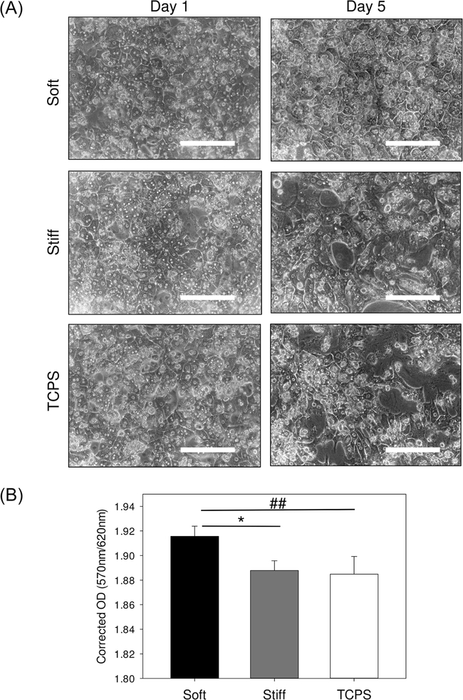
We investigated the effect of substrate stiffness on morphology and viability of primary hepatocytes cultured on PDMS substrates coated with collagen and compared the results with TCPS coated with collagen (Fig. 1). We observed that on day 1 primary hepatocytes displayed similar morphology on soft, stiff and TCPS substrates (Fig. 1A). The cells demonstrated tight cell–cell junctions, visible cell boundaries and also displayed typical polygonal shape, all of which are indicative of the typical hepatocyte epithelial morphology. On day 5, the hepatocytes on TCPS displayed considerable increase in cell-spreading and a fibroblast-like morphology. Similar trend was observed, but to a lesser magnitude, in cells cultured on the stiff substrate. On the contrary, cells on the soft substrate retained the initial polygonal shape and visible tight junctions between the cells. Following changes in morphology, we studied cell viability using MTT assay (Fig. 1B). We observed that the cells grown on soft substrates had a higher viability as compared to hepatocytes on stiff and TCPS substrates indicating a potential effect of stiffness on hepatocytes.
Fig. 1.
(A) Characterization of primary hepatocyte morphology when cultured on soft, stiff and TCPS substrates; (B) quantification of primary hepatocyte viability using MTT assay on day 5 in culture, scale bar = 200 microns, significant difference between the soft/stiff group and TCPS group is denoted by ##p value < 0.005, significant difference between the soft and stiff group is denoted by *p value < 0.05.
Primary hepatocyte cell attachment and cell spreading area
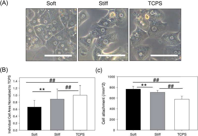
We investigated the effect of substrate stiffness on individual primary hepatocytes when seeded at a sub-confluent density of 50 000 cells per cm2. As seen in Fig. 2, the individual cell morphology displays a significant difference between soft, stiff and TCPS substrates. The individual cells on the soft substrate have significantly lower cell spreading area (66.1 ± 18.7%) compared to the cells attached on stiff (88.7 ± 28.9%) and TCPS control (100 ± 27.7%). We further measured the cell attachment on these substrates and the total number of cells attached per unit area was highest in soft substrates (768.1 ± 50.1 cells per mm2) compared to stiff (707.8 ± 33.9 cells per mm2) and TCPS control (577.4 ± 56.8 cells per mm2).
Fig. 2.
(A) Phase contrast images primary hepatocytes on soft, stiff and TCPS substrates; (B) quantification of primary hepatocyte area normalized with respect to cells on TCPS; (C) quantification of total primary hepatocyte attachment per unit area after 24 hours in culture, scale bar = 100 microns, significant difference between the soft/stiff group and TCPS group is denoted by ##p value < 0.005, significant difference between the soft and stiff group is denoted by **p value < 0.005.
Effect of stiffness on urea production
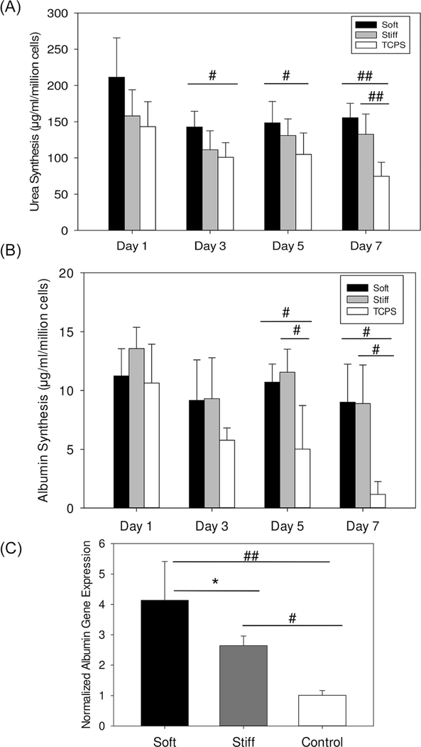
We investigated the effect of substrate stiffness on urea production, a key functional marker for primary hepatocytes and an indicator of intact nitrogen metabolism and detoxification, over a duration of 7 days (Fig. 3A).20 Hepatocytes cultured on soft substrates produced 155.4 ± 20.0 μg per ml per million cells urea on day 7 compared to 132.4 ± 28.2 μg per ml per million cells urea and 74.5 ± 19.3 μg per ml per million cells urea by hepatocytes cultured on stiff and TCPS substrates on day 7, respectively. Interestingly, primary hepatocytes cultured on stiff substrates recreating the diseased liver microenvironment produced significantly more urea as compared to those on TCPS, which is considered the gold standard culture substrate.
Fig. 3.
(A) Quantification of urea synthesis by primary hepatocytes on soft, stiff and TCPS substrates; (B) quantification of albumin synthesis primary hepatocytes cultured on soft, stiff and TCPS substrates using ELISA; and (C) gene expression analysis albumin of primary hepatocytes cultured on soft, stiff and TCPS substrates on day 7, significant difference between the soft/stiff group and TCPS group is denoted by #p value < 0.05, ##p value < 0.005, significant difference between the soft and stiff group is denoted by *p value < 0.05.
Effect of substrate stiffness on albumin
Albumin synthesis is a widely accepted marker of hepatocyte synthetic function.19,20 The long term metabolic response of continuous hepatocyte culture on soft and stiff substrates was compared with collagen coated surfaces by measuring albumin production as shown in Fig. 3B and C. Fig. 3B and C illustrate the rate of albumin production and albumin gene expression, respectively, for cultures up to one week. The daily production of albumin on soft and stiff surfaces were comparable and significantly higher than collagen coated TCPS surfaces. By day 7, the liver specific albumin production was approximately 8.9 μg per ml per million cells albumin in both soft and stiff compared to 1 μg per ml per million cells albumin on TCPS surface. We measured the albumin gene expression of hepatocytes cultured on these substrates on day 7 and observed that the soft and stiff substrates showed a 4 fold and 2.5 fold higher albumin gene expression, respectively, compared to TCPS. The hepatocytes in the soft surfaces mimicking healthy liver environment had a significantly higher albumin gene expression when compared to stiff surfaces mimicking the diseased liver environment.
Effect of stiffness on cytochrome P450 activity
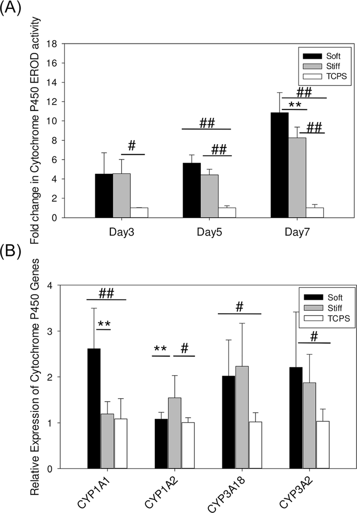
Cytochrome P450 enzymatic activity is a critical function of hepatocytes that plays an important role in the metabolism of multiple toxins, xenobiotics and pharmaceuticals.17 Cytochrome P450 1A1/2 (CYP1A1/2) activity was monitored in hepatocytes cultured on soft, stiff, and TCPS control on days 3, 5, and 7 (Fig. 4A). Our goal was to investigate the effect of stiffness on the enzymatic kinetics over the observation period, since decrease in enzymatic activity is usually indicative of a deteriorating phenotype. Thus, we report the difference in CYP enzyme activity on soft and stiff substrates as a fold change compared to TCPS on days 3, 5, and 7. CYP1A1/2 activity on soft substrates on days 3, 5, 7 was 4.5, 5.6, and 10.8 fold higher than TCPS. CYP1A1/2 activity on stiff substrates on day 3, 5, 7 was 4.5, 4.4, and 8.2 fold higher than TCPS. Furthermore, the CYP activity of hepatocytes on soft substrates on day 7 was significantly higher than the cells on stiff substrates.
Fig. 4.
(A) Quantification of cytochrome P450 activity of primary hepatocytes when cultured on soft, stiff and TCPS substrates; (B) gene expression analysis of cytochrome P450 gene markers of primary hepatocytes cultured on soft, stiff and TCPS substrates on day 7, significant difference between the soft/stiff group and TCPS group is denoted by #p value < 0.05, ##p value < 0.005, significant difference between the soft and stiff group is denoted by **p value < 0.005.
We also probed the gene expression levels of CYP1A1, CYP3A2 (rat species equivalent to the human CYP3A4 46), CYP3A18, and CYP1A2 (Fig. 4B). We observed that hepatocytes cultured on soft substrates demonstrated 2.7, 1.9, and 2.1-fold up regulation of CYP1A1, CYP3A2 and CYP3A18 gene expression, respectively, compared to TCPS. Cells cultured on stiff substrates had 1.2-fold increase in CYP1A1 gene expression and no significant change in CYP3A2 and CYP3A18 gene expression compared to TCPS. This indicates that the hepatocytes on soft substrates representative of healthy liver environment have better maintenance of drug metabolizing enzymes compared to disease like stiff substrates. Interestingly, CYP1A2 gene expression did not demonstrate up regulation on the soft substrate but showed 1.5 fold up regulation in the stiff substrate, compared to TCPS.
Effect of stiffness on the maintenance of hepatocyte specific markers
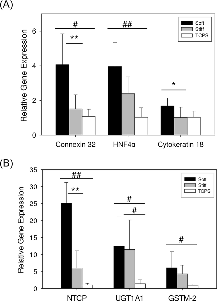
We evaluated the effect of stiffness on the differentiated epithelial-like phenotype of the hepatocytes cultured on the soft, stiff and TCPS substrates by probing the gene expression of hepatocyte nuclear factor 4α (HNF4α), cytokeratin 18 (CK18) and connexin 32 (Fig. 5A). We observed that hepatocytes cultured on the soft substrates demonstrated the highest degree of epithelial-like phenotype. Hepatocytes on soft and stiff surfaces had a 4-fold and 2-fold higher HNF4α gene expression, respectively, compared to TCPS. Hepatocytes cultured on soft substrates had 1.6-fold higher CK18 gene expression compared to TCPS while cells on stiff substrates had comparable CK18 gene expression to TCPS. We also observed that hepatocytes cultured on soft substrates had 4-fold higher gene expression of connexin 32 compared to TCPS while cells cultured on stiff substrates had no significant change in connexin 32 gene expression compared to TCPS.
Fig. 5.
(A) Gene expression analysis of epithelial cell markers of primary hepatocytes cultured on soft, stiff and TCPS substrates on day 7; (B) gene expression analysis of hepatic functional markers on day 7, significant difference between the soft/stiff group and TCPS group is denoted by #p value < 0.05, ##p value < 0.005, significant difference between the soft and stiff group is denoted by *p value < 0.05 and **p value < 0.005.
We also evaluated the role of matrix stiffness on primary hepatocyte functional gene markers-NTCP (sodium dependent bile acid transporter), GSTM-2 (glutathione S-transferase mu 2) and UGT1A1 (UDP glycosyltransferase) (Fig. 5B). We observed that hepatocytes cultured on soft substrates resulted in 12-fold, 6-fold, and 25-fold upregulation of UGT1A1, GSTM-2, and NTCP gene expression, respectively, compared to TCPS. Cells cultured on stiff substrates had no significant change in GSTM-2 and NTCP gene expression compared to TCPS.
Discussion
Liver damage as a consequence of liver injury or disease (e.g., chronic hepatitis C virus [HCV] infection, alcohol abuse, and nonalcoholic steatohepatitis [NASH]) is extremely prevalent and results in a substantial economic burden on patients.47,48 Several liver diseases can lead to fibrosis, which results from an imbalance between production and resorption of extracellular matrix (ECM) and a restructuring of the liver microenvironment (LME). The earliest changes in LME as a result of liver disease occur in response to ECM remodeling, resulting in accumulation of ECM proteins and an increase in liver stiffness. Furthermore, the distorted balance between matrix production and degradation leads to deleterious effects on the liver function. Clinical studies suggest that the altered LME provides a permissive milieu for the development of cellular dysplasia and is a key feature of liver dysfunction that leads to cirrhosis and hepatocellular carcinoma (HCC).49–51 There is a range of underlying mechanisms that contribute to liver diseases; however, despite extensive research over many decades, the precise molecular mechanisms of how the changes in liver stiffness affects liver function remain poorly understood. In this study, we utilized PDMS substrate with tunable elastic modulus for studying the stiffness-mediated effect on primary hepatocyte behavior. We identified two substrates with stiffness of 2 kPa (soft) and 55 kPa (stiff) as mimics for the LME of the healthy and the diseased liver.42
Recent reports have demonstrated the effect of stiffness on hepatocytes function.12,24 However, the stiffness ranges employed in these studies have limitations with respect to recreating physiologically relevant conditions for healthy and diseased liver. In the platform used in this study, we adapted the method developed by Palchesko and co-workers by using different ratios Sylgard 527 and Sylgard 184 to tune the elastic modulus of PDMS.44 Although not many cell culture studies have utilized Sylgard 527 as a substrate, we observed that the use of Sylgard 527 does not lead to any compromise in the cellular viability. PDMS mixtures of 527 and 184 also retain optical transparency that makes them an ideal substrate material for imaging cells. Although the material is inherently hydrophobic, studies have shown that oxygen plasma treatment temporarily modifies the surface Si–OH groups to render hydrophilicity.52 We utilized this surface modification to coat the substrates with collagen, which is commonly used for facilitating the adhesion of primary hepatocytes. The typical polygonal morphology of primary hepatocytes was retained in soft substrates and significantly lost on stiff and TCPS substrates after day 5 in culture. The cells also show some degree of aggregation in the TCPS substrates, which is a strong indication of de-differentiation (loss of function) of hepatocytes. The polygonal shape and presence of visible boundaries between the cells are generally attributed to the differentiated state of the cells and the elongated fibroblast like morphology is associated with dedifferentiation and can also be associated with loss in membrane integrity.53 The viability of the cells was also significantly lowered in stiff and TCPS substrates when compared to soft substrates after day 5 in culture demonstrating the potential compromise in hepatocytes viability in stiff (diseased liver mimic) surfaces. Together, these results indicate that the primary hepatocytes appear to be more differentiated on the soft substrate (healthy liver mimic), as compared to stiff and TCPS. Furthermore, the cells were more circular and had lesser spreading area in the soft substrate compared to stiff and TCPS surfaces. This observation is consistent with studies carried out by groups on other adherent cell types.54 The higher cell attachment of hepatocytes on soft compared to stiff substrates also demonstrates that hepatocytes when exposed to diseased liver like environment lose their viability akin to liver diseases.
A significant finding of our study was the higher production of urea and albumin on soft substrates compared to stiff substrates. Hepatocytes on both soft and stiff substrates displayed higher production of urea and albumin compared to TCPS, thus indicating that culturing hepatocytes on TCPS is a poor in vitro model to study hepatocyte function. These trends clearly indicate that hepatocellular functions are also maintained, since urea and albumin are two of the key markers of hepatic function. In addition, we observed that the albumin gene expression was significantly higher in soft surfaces compared to both stiff and TCPS surfaces. Clinical studies have demonstrated that the expression of albumin mRNA in acute hepatic failure and decompensated liver cirrhosis was reduced significantly compared to normal control liver in human liver samples.55,56 These studies suggest that albumin concentration is mainly regulated at albumin mRNA level in the liver despite the presence of other regulatory mechanisms and that expression of albumin mRNA level is correlated with disease severity. We hypothesized that the hepatocytes on soft environment maintains the hepatic function longer compared to stiffer and TCPS surfaces as the soft substrates mimics the healthy LME while the stiff surfaces recreates the diseased LME. Our in vitro model recreates this phenomenon, thus increasing our confidence in our platform to mimic the clinical aspects of liver fibrosis. These data further indicate the need for tissue engineered liver models that recapitulate the mechanical environment found in vivo.55,56
Cytochrome P450 enzymatic activity in hepatocytes is critical for the liver to conduct detoxification of a wide range of toxins, drugs and xenobiotics. However, this function is lost in several liver diseases.57,58 Peterson and co-workers induced hepatic fibrosis in mice models and found that fibrotic livers had significant reduction in CYP1A subgroup levels.58 Since liver stiffness increases with varying stages of liver disease, the understanding of how stiffness affects enzymatic activity is key for potential therapeutic strategies in the future. We focused our attention on a major class of CYP enzymes, CYP1A1/2, since this enzyme typically mediates the metabolism of drugs. The soft substrates exhibited higher enzymatic activity over seven-day observation period compared to stiff and TCPS surfaces. These trends were mirrored in the gene expressions of CYP1A1, CYP3A2 and CYP3A18. This indicates that substrate stiffness acts as an important cue in preserving the clearance functions of hepatocytes. These results are of prime interest when modeling hepatoxicity or drug screening studies in vitro. In a similar stiffness-mediated drug metabolism study, Zustiak and co-workers, demonstrated that various types of cells when cultured on different stiffness substrates showed varying drug resistance and thus, the substrate stiffness affects the reliability of the in vitro drug screening platforms.59 We also observed that hepatocytes on soft substrates have higher gene expression of key phase II transporters (NTCP, GSTM-2 and UGT1A1).
Maintenance of epithelial phenotype and repression of dedifferentiation in hepatocytes is an important event in the progression of liver diseases.60 Homotypic hepatocyte interactions and gap junctions are marked by the expression of connexin 32.61 Connexin 32 is a vital gap junction protein expressed in hepatocytes that regulates the signal transduction related to cell function, growth and dedifferentiation in the liver.62 Clinical studies have reported significant decrease in the mRNA levels of connexin 32 in chronic liver diseases and cirrhosis.63 Cytokeratin 18 is an intermediate filament present in epithelial cells such as hepatocytes and the decrease in the cytokeratin 18 expression levels is indicative of dedifferentiating epithelial phenotype.60 Hepatic nuclear factor 4 alpha (HNF4α) plays an active role in the maintenance of the differentiated epithelial phenotype and in the repression of dedifferentiation in hepatocytes. 64 HNF4α is the regulator of multiple signaling cascades and hence directly mediates the expression levels of a broad spectrum of genes that are involved in the maintenance of hepatocyte homeostasis.65 Clinical reports have demonstrated that cirrhosis and fibrotic liver have decreased levels of HNF4α expression, indicating the role they play in the altered hepatic functions such as lipid and carbohydrate metabolism, and bile acid synthesis in the diseased hepatocytes.66,67 Overall, these markers can be the vital mechanistic regulators of the maintenance of the phenotypic traits of hepatocytes. We observed that the soft substrate supports the highest level of differentiated hepatocyte maintenance as demonstrated by the expression of these key epithelial markers. Analogous to the maintenance of the differentiated phenotype in the soft substrate, we observed a more disease-like phenotype induction in the stiff substrate, entirely based on the mechano-signaling changes using the substrates. Therefore, together these data suggest that stiffness has a strong impact on hepatocytes function and substrates of varying stiffness might provide a method to recreate various stages of liver diseases.
In summary, we investigated the function of primary hepatocytes cultured on PDMS substrates that can be tuned to demonstrate varying stiffness. Our study demonstrates that stiffness of the culture substrate plays a crucial role in determining the phenotypic maintenance of primary hepatocytes. We observed that hepatocytes grown on the healthy liver stiffness (2 kPa) displayed a consistently more differentiated and functional phenotype for a longer duration as compared to the hepatocytes that were cultured on the stiff substrate, which represents the diseased liver (55 kPa) and, TCPS. Increase in liver stiffness is a strong indication of the progression of liver fibrosis. There is a critical need to engineer in vitro models that will mimic the various stages of liver disease to serve as accurate models for studying disease mechanism and drug and toxicity testing. Such models need to incorporate the dynamic changes in LME including the change in liver stiffness. In the current study, we demonstrate that hepatocytes phenotype and function is strongly modulated based on the stiffness the cells are exposed. Hepatocytes on environment comparable to healthy liver resulted in the simultaneous maintenance of their epithelial phenotype and increased hepatocellular functions compared to the cells on environment similar to the diseased liver. The intricate LME-hepatocytes signaling pathways through which these cells communicate within the LME will be further investigated in our future studies.
Supplementary Material
Acknowledgements
We thank Dr Edward Harris for kindly helping us with the isolation of primary hepatocytes for our experiments. We thank Dr William H. Velander for access to the microplate spectrophotometer. We thank Dr Concetta DiRusso for access to the fluorescent plate reader. This work was funded by the startup funds from UNL, Layman’s Award, MRSEC Seed Grant, UNL CIBC Research Cluster Development Grant, and UNL Interdisciplinary Award.
Footnotes
Electronic supplementary information (ESI) available. See DOI: 10.1039/c5ra15208a
References
- 1.Foucher J, Chanteloup E, Vergniol J, Castera L, le Bail B, Adhoute X, Bertet J, Couzigou P and de Ledinghen V, Gut, 2006, 55, 403–408. [DOI] [PMC free article] [PubMed] [Google Scholar]
- 2.Georges PC, Hui J-J, Gombos Z, McCormick ME, Wang AY, Uemura M, Mick R, Janmey PA, Furth EE and Wells RG, Am. J. Physiol, 2007, 293, G1147–G1154. [DOI] [PubMed] [Google Scholar]
- 3.Yin M, Kolipaka A, Woodrum DA, Glaser KJ, Romano AJ, Manduca A, Talwalkar JA, Araoz PA, McGee KP, Anavekar NS and Ehman RL, Chin. J. Magn. Reson. Imaging, 2013, 38, 809–815. [DOI] [PMC free article] [PubMed] [Google Scholar]
- 4.Yin M, Woollard J, Wang X, Torres VE, Harris PC, Ward CJ, Glaser KJ, Manduca A and Ehman RL, Magnetic resonance in medicine : official journal of the Society of Magnetic Resonance in Medicine/Society of Magnetic Resonance in Medicine, 2007, 58, 346–353. [DOI] [PubMed] [Google Scholar]
- 5.Henderson NC and Forbes SJ, Toxicology, 2008, 254, 130–135. [DOI] [PubMed] [Google Scholar]
- 6.Lozoya OA, Wauthier E, Turner RA, Barbier C, Prestwich GD, Guilak F, Superfine R, Lubkin SR and Reid LM, Biomaterials, 2011, 32, 7389–7402. [DOI] [PMC free article] [PubMed] [Google Scholar]
- 7.Wells RG, Hepatology, 2008, 47, 1394–1400. [DOI] [PubMed] [Google Scholar]
- 8.Kidambi S, Yarmush RS, Novik E, Chao P, Yarmush ML and Nahmias Y, Proc. Natl. Acad. Sci. U. S. A, 2009, 106, 15714–15719. [DOI] [PMC free article] [PubMed] [Google Scholar]
- 9.Kidambi S, Sheng LF, Yarmush ML, Toner M, Lee I and Chan C, Macromol. Biosci, 2007, 7, 344–353. [DOI] [PubMed] [Google Scholar]
- 10.Kidambi S, Lee I and Chan C, J. Am. Chem. Soc, 2004, 126, 16286–16287. [DOI] [PubMed] [Google Scholar]
- 11.Stevens KR, Ungrin MD, Schwartz RE, Ng S, Carvalho B, Christine KS, Chaturvedi RR, Li CY, Zandstra PW, Chen CS and Bhatia SN, Nat. Commun, 2013, 4, 1847. [DOI] [PMC free article] [PubMed] [Google Scholar]
- 12.Chen AA, Khetani SR, Lee S, Bhatia SN and van Vliet KJ, Biomaterials, 2009, 30, 1113–1120. [DOI] [PMC free article] [PubMed] [Google Scholar]
- 13.Khetani SR and Bhatia SN, Nat. Biotechnol, 2008, 26, 120–126. [DOI] [PubMed] [Google Scholar]
- 14.Larkin AL, Rodrigues RR, Murali T and Rajagopalan P, Tissue Eng., Part C, 2013, 19, 875–884. [DOI] [PMC free article] [PubMed] [Google Scholar]
- 15.Zheng MH, Ye C, Braddock M and Chen YP, Cytotherapy, 2010, 12, 349–360. [DOI] [PubMed] [Google Scholar]
- 16.Mehta G, Williams CM, Alvarez L, Lesniewski M, Kamm RD and Griffith LG, Biomaterials, 2010, 31, 4657–4671. [DOI] [PMC free article] [PubMed] [Google Scholar]
- 17.Arias I, Wolkoff A, Boyer J, Shafritz D, Fausto N, Alter H and Cohen D, The liver: biology and pathobiology, John Wiley & Sons, 2011. [Google Scholar]
- 18.Huang X, Hang R, Wang X, Lin N, Zhang X and Tang B, Artif. Organs, 2013, 37, 166–174. [DOI] [PubMed] [Google Scholar]
- 19.Ben-Ze’ev A, Robinson GS, Bucher N and Farmer SR, Proc. Natl. Acad. Sci. U. S. A, 1988, 85, 2161–2165. [DOI] [PMC free article] [PubMed] [Google Scholar]
- 20.Mooney D, Hansen L, Vacanti J, Langer R, Farmer S and Ingber D, J. Cell. Physiol, 1992, 151, 497–505. [DOI] [PubMed] [Google Scholar]
- 21.Hansen LK, Wilhelm J and Fassett JT, Curr. Top. Dev. Biol, 2006, 72, 205–236201 plate. [DOI] [PubMed] [Google Scholar]
- 22.Fassett J, Tobolt D and Hansen LK, Mol. Biol. Cell, 2005, 17, 345–356. [DOI] [PMC free article] [PubMed] [Google Scholar]
- 23.Semler EJ, Lancin PA, Dasgupta A and Moghe PV, Biotechnol. Bioeng, 2005, 89, 296–307. [DOI] [PubMed] [Google Scholar]
- 24.You J, Park SA, Shin DS, Patel D, Raghunathan VK, Kim M, Murphy CJ, Tae G and Revzin A, Tissue Eng., Part A, 2013, 19, 2655–2663. [DOI] [PMC free article] [PubMed] [Google Scholar]
- 25.Marois Y, Sigot-Luizard M-F and Guidoin R, ASAIO J, 1999, 45, 272–280. [PubMed] [Google Scholar]
- 26.Park JH, Park KD and Bae YH, Biomaterials, 1999, 20, 943–953. [DOI] [PubMed] [Google Scholar]
- 27.Sherman MA, Kennedy JP, Ely DL and Smith D, J. Biomater. Sci., Polym. Ed, 1999, 10, 259–269. [DOI] [PubMed] [Google Scholar]
- 28.Bordenave L, Bareille R, Lefebvre F, Caix J and Baquey C, J. Biomater. Sci., Polym. Ed, 1992, 3, 409–416. [DOI] [PubMed] [Google Scholar]
- 29.van Kooten TG, Whitesides JF and von Recum AF, J. Biomed. Mater. Res, 1998, 43, 1–14. [DOI] [PubMed] [Google Scholar]
- 30.Ertel SI, Ratner BD, Kaul A, Schway MB and Horbett TA, J. Biomed. Mater. Res, 1994, 28, 667–675. [DOI] [PubMed] [Google Scholar]
- 31.Dahrouch M, Schmidt A, Leemans L, Linssen H and Goetz H, Macromol. Symp, 2003, 199, 147–162. [Google Scholar]
- 32.Interrante LV, Shen Q and Li J, Macromolecules, 2001, 34, 1545–1547. [Google Scholar]
- 33.Ng JMK, Gitlin I, Stroock AD and Whitesides GM, Electrophoresis, 2002, 23, 3461–3473. [DOI] [PubMed] [Google Scholar]
- 34.McDonald JC and Whitesides GM, Acc. Chem. Res, 2002, 35, 491–499. [DOI] [PubMed] [Google Scholar]
- 35.Sia SK and Whitesides GM, Electrophoresis, 2003, 24, 3563–3576. [DOI] [PubMed] [Google Scholar]
- 36.Ostuni E, Chapman RG, Liang MN, Meluleni G, Pier G, Ingber DE and Whitesides GM, Langmuir, 2001, 17, 6336–6343. [Google Scholar]
- 37.Folch A, Jo B-H, Hurtado O, Beebe DJ and Toner M, J. Biomed. Mater. Res, 2000, 52, 346–353. [DOI] [PubMed] [Google Scholar]
- 38.Daverey A, Mytty AC and Kidambi S, Nano LIFE, 2012, 2, 1241009. [Google Scholar]
- 39.Kidambi S, Udpa N, Schroeder SA, Findlan R, Lee I and Chan C, Tissue Eng, 2007, 13, 2105–2117. [DOI] [PMC free article] [PubMed] [Google Scholar]
- 40.Castera L, Hepatology International, 2011, 5, 625–634. [DOI] [PMC free article] [PubMed] [Google Scholar]
- 41.Mueller S, Clin. Use Drugs Patients Kidney Liver Dis, 2013, 2, 68–71. [DOI] [PMC free article] [PubMed] [Google Scholar]
- 42.Mueller S and Sandrin L, Hepatic Med, 2010, 2, 49. [DOI] [PMC free article] [PubMed] [Google Scholar]
- 43.Seglen PO, Exp. Cell Res, 1973, 82, 391–398. [DOI] [PubMed] [Google Scholar]
- 44.Palchesko RN, Zhang L, Sun Y and Feinberg AW, PLoS One, 2012, 7, e51499. [DOI] [PMC free article] [PubMed] [Google Scholar]
- 45.Yang C, Tibbitt MW, Basta L and Anseth KS, Nat. Mater, 2014, 13, 645–652. [DOI] [PMC free article] [PubMed] [Google Scholar]
- 46.Wójcikowski J, Haduch A and Daniel WA, Pharmacol. Rep, 2012, 64, 1411–1418. [DOI] [PubMed] [Google Scholar]
- 47.Asrani SK, Larson JJ, Yawn B, Therneau TM and Kim WR, Gastroenterology, 2013, 145, 375–382, e372. [DOI] [PMC free article] [PubMed] [Google Scholar]
- 48.Lancet T, Lancet, 2013, 381, 508. [Google Scholar]
- 49.Iredale JP, Thompson A and Henderson NC, Biochim. Biophys. Acta, 2013, 1832, 876–883. [DOI] [PubMed] [Google Scholar]
- 50.Mederacke I, Zeitschrift für Gastroenterologie, 2013, 51, 55–62. [DOI] [PubMed] [Google Scholar]
- 51.Ramachandran P and Iredale JP, J. Assoc. Physicians India, 2012, 105, 813–817. [DOI] [PMC free article] [PubMed] [Google Scholar]
- 52.Bodas D and Khan-Malek C, Sens. Actuators, B, 2007, 123, 368–373. [Google Scholar]
- 53.DiPersio C, Jackson D and Zaret K, Mol. Cell. Biol, 1991, 11, 4405–4414. [DOI] [PMC free article] [PubMed] [Google Scholar]
- 54.Yeung T, Georges PC, Flanagan LA, Marg B, Ortiz M, Funaki M, Zahir N, Ming W, Weaver V and Janmey PA, Cell Motil. Cytoskeleton, 2005, 60, 24–34. [DOI] [PubMed] [Google Scholar]
- 55.Ozaki I, Motomura M, Setoguchi Y, Fujio N, Yamamoto K, Kariya T and Sakai T, Gastroenterol. Jpn, 1991, 26, 472–476. [DOI] [PubMed] [Google Scholar]
- 56.Natsume M, Tsuji H, Harada A, Akiyama M, Yano T, Ishikura H, Nakanishi I, Matsushima K, Kaneko S.-i. and Mukaida N, J. Leukocyte Biol, 1999, 66, 601–608. [PubMed] [Google Scholar]
- 57.Frye RF, Zgheib NK, Matzke GR, Chaves-Gnecco D, Rabinovitz M, Shaikh OS and Branch RA, Clin. Pharmacol. Ther, 2006, 80, 235–245. [DOI] [PubMed] [Google Scholar]
- 58.Peterson TC, Hodgson P, Fernandez-Salguero P, Neumeister M and Gonzalez FJ, Hepatol. Res, 2000, 17, 112–125. [DOI] [PubMed] [Google Scholar]
- 59.Zustiak S, Nossal R and Sackett DL, Biotechnol. Bioeng, 2014, 111, 396–403. [DOI] [PMC free article] [PubMed] [Google Scholar]
- 60.Caja L, Bertran E, Campbell J, Fausto N and Fabregat I, J. Cell. Physiol, 2011, 226, 1214–1223. [DOI] [PubMed] [Google Scholar]
- 61.Bhatia S, Balis U, Yarmush M and Toner M, FASEB J, 1999, 13, 1883–1900. [DOI] [PubMed] [Google Scholar]
- 62.Seo S-J, Choi Y-J, Akaike T, Higuchi A and Cho C-S, Tissue Eng, 2006, 12, 33–44. [DOI] [PubMed] [Google Scholar]
- 63.Yamaoka K, Nouchi T, Kohashi T, Marumo F and Sato C, Liver, 2000, 20, 104–107. [DOI] [PubMed] [Google Scholar]
- 64.Santangelo L, Marchetti A, Cicchini C, Conigliaro A, Conti B, Mancone C, Bonzo JA, Gonzalez FJ, Alonzi T and Amicone L, Hepatology, 2011, 53, 2063–2074. [DOI] [PMC free article] [PubMed] [Google Scholar]
- 65.Lazarevich N, Shavochkina D, Fleishman D, Kustova I, Morozova O, Chuchuev E and Patyutko Y, Exp. Oncol, 2010, 32, 167–171. [PubMed] [Google Scholar]
- 66.Takahara Y, Takahashi M, Zhang Q-W, Wagatsuma H, Mori M, Tamori A, Shiomi S and Nishiguchi S, World J. Gastroenterol, 2008, 14, 2010. [DOI] [PMC free article] [PubMed] [Google Scholar]
- 67.Omori K, Terai S, Ishikawa T, Aoyama K, Sakaida I, Nishina H, Shinoda K, Uchimura S, Hamamoto Y and Okita K, FEBS Lett, 2004, 578, 10–20. [DOI] [PubMed] [Google Scholar]
Associated Data
This section collects any data citations, data availability statements, or supplementary materials included in this article.


