Abstract
Context:
Polycystic ovarian syndrome (PCOS) is a common endocrine system disorder among the women of reproductive age, yet the etiology of PCOS remains unclear. Infertility in females with PCOS can be caused by anovulation, high luteinizing hormone levels, and hyperandrogenism.
Aims:
This research analyzed the role of the aromatase gene (CYP19A1) in PCOS pathogenesis.
Settings and Design:
This study used an observational, cross-sectional design.
Subjects and Methods:
A total of 110 research participants (55 PCOS patients and 55 non-PCOS patients) were included in the study.
Statistical Analysis Used:
A real-time quantitative polymerase chain reaction was used to analyze the mRNA expression for aromatase in granulosa cells.
Results:
The relative expression of aromatase mRNA is lower in women with PCOS compared to those without PCOS (P < 0.05). Relative expression of CYP19A1 (aromatase) mRNA in PCOS group was 0.38 ± 0.25, whereas in non-PCOS group was 1.00 ± 0.00. The decline in aromatase activity contributes to an increase in testosterone level. This condition has a role in hyperandrogenism which is a typical characteristic of PCOS women. Granulosa cells in polycystic ovary undergo disturbance in the development and cannot respond to follicle-stimulating hormone (FSH) stimulation. Lack of stimulation of FSH causes induction inadequacy to aromatase enzyme activity in the aromatization process. The decline in FSH activity is caused by various factors that are associated with typical characteristics of PCOS.
Conclusions:
There is a decrease in the relative expression rate of granulosa cells’ aromatase mRNA in women with PCOS compared to the non-PCOS.
KEYWORDS: Aromatase, granulosa cells, polycystic ovarian syndrome
INTRODUCTION
Polycystic ovarian syndrome (PCOS) is a common endocrine system disorder among the women of reproductive age, yet the etiology of PCOS remains unclear. Infertility in females with PCOS can be caused by anovulation, high luteinizing hormone (LH) levels, and hyperandrogenism. In PCOS, follicle arrest occurs, and it prevents full maturation of ovum. Granulosa cells are essential in the production of steroid hormones, providing nutrition and other growth factors that may interact with the oocyte during its development in the ovarian follicle. Follicle-stimulating hormone (FSH) has a role in inducing granulosa cell proliferation, recruitment of secondary follicles, and selection of the dominant follicle and regulates the aromatase activity in the granulosa cells. This research analyses the role of the aromatase gene (CYP19A1) in PCOS pathogenesis.[1,2,3,4,5,6,7,8,9,10]
SUBJECTS AND METHODS
The study protocols were reviewed and approved by the Ethics Committee of the Faculty of Medicine, University of Indonesia (No. 106/H2.F1/ETIK/2013), and all participants provided written informed consent. The design of the study used an analytical, observational, cross-sectional study which was carried out to reach the target of the study that is along with the purpose of the study. The participants of the study that fulfill the criteria of inclusion to analyze the expression of mRNA were 110 people consisting of 55 women with PCOS and 55 women non-PCOS.
The number of participants in this study fulfilled the minimum number of samples needed for research in a health study of 30 samples. So, the data generated is expected to be normally distributed in calculating statistics. As a consideration, the inclusion criteria for the participants of this study were women with PCOS aged 18–40 years who underwent an in vitro fertilization (IVF) program. This is a factor that limits the acquisition of samples in this study because there are not many PCOS women who can undergo IVF programs.
Follicular fluid was collected from the women with PCOS and non-PCOS who were carrying out the fertility program. The granulosa cell samples were obtained from follicle fluid that was aspirated during ovum pick-up (OPU) procedure. The sample of follicle fluid is about 0.5–1.5 ml. Granulosa cell is the excess from the OPU process which is not used for the next IVF steps. However, the granulosa cell is important for research in support of ovum quality.
OPU procedure is an standard operating procedure for the stages of the process that must be undertaken by the patients undergoing IVF. Those who carry out the procedure are medical personnel and embryologists who are competent in their field and have certificates to carry out these actions. The researchers only took and examined the sucking fluid follicles left in the aspiration process during the OPU. While the ovum obtained in the OPU process will be treated for the next IVF process.
Granulosa cell is isolated from follicle fluid by using Ficoll solution. Then, it was multiplied by the cell culture method by using the combination of DMEM F12 media (Gibco) and fetal bovine serum (Gibco). The measurement of the expression mRNA aromatase in the granulosa cell uses real-time quantitative polymerase chain reaction (RT-qPCR).
The analysis of the expression gene was measured in relative quantification by using the Livak method from the value of cycle threshold. Level of mRNA expression was obtained. The ratio of mRNA CYP19A1 expression is rated based on the Livak formula: n = 2-AACt.[11]
The analysis of statistics is to determine the comparison between the relative expression of mRNA CYP19A1 gene. It was done on the sample of granulosa cells in PCOS patients and non-PCOS patients with a t-test independently upon the limit of significance of P ≤ 0.05 for data which are normal. Meanwhile, the Mann–Whitney U test was used for the data which are not normal (SPSS program version 17, IBM Statistics, New York, USA).[12]
RESULTS
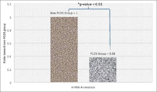
The measurement of mRNA CYP19A1 expression (aromatase) in the PCOS group and the non-PCOS group used RT-qPCR technique and Livak method. The result of the ratio measurement of mRNA aromatase in granulosa cells in women with PCOS group which was normalized toward non-PCOS (comparison) is shown in Figure 1.
Figure 1.

mRNA CYP19A1 expression ratio in women with polycystic ovarian syndrome group toward the nonpolycystic ovarian syndrome group
The average expression of mRNA aromatase in granulosa cells in women with the PCOS group is 0.38 ± 0.25, while non PCOS group is 1.00 ± 0.00. The result using the Mann–Whitney test shows the difference (P < 0.01) between the average of mRNA aromatase expression of granulosa cells in women with PCOS compared with women of non-PCOS. The result of this study showed that the average mRNA expression in granulosa cells was lower than the mRNA expression of aromatase in granulosa cells in women with non-PCOS.
Follicle granulosa cells are cells which express the aromatase. One of the main functions of FSH is regulating the activity of enzyme aromatase in granulosa cells through the mechanism which is mediated by cyclic adenosine monophosphate (cAMP) which will stimulate the transcription of aromatase in granulosa cells. FSH acts in granulosa cells to aromatize the androgens from theca cells into estrogen, via its action through follicle stimulating hormone receptor (FSHR). In PCOS cases, Asn680Ser genes possibly affects the regulation of aromatase enzyme in granulosa cells. The aromatase expression is regulated by signaling FSHR. The variety of genotypes in FSHR affects the role of FSHR in regulating the aromatase gene expression which depends on the FSHR signal.
DISCUSSION
The subject of this study were PCOS women in Indonesia who were undergoing an IVF program aged 18–40 years as compared to those women who underwent IVF for male factor infertility, tubal factor or unexplained infertility.
This study has differences from previous studies on the topic of the CYP 19A1 gene from its purpose of analyzing the role of granulosa cells in the pathogenesis of PCOS in terms of the expression of the CYP19A1 gene mRNA which encodes the aromatase enzyme. This enzyme plays a role in the estrogen biosynthesis in granulosa cells. The ability of granulosa cells to carry out their functions determines the quality of oocytes production which will determine the quality of the embryo.
The average mRNA CYP19A1 expression for aromatase in granulosa cells in women with PCOS was lower than women with non-PCOS [Figure 1]. CYP19A1 gene was located on chromosome 15q21.1, encodes the aromatase (P450arom), which is steroidogenesis enzyme key that catalyzes the last steps of estrogen biosynthesis from androgens. It transforms androstenedione to estrone and testosterone to estradiol (E2) and estrone in gonads and extragonadal tissue (brain and adipose tissue).[13,14,15] The previous result also showed that the ratio of mRNA expression for aromatase in granulosa cells in women with PCOS was lower than non-PCOS women.
The difference of mRNA expression for aromatase in granulosa cells between both groups was index significant statistically of P < 0.05. It can be assumed that the protein in the aromatase of PCOS group declines because of the excretion compared to the non-PCOS group. It also occurred in the previous result study that states that there was a significant relationship between the expression of mRNA and protein expression. It showed that gene which was expressed high would increase the production of protein more highly than the gene which was expressed lower.
The biological factors which affect the relationship between mRNA expression and abundance of protein are, protein synthesis, degradation speed of m RNA and protein as well as posttranscriptional m RNA regulation. The involvement of posttranscriptional mechanism and complicated posttranslation techniques possibly determines the abundance of protein.[16]
The decrease in aromatase activity contributes to the increase of testosterone. This situation has a role in hyperandrogenism which is a typical characteristic of PCOS. In PCOS, as the consequence of the increased LH secretion, it stimulates theca cells to synthesize the testosterone abundantly, while the decrease of aromatase activity causes disorder in the aromatization process. Granulosa cells are unable to aromatize the androgen into estrogen. Hence there is inadequate amount of estrogen for oocyte maturation resulting in chronic anovulation, typical of PCOS.
In this study, no androgen profile was examined. Previous study stated that the most common phenotype encountered for PCOS was normoandrogenic with the phenotype of anovulation and the picture of polycystic ovaries.[17] Anti-Müllerian hormone (AMH) levels of women in the PCOS group was almost twice as high as those of women who were not PCOS. This condition is caused by follicular arrest in small preantral and antral stages, where AMH is expressed in high concentrations. Such condition ultimately causes an increase in AMH levels. The research subjects have fulfilled the criteria for the characteristics of patients with PCOS, including AMH levels in the PCOS group, almost twice higher than those in the non-PCOS group. This is also a characteristic of PCOS.
The result of the study is in line with the report of Naessen et al.[18] which states that the activity of aromatase enzyme is lower in women with the PCOS group. The converting activity of testosterone into estrogen by aromatase is lower in follicle fluid for PCOS compared to the control group, and then granulosa cells which were obtained from antral follicles of PCOS patients show low aromatase activity.[19,20] It gets more interesting to analyze forward about the role of CYP19A1 gene with pathogenesis PCOS. Mainly, it is compared to hyperandrogenism in PCOS women. Aromatase has a closer relationship with the concentration of androgen and estrogen, and it is essential to develop the ovary in teenage. The lack of stimulation by FSH will cause inadequacy induction toward enzyme aromatase activity which is very important for synthesizing estrogen, and also it will cause anovulation. FSH can induce aromatase activity which has a positive correlation with E2. Therefore, inadequate E2 can stimulate to produce FSH as a negative feedback. High FSH shows lower aromatase activity.[5,21,22]
The role of the CYP19A1 gene is possibly involved in pathophysiology syndrome. It can be explained that the relation between FSH and the receptor in the membrane of granulosa cells causes the stimulation of promoter II activity by FSH. The intercellular cAMP will increase, and it will give more significant chance to bind both critical transcription factors that are steroidogenic factor-1 and cAMP response element-binding protein, in proximal promoter II gene aromatase. Aromatase activity gets expressed so that it causes the secretion of estrogen from the preovulatory follicle.
CONCLUSION AND SUGGESTION
Expression of mRNA CYP19A1 in granulosa cells of PCOS patients is lower than non PCOS patient. Further we need to analyze aromatase expression in protein to support the explanation about the role of CYP19A1 towards pathogenesis PCOS.
Financial support and sponsorship
Nil.
Conflicts of interest
There are no conflicts of interest.
Acknowledgment
My appreciation and thanks for Rector of Lambung Mangkurat University Kalimantan Selatan, Dean of Medical Faculty Lambung Mangkurat University Kalimantan Selatan and also Universitas Indonesia, Jakarta and lecturers who have given their knowledge during this time, my friends; Maman Saputra who helped in publication process and also Yasmin Clinic-RSCM Kencana with permission and willingness in this study.
REFERENCES
- 1.Alvero R, Schlaff WD. Reproductive Endrocrinology and Infertility: The Requisites in Obstretrics and Gynecology. USA: Elsevier Inc; 2007. pp. 65–76. [Google Scholar]
- 2.Pramayadi CT, Hestiantoro A, Hadisaputra W, Prasmusinto D. Effect of laparoscopic ovarian drilling on changes in blood flow of ovarian stroma and ratio of LH: FSH to polycystic ovary syndrome. J Obstet Gynecol Indonesia. 2008;32:3–10. [Google Scholar]
- 3.Xita N, Georgiou I, Tsatsoulis A. The genetic basis of polycystic ovary syndrome. Eur J Endocrinol. 2002;147:717–25. doi: 10.1530/eje.0.1470717. [DOI] [PubMed] [Google Scholar]
- 4.Norman RJ, Dewailly D, Legro RS, Hickey TE. Polycystic ovary syndrome. Lancet. 2007;370:685–97. doi: 10.1016/S0140-6736(07)61345-2. [DOI] [PubMed] [Google Scholar]
- 5.Simoni M, Tempfer CB, Destenaves B, Fauser BC. Functional genetic polymorphisms and female reproductive disorders: Part I: Polycystic ovary syndrome and ovarian response. Hum Reprod Update. 2008;14:459–84. doi: 10.1093/humupd/dmn024. [DOI] [PMC free article] [PubMed] [Google Scholar]
- 6.Brůcková L, Soukup T, Moos J, Moosová M, Pavelková J, Rezábek K, et al. The cultivation of human granulosa cells. Acta Medica (Hradec Kralove) 2008;51:165–72. doi: 10.14712/18059694.2017.19. [DOI] [PubMed] [Google Scholar]
- 7.Jacob TZ. Aromatase in gynecology Collection of Papers for Seminar on New Non-invasive. Diagnostic Tips and Medical Treatment for Endometriosis. 2005:1–21. [Google Scholar]
- 8.Petry CJ, Ong KK, Michelmore KF, Artigas S, Wingate DL, Balen AH, et al. Association of aromatase (CYP 19) gene variation with features of hyperandrogenism in two populations of young women. Hum Reprod. 2005;20:1837–43. doi: 10.1093/humrep/deh900. [DOI] [PubMed] [Google Scholar]
- 9.Power MR, Wilson AL, Cardia SR, Chu J, Ferrell R. Aromatase gene (CYP19) polymorphism and endogenous androgen concentration in multiracial/multiethnic, multisite study of women at midlife. Am J Med. 2006;119:23–30. doi: 10.1016/j.amjmed.2006.07.003. [DOI] [PubMed] [Google Scholar]
- 10.Ermann DA. Polycystic ovary syndrome. N Engl J Med. 2005;325:1223–36. doi: 10.1056/NEJMra041536. [DOI] [PubMed] [Google Scholar]
- 11.Livak KJ, Schimttgen TD. Analysis of relative gene expression data using real-time quantitative PCR and the 2-tCt Method. Methods. 2001;25:402–8. doi: 10.1006/meth.2001.1262. [DOI] [PubMed] [Google Scholar]
- 12.Now MS. Statistics for Medicine and Health: Descriptive, Bivariate, and Multivariate Equipped with Application using SPSS. Jakarta: Publisher of Salemba Medika; 2003. pp. 61–86. 167-79. [Google Scholar]
- 13.Jin JL, Sun J, Ge HJ, Cao YX, Wu XK, Liang FJ, et al. Association between CYP19 gene SNP rs2414096 polymorphism and polycystic ovary syndrome in Chinese women. BMC Med Genet. 2009;10:139. doi: 10.1186/1471-2350-10-139. [DOI] [PMC free article] [PubMed] [Google Scholar]
- 14.Gharani N, Waterworth DM, Batty S, White D, Gilling-Smith C, Conway GS, et al. Association of the steroid synthesis gene CYP11a with polycystic ovary syndrome and hyperandrogenism. Hum Mol Genet. 1997;6:397–402. doi: 10.1093/hmg/6.3.397. [DOI] [PubMed] [Google Scholar]
- 15.Guo Y, Xiong DH, Yang TL, Guo YF, Recker RR, Deng HW. Polymorphisms of estrogen-biosynthesis genes CYP17 and CYP19 may influence age at menarche: A genetic association study in Caucasian females. Hum Mol Genet. 2006;15:2401–8. doi: 10.1093/hmg/ddl155. [DOI] [PMC free article] [PubMed] [Google Scholar]
- 16.Yu EZ, Burba AE, Gerstein M. PARE: A tool for comparing protein abundance and mRNA expression data. BMC Bioinformatics. 2007;8:309. doi: 10.1186/1471-2105-8-309. [DOI] [PMC free article] [PubMed] [Google Scholar]
- 17.Wiweko B, Maidarti M, Priangga MD, Shafira N, Fernando D, Sumapraja K, et al. Anti-mullerian hormone as a diagnostic and prognostic tool for PCOS patients. J Assist Reprod Genet. 2014;31:1311–6. doi: 10.1007/s10815-014-0300-6. [DOI] [PMC free article] [PubMed] [Google Scholar]
- 18.Naessen T, Kushnir MM, Chaika A, Nosenko J, Mogilevkina I, Rockwood AL, et al. Steroid profiles in ovarian follicular fluid in women with and without polycystic ovary syndrome, analyzed by liquid chromatography-tandem mass spectrometry. Fertil Steril. 2010;94:2228–33. doi: 10.1016/j.fertnstert.2009.12.081. [DOI] [PubMed] [Google Scholar]
- 19.Wang H, Li Q, Wang T, Yang G, Wang Y, Zhang X, et al. A common polymorphism in the human aromatase gene alters the risk for polycystic ovary syndrome and modifies aromatase activity in vitro. Mol Hum Reprod. 2011;17:386–91. doi: 10.1093/molehr/gar007. [DOI] [PubMed] [Google Scholar]
- 20.Magoffin DA. Ovarian enzyme activities in women with polycystic ovary syndrome. Fertil Steril. 2006;86(Suppl 1):S9–S11. doi: 10.1016/j.fertnstert.2006.03.015. [DOI] [PubMed] [Google Scholar]
- 21.Simpson ER, Michael MD, Agarwal VR, Hinshelwood MM, Bulun SE, Zhao Y. Cytochromes P450 11: Expression of the CYP19 (aromatase) gene: An unusual case of alternative promoter usage. FASEB J. 1997;11:29–366. doi: 10.1096/fasebj.11.1.9034163. [DOI] [PubMed] [Google Scholar]
- 22.Larsen PR, Kronenberg HM, Melmed S, Polonsky KS. William Textbook of Endocrynology. 10th ed. Pennsylvania: WB Saunders; 2002. [Google Scholar]


