Abstract
Detection of minimal disseminated disease is a validated prognostic factor in ALK-positive anaplastic large cell lymphoma. We previously reported that quantification of minimal disease by quantitative real-time polymerase chain reaction (RQ-PCR) in bone marrow applying a cut-off of 10 copies NPM-ALK/104 copies of ABL1 identifies very high-risk patients. In the present study, we aimed to confirm the prognostic value of quantitative minimal disseminated disease evaluation and to validate digital polymerase chain reaction (dPCR) as an alternative method. Among 91 patients whose bone marrow was analyzed by RQ-PCR, more than 10 normalized copy-numbers correlated with stage III/IV disease, mediastinal and visceral organ involvement and low anti-ALK antibody titers. The cumulative incidence of relapses of 18 patients with more than 10 normalized copy-numbers of NPM-ALK was 61±12% compared to 21±5% for the remaining 73 patients (P=0.0002). Results in blood correlated with those in bone marrow (r=0.74) in 70 patients for whom both materials could be tested. Transcripts were quantified by RQ-PCR and dPCR in 75 bone marrow and 57 blood samples. Copy number estimates using dPCR and RQ-PCR correlated in 132 samples (r=0.85). Applying a cut-off of 30 copies NPM-ALK/104 copies ABL1 for quantification by dPCR, almost identical groups of patients were separated as those separated by RQ-PCR. In summary, the prognostic impact of quantification of minimal disseminated disease in bone marrow could be confirmed for patients with anaplastic large cell lymphoma. Blood can substitute for bone marrow. Quantification of minimal disease by dPCR provides a promising tool to facilitate harmonization of minimal disease measurement between laboratories and for clinical studies.
Introduction
ALK-positive anaplastic large cell lymphomas (ALCL) in children and adolescents are characterized by translocations involving the ALK gene on chromosome 2p23.1 About 90% of ALK-positive ALCL carry the translocation t(2;5)(p23;q35) resulting in the fusion gene NPM-ALK.2–4 Between 25% and 35% of patients relapse with current treatment protocols.5–9
Detection of blasts in bone marrow (BM) and blood by cytomorphological analysis is a rare event in ALCL.6,7 The chimeric fusion gene transcript NPM-ALK has been used to investigate the prognostic value of submicroscopic minimal disseminated disease (MDD) in BM and blood at diagnosis in independent cohorts of patients.10–13 Polymerase chain reaction (PCR) analysis allows the reliable detection of one circulating tumor cell among 100,000 normal cells.10 The detection of MDD by qualitative PCR in BM or blood (55% of patients) conferred a relapse risk of 50% in several studies.10–12,14 Measurement of minimal residual disease (MRD) using the qualitative PCR assay enabled identification of a very high-risk group of patients.14
We previously reported the possibility of identifing patients bearing a very high risk of relapse already at diagnosis by quantification of fusion gene transcripts using an NPM-ALK-specific quantitative real-time (RQ)-PCR assay. Applying a cut off of 10 copies NPM-ALK per 104copies of the reference transcript ABL1 (normalized copy numbers, NCN), 16 patients (22%) with more than 10 NCN NPM-ALK in the BM at diagnosis had a 5-year probability of event-free survival of 23±11% compared to 78±6% for the 58 patients with NCN below the cut-off. MDD levels measured in blood provided comparable results.12
The Japanese NHL study group recently reported the outcomes of 60 ALCL-patients according to MDD in blood or BM using the same cut-off of 10 NCN NPM-ALK.13 The patients received comparable therapies. Compared to the Berlin-Frankfurt-Münster (BFM) group, however, more patients showed MDD levels above 10 NCN. The progression-free survival of 37 patients with >10 NCN NPM-ALK in blood or BM was 58±12% compared to 85±6% for the 22 patients with ≤10 NCN.13
The differences regarding the prognostic value of MDD assessment by RQ-PCR in these two studies illustrate the need for standardization before the implementation of quantification of NPM-ALK transcripts for initial risk assessment or MRD evaluation in clinical studies. Currently, quantitative values from different laboratories cannot be directly compared to each other, whereas MRD quantification within one laboratory has been reported to enable the course of the disease to be monitored.15–17
To achieve comparability of MRD quantification for NPM-ALK obtained by RQ-PCR in different reference laboratories, extensive protocol harmonization is necessary, as was done for the quantification of BCR-ABL1 fusion gene transcripts in acute lymphoblastic leukemia and chronic myelogenous leukemia.18,19 Since quantification is performed at the lowest end of the necessary standard curve in NPM-ALK-specific RQ-PCR, a quantitative PCR approach with improved reproducibility without the need for a standard curve would be advantageous. Digital PCR (dPCR) may represent a quantitative PCR method that could be used as a replacement for RQ-PCR for NPM-ALK copy number estimation in ALK-positive ALCL. dPCR is a quantitative PCR method based on the distribution of the target RNA or DNA molecules in many partitions.20 The amount of partitions with a positive PCR results allows the concentration of a given target to be determined without the need for standard curve calibration.21
The aim of this work was to validate the prognostic meaning of quantitative MDD measurements of NPM-ALK fusion gene transcripts by RQ-PCR in an independent cohort of uniformly treated ALK-positive ALCL patients of the BFM group. In addition, in an effort to facilitate quality-controlled quantification between different laboratories, a dPCR assay for quantification of NPM-ALK transcripts was developed and validated.
Methods
Patients
Patients with ALCL from Austria, Germany and Switzerland enrolled in the ALCL99 trial or the NHL-BFM registry 2012 between January 2006 and December 2016 were eligible after confirmation of NPM-ALK positivity of the ALCL. Both studies were approved by the institutional ethics committee of the primary investigators. Informed consent from the patients/caregivers to the studies included consent for future research on MDD.
Controls and cell lines
Blood from 20 healthy donors and eight ALK-negative ALCL patients included in ALCL99 or the NHL-BFM registry served as negative controls after written informed consent.
The cell lines HL-60 (acute myeloid leukemia), SU-DHL-1 and Karpas-299 (NPM-ALK-positive ALCL) were obtained from the Deutsche Sammlung von Mikroorganismen und Zellkulturen (DSMZ, Braunschweig, Germany).
Complementary DNA synthesis and quantitative real-time polymerase chain reaction
Complementary DNA (cDNA) synthesis and RQ-PCR were performed as described previously.12 In total four replicates were analyzed (two with undiluted cDNA and two with 1+1 diluted cDNA, as an additional control for RQ-PCR inhibition). Samples which were positive for NPM-ALK in one to three of four replicates only or had NCN below one copy were considered as low positive not quantifiable. Negativity for NPM-ALK in all four replicates was considered as negative.
Digital polymerase chain reaction assay
Primer and probe sequences for the NPM-ALK- and ABL1-specific dPCR assay were identical to those used for the RQ-PCR assay.12 Probes used for dPCR were ordered with 5’FAM™ as the reporter dye and the double quencher dyes ZEN™ and 3’Iowa-Black®FQ (IDT, Leuven, Belgium).
Ten microliters of dPCR™ supermix for probes (no dUTP; BIO-RAD, Munich, Germany), 0.6 μL forward primers, 0.6 μL reverse primers (10 μM, final concentration 300 pM) and 1 μL probe (final concentration 250 pM) were used in a reaction volume of 20 μL. Droplets were generated with the QX-200 droplet generator (BIO-RAD, Munich, Germany). The PCR was performed at 95°C for 10 min for enzyme activation, 44 cycles at 94°C for 30 s, followed by 1 min at 54.1°C for annealing and extension, and enzyme inactivation at 98°C for 10 min. Droplets were measured with the QX200 droplet reader and were analyzed with Quanta Soft pro analysis software (BIO-RAD, Munich, Germany). Four replicates per sample were measured. Only replicates with ≥10,000 accepted droplets were included in the analysis. The threshold for discrimination between positive and negative droplets was set manually with an adequate distance from the background. cDNA from the ALK-positive cell line SU-DHL-1 (positive control), HL-60 (negative control) and no template controls were included in each measurement. Copy numbers were normalized to 10,000 copies of the reference gene ABL1 (NCN). Samples with <1,000 copies of ABL1 were excluded. Samples with detectable fusion gene transcripts in one to three, but not in all four replicates were defined as low positive, not quantifiable. Samples were defined as negative if no positive droplets were observed.
Statistical analysis
Event-free survival and overall survival were analyzed using the Kaplan-Meier method with differences compared by the log-rank test. Cumulative incidence functions for relapse were constructed using the method of Kalbfleisch and Prentice. Functions were compared with the Gray test. Quantification by RQ-PCR and dPCR was compared using Spearman correlation. All analyses were performed using SAS (SAS-PC, version 9.4, SAS Institute Inc., Cary, NC, USA).
Results
Quantification of NPM-ALK fusion gene transcripts by quantitative real-time polymerase chain reaction (validation cohort)
Patients’ characteristics
MDD was quantified by RQ-PCR in initial BM samples from 91 NPM-ALK-positive ALCL patients. Parallel blood samples for quantification were available from 70 of those patients. The clinical and biological characteristics of the 91 patients are shown in Table 1. Twenty-six of the 91 patients relapsed, one patient died from initial tumor complications. The cumulative incidence of relapse at 3 years of the 91 patients was 29±5%, the event-free survival at 3 years was 70±5% and the overall survival 92±3%. More than 10 NCN NPM-ALK were measured in the BM of 18 patients and ≤10 NCN NPM-ALK were detected in the remaining 73 patients.
Table 1.
Association of the quantity of NPM-ALK transcripts in bone marrow with patients’ characteristics, clinical and biological risk factors in the validation cohort.
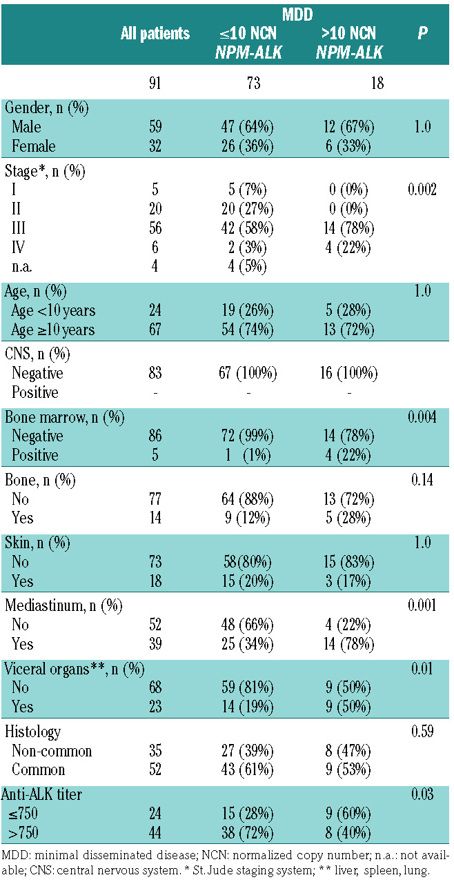
The detection of >10 NCN NPM-ALK in BM correlated with stage III/IV disease, mediastinal and visceral organ involvement, as well as low anti-ALK antibody titers (Table 1). No association of NPM-ALK copy numbers above 10 NCN and histological subtype was observed (Table 1).
Prognostic impact of quantitative minimal disseminated disease in bone marrow
The cumulative incidence of relapse of 18 patients with more than 10 NCN NPM-ALK in BM was 61±12% compared to 21±5% for the remaining 73 patients (P=0.0002), The event-free survival rates at 3 years were 33±11% and 79±5%, respectively (P<0.0001), the overall survival rates were 83±9% and 94±3%, respectively (P=0.099) (Online Supplementary Figure S1). Application of the cut-off of 10 NCN NPM-ALK allowed the separation of a group of patients with a very high risk of relapse in the validation cohort.
Prognostic impact of quantitative minimal disseminated disease in blood
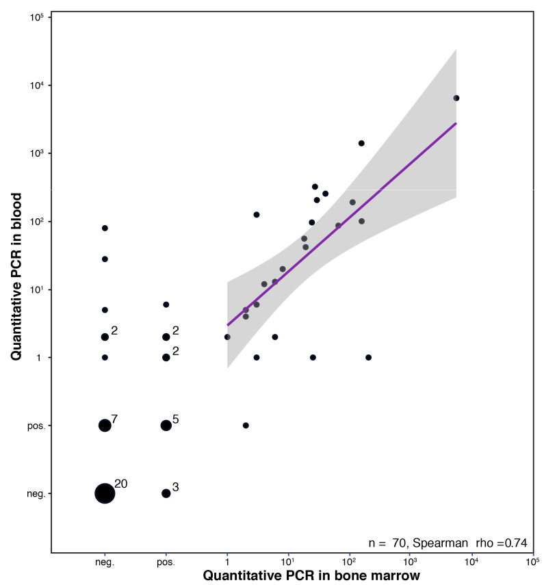
In 70 of the 91 patients for whom MDD was measured in the BM, NPM-ALK transcripts could be measured in blood, as well. The results for blood and BM in the same patients correlated (r=0.74). Notably, more patients were MDD-positive and showed higher copy numbers in blood compared to BM (Figure 1).
Figure 1.
Normalized copy numbers of NPM-ALK in blood and bone marrow measured by quantitative real-time polymerase chain reaction. Copy numbers of NPM/ALK/104 copy numbers of ABL1 were measured in 70 patients in initial blood and bone marrow samples. PCR: polymerase chain reaction.
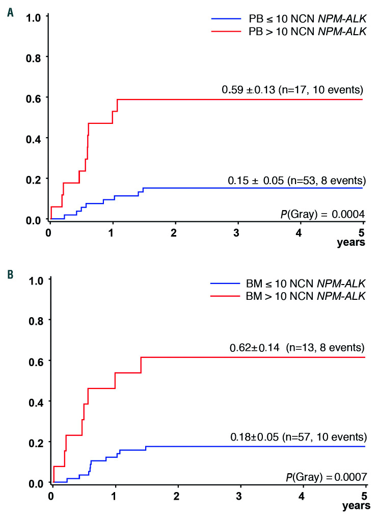
At 3 years the cumulative incidence of relapse of the 70 patients for whom MDD measurements were available in both BM and blood was 26±5%, the event-free survival was 74±5% and the overall survival 94±3%. To analyze a possible influence of the biological medium used for the quantitative MDD measurement on the detection of very high-risk patients, outcome was compared according to quantitative MDD in blood and BM using the same cut off among these 70 patients. The cumulative incidence of relapse of the 17 patients with >10 NCN NPM-ALK measured in blood was 59±13% compared to 15±5% in 53 patients with ≤10 NCN NPM-ALK (P=0.0004). In comparison, the cumulative incidence of relapse of the 13 patients with >10 NCN NPM-ALK measured in BM was 62±14% compared to 18±5% in the 57 patients with ≤10 NCN (P=0.0007) (Figure 2).
Figure 2.
Outcome according to NPM-ALK copy numbers measured by quantitative real-time polymerase chain reaction in blood and bone marrow. Cumulative incidence of relapse (according to a cut-off of 10 normalized copy numbers of NPM/ALK/104 copy numbers of ABL1) measured in initial (A) blood and (B) bone marrow samples in 70 patients. PB: blood; BM: bone marrow; NCN: normalized copy number.
Establishment and validation of a digital droplet polymerase chain reaction assay
To overcome some limitations of RQ-PCR we tested an NPM-ALK-specific dPCR assay for fusion gene and reference gene quantification.
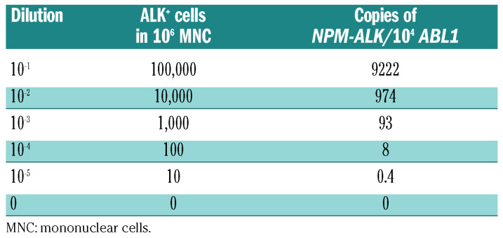
A gradient PCR was performed to optimize the performance of the dPCR assays for NPM-ALK and ABL1.12 The amplification and elongation temperature was set to 54.1°C after optimization. A serial limited 10-fold dilution of the ALK-positive cell line Karpas 299 in peripheral blood mononuclear cells from 10-1 to 10-5 was performed to evaluate the sensitivity and specificity of the dPCR assay (Table 2). Normalized copies of NPM-ALK were detected in cDNA prepared from dilutions of 10 ALK-positive cells in 1,000,000 normal cells in one out of four replicates. The peripheral blood mononuclear cells used for dilution had no detectable copies of NPM-ALK.
Table 2.
Normalized copy numbers of NPM-ALK measured by digital polymerase chain reaction analysis in a 10-fold serial dilution of a NPM-ALK anaplastic large cell lymphoma cell line in 106 mononuclear cells.
To estimate the rate of false positivity, blood from 20 healthy controls and BM or blood from eight ALK-negative ALCL patients was analyzed. No positive droplets were detected in BM or blood from ALK-negative ALCL patients or 20 healthy controls. All samples were negative by RQ-PCR. Since higher copy numbers of NPM-ALK were measured by dPCR compared to RQ-PCR, the cut-off for outcome analysis was set at 30 NCN for the dPCR.
Comparison of NPM-ALK quantification by digital and quantitative real-time polymerase chain reaction analyses in bone marrow and blood samples
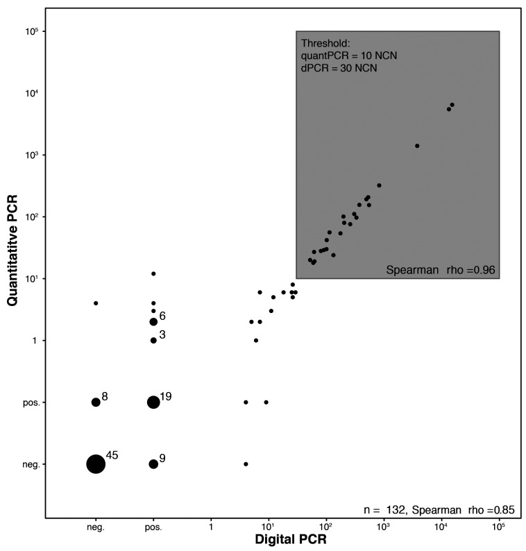
Measurement of circulating tumor cells using RQ-PCR and dPCR was possible in 132 initial BM (n=75) or blood (n=57) samples from ALCL patients. Forty-five samples were negative by both RQ-PCR as well as dPCR (31/75 BM, 14/57 blood samples). Of the 75 BM samples, 19 and 15 were low positive, not quantifiable by dPCR and RQ-PCR, respectively. Of the 57 blood samples, 21 and 14 were low positive by dPCR and RQ-PCR, respectively. The results of dPCR and RQ-PCR correlated with a correlation coefficient of r=0.85 (Figure 3). Above the threshold of 10 NCN NPM-ALK measured by RQ-PCR the copy numbers obtained by both methods were highly correlated with a correlation coefficient of r=0.96, but generally higher with dPCR.
Figure 3.
Comparison of NPM-ALK copy numbers measured by quantitative real-time polymerase chain reaction and digital polymerase chain reaction. Normalized copy numbers of NPM/ALK/104 copy numbers of ABL1 measured in 132 blood and bone marrow samples. quantPCR: quantitative real-time polymerase chain reaction; dPCR: digital polymerase chain reaction; NCN: normalized copy number.
Comparison of patient stratification according to the results of quantitative real-time and droplet polymerase chain reaction analyses in bone marrow and blood
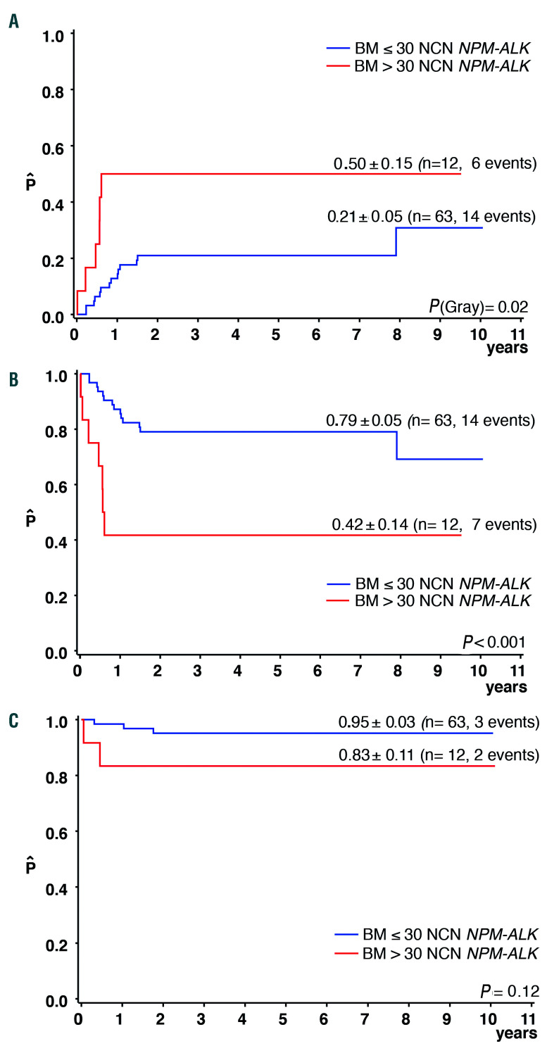
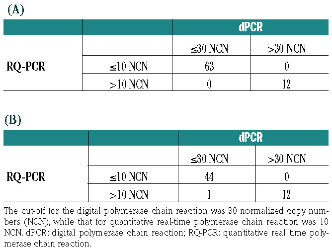
The cumulative incidence of relapse, event-free survival and overall survival of patients with dPCR quantification of MDD in BM (n=75) and blood (n=57) by dPCR using a threshold of 30 NCN are shown in Figure 4. The comparable data according to RQ-PCR, using the cut-off of 10 NCN, were almost identical. The patients’ distribution according to MDD measured in BM was concordant with both quantification methods (Table 3). Among the 57 patients for whom MDD was quantified in blood, only one patient had a discordant result with ≤30 (dPCR) but >10 NCN (RQ-PCR) (Table 3b).
Figure 4.
Outcome according to NPM-ALK copy numbers measured by digital polymerase chain reaction in bone marrow. (A) Cumulative incidence of relapse, (B) event-free survival and (C) overall survival at 3 years according to a cut-off of 30 normalized copy numbers of NPM/ALK/104 copy numbers of ABL1. BM: bone marrow, NCN: normalized copy numbers.
Table 3.
Concordance of patients’ stratification according to quantity of NPM-ALK transcripts determined using digital polymerase chain reaction and quantitative real-time polymerase chain reaction in (A) bone marrow and (B) blood.
Discussion
In our previous study, quantitative measurement of NPM-ALK fusion gene transcripts in blood or BM using a cut-off of 10 NCN in the RQ-PCR analysis allowed us to identify the group of patients with the highest risk of relapse. We were able to confirm these results in the validation cohort of uniformly treated NPM-ALK-positive ALCL patients. As in our previous analysis, only 20% of patients had more than 10 NCN NPM-ALK detectable in BM. Two-thirds of those patients relapsed in both series of altogether 175 ALCL patients.
When comparing the event-free survival of the very high-risk group determined by quantification of MDD in blood between the two cohorts, the EFS of high-risk patients was somewhat higher in the validation cohort than in the earlier cohort. This difference might be attributable to a selection bias with a higher event-free survival in the current cohort analyzed in blood compared to the previously reported cohort (previous cohort 61±6%, current cohort 74±5%). In the current validation cohort, MDD results measured in blood and BM of the same patients were comparable and had the same prognostic impact. For future studies, investigation of peripheral blood could therefore be sufficient for quantitative MDD evaluation. This is especially helpful bearing in mind the application of MRD to follow the course of disease in very high-risk patients or after relapse.
Compared to the earlier cohort including patients diagnosed until 2005, the survival of the very high-risk patients, as defined by more than 10 NCN in BM, improved in the validation cohort (83% compared to 46%). New therapeutic options became available for patients with relapsed ALCL, ranging from vinblastine monotherapy, brentuximab vedotin, ALK kinase inhibitors to PD-L1 checkpoint inhibitors.15,17,22–28 In addition, allogeneic blood stem cell transplantation was increasingly used for consolidation in relapse.29–32
Our results show that separation of patients with a high risk of relapse can be achieved by quantification of MDD in patients with ALCL with the prerequisites that quantitative PCR evaluation is performed in the same laboratory by the same persons, according to standard operating procedures for RQ-based MDD measurement and analysis. However, there are still significant inter-laboratory differences and quantification of minimal disease in patients with ALCL can currently not be compared between laboratories.12,13,17,33 This is exemplified by the comparison of the Japanese study group’s data with our data. Both groups used the same therapy and the same RQ-PCR assay for quantification. Twenty per cent of patients showed >10 NCN NPM-ALK in our cohorts and 37% of patients had high copy numbers in the Japanese cohort even though the relapse rate in the Japanese cohort was somewhat lower. Accordingly, the relapse risk of patients with >10 NCN NPM-ALK was higher in our cohorts (65%) than in the Japanese cohort (40%). In order to use quantification of copy numbers for patient stratification or to follow the course of individual patients in multinational studies, the RQ-PCR method needs very strict protocol harmonization and quality control. The experiences from quantification of BCR-ABL1 transcripts can partly guide this development.34 The introduction of calibrators, specific conversion factors to the calibrators for each laboratory and calibrated reference material led to a high standardization of BCR-ABL1 measurements.19,35–37 In Philadelphia-positive acute leukemia the optimization and standardization process for RQ-PCR-based measurement of m-BCR- ABL1 transcripts underscores the importance of standardization of all steps for quantitative PCR, including data interpretation and quality controls.38 In the standardization process of MRD assessment of m-BCR-ABL1 fusion gene transcripts organized by the Euro MRD consortium, the usage of a common primer and probe set as well as a centrally distributed plasmid standard curve had the greatest impact on overcoming inter-laboratory variability.38
Since the same primer/probe sets and the same RQ-PCR protocol were applied by the Japanese and BFM study groups, the differences in results might be related to the use of standard curves that were not centrally distributed and to the fact that the cut-off at 10 NCN NPM-ALK is close to the detection limit of the assay. The latter point is an unchangeable limitation to inter-laboratory comparability for quantification of NPM-ALK transcripts and a major difference from MRD quantification in leukemia.
In order to overcome some of the technical problems inherent to RQ-PCR we developed a dPCR method for the quantification of NPM-ALK transcripts and compared the results obtained with this method to those obtained with RQ-PCR in a large cohort of patients. Using dPCR with a cut-off at 30 NCN NPM-ALK for quantitative measurements of MDD in blood and BM in the presented study allowed measurements near to the detection limit without needing standard curve calibration. The dPCR assay might be more suitable for quantitative measurements of NPM-ALK in a multinational setting, because it overcomes several limitations of the RQ-PCR assay. First, it is independent of a calibration curve, thereby excluding the impact of standard curve differences for inter-laboratory comparisons. Second, partitioning of target molecules leads to a more precise detection especially of rare events so that the assay is able to detect low copy numbers more accurately than RQ-PCR.39,40 To exclude that templates are not amplifiable during the dPCR reaction it is still unavoidable to verify the quality of a given cDNA by parallel estimation of a reference gene. Furthermore, for measurement of clinical samples, appropriate positive and negative controls must be included in order to control for the overall performance of a given dPCR experiment.
The applicability of dPCR for MRD measurement has already been shown for several hematologic malignancies. The accuracy of dPCR and high concordance with RQ-PCR was demonstrated for DNA-based MRD measurements of the BCL2/IGH rearrangement in blood and BM from patients with low stage follicular lymphoma41 and immunoglobulin/T-cell receptor rearrangements in patients with acute lymphoblastic leukemia.42 dPCR was shown to be reliable for quantifying BCR-ABL1 transcripts for MRD monitoring in chronic myeloid leukemia and Philadelphia-positive acute lymphoblastic leukemia.43–46 Altogether, dPCR is a valuable tool for highly reproducible quantification of minimal disease at both DNA and RNA levels in patients’ samples without requiring standard curves. In addition, for MDD and MRD in ALCL, it might have the advantage over RQ-PCR that the quantification is more accurate at lower copy numbers. Stringent protocol standardization and quality control are needed for this technique, as well.47,48
In summary, our data validate that quantification of NPM-ALK transcripts by RQ-PCR using a cut-off of 10 NCN identifies very high-risk patients if performed in one laboratory. Quantification of MRD is indicated to follow the course of disease and response to treatment modules in MDD-positive or relapsed ALCL patients. In a rare disease such as ALCL, with planned and ongoing international trials, both methods for transcript quantification require inter-laboratory comparability of measurements. Since harmonization is difficult and expensive with RQ-PCR, we developed and validated a dPCR assay enabling reliable quantification of NPM-ALK transcripts at very low copy numbers without the need for standard calibration curves.
Acknowledgments
The authors would like to thank Claudia Keller for excellent technical assistance and Ulrike Meyer for excellent assistance with data management. The NHL-BFM Registry 2012 is supported by the Deutsche Kinderkrebsstiftung. CDW, NK, JS and WW were additionally supported by Forschungshilfe Peiper. The pediatric lymphoma research of IO and WK is supported by the KinderKrebsInitiative Buchholz, Holm-Seppensen.
Footnotes
Check the online version for the most updated information on this article, online supplements, and information on authorship & disclosures: www.haematologica.org/content/105/8/2141
References
- 1.Morris SW, Kirstein MN, Valentine MB, et al. Fusion of a kinase gene, ALK, to a nucleolar protein gene, NPM, in non-Hodgkin’s lymphoma. Science.1994;263(5151):1281–1284. [DOI] [PubMed] [Google Scholar]
- 2.Perkins SL, Pickering D, Lowe EJ, et al. Childhood anaplastic large cell lymphoma has a high incidence of ALK gene rearrangement as determined by immunohistochemical staining and fluorescent in situ hybridisation: a genetic and pathological correlation. Br J Haematol. 2005;131(5):624–627. [DOI] [PubMed] [Google Scholar]
- 3.Damm-Welk C, Klapper W, Oschlies I, et al. Distribution of NPM1-ALK and X-ALK fusion transcripts in paediatric anaplastic large cell lymphoma: a molecular-histological correlation. Br J Haematol. 2009;146 (3):306–309. [DOI] [PubMed] [Google Scholar]
- 4.Lamant L, McCarthy K, D’Amore E, et al. Prognostic impact of morphologic and phenotypic features of childhood ALK-positive anaplastic large-cell lymphoma: results of the ALCL99 study. J Clin Oncol. 2011;29 (35):4669–4676. [DOI] [PubMed] [Google Scholar]
- 5.Brugieres L, Deley MC, Pacquement H, et al. CD30(+) anaplastic large-cell lymphoma in children: analysis of 82 patients enrolled in two consecutive studies of the French Society of Pediatric Oncology. Blood. 1998;92(10):3591–3598. [PubMed] [Google Scholar]
- 6.Brugieres L, Le Deley MC, Rosolen A, et al. Impact of the methotrexate administration dose on the need for intrathecal treatment in children and adolescents with anaplastic large-cell lymphoma: results of a randomized trial of the EICNHL Group. J Clin Oncol. 2009;27(6):897–903. [DOI] [PubMed] [Google Scholar]
- 7.Seidemann K, Tiemann M, Schrappe M, et al. Short-pulse B-non-Hodgkin lymphoma-type chemotherapy is efficacious treatment for pediatric anaplastic large cell lymphoma: a report of the Berlin-Frankfurt-Munster Group Trial NHL-BFM 90. Blood. 2001;97(12):3699–3706. [DOI] [PubMed] [Google Scholar]
- 8.Williams DM, Hobson R, Imeson J, Gerrard M, McCarthy K, Pinkerton CR. Anaplastic large cell lymphoma in childhood: analysis of 72 patients treated on the United Kingdom Children’s Cancer Study Group chemotherapy regimens. Br J Haematol. 2002;117(4):812–820. [DOI] [PubMed] [Google Scholar]
- 9.Rosolen A, Pillon M, Garaventa A, et al. Anaplastic large cell lymphoma treated with a leukemia-like therapy: report of the Italian Association of Pediatric Hematology and Oncology (AIEOP) LNH-92 protocol. Cancer. 2005;104(10):2133–2140. [DOI] [PubMed] [Google Scholar]
- 10.Mussolin L, Pillon M, d’Amore ES, et al. Prevalence and clinical implications of bone marrow involvement in pediatric anaplastic large cell lymphoma. Leukemia. 2005;19(9): 1643–1647. [DOI] [PubMed] [Google Scholar]
- 11.Mussolin L, Damm-Welk C, Pillon M, et al. Use of minimal disseminated disease and immunity to NPM-ALK antigen to stratify ALK-positive ALCL patients with different prognosis. Leukemia. 2013;27(2):416–422. [DOI] [PubMed] [Google Scholar]
- 12.Damm-Welk C, Busch K, Burkhardt B, et al. Prognostic significance of circulating tumor cells in bone marrow or peripheral blood as detected by qualitative and quantitative PCR in pediatric NPM-ALK-positive anaplastic large-cell lymphoma. Blood. 2007;110(2):670–677. [DOI] [PubMed] [Google Scholar]
- 13.Iijima-Yamashita Y, Mori T, Nakazawa A, et al. Prognostic impact of minimal disseminated disease and immune response to NPM-ALK in Japanese children with ALK-positive anaplastic large cell lymphoma. Int J Hematol. 2018;107(2):244–250. [DOI] [PubMed] [Google Scholar]
- 14.Damm-Welk C, Mussolin L, Zimmermann M, et al. Early assessment of minimal residual disease identifies patients at very high relapse risk in NPM-ALK-positive anaplastic large-cell lymphoma. Blood. 2014;123(3): 334–337. [DOI] [PubMed] [Google Scholar]
- 15.Hebart H, Lang P, Woessmann W. Nivolumab for refractory anaplastic large cell lymphoma: a case report. Ann Intern Med. 2016;165(8):607–608. [DOI] [PubMed] [Google Scholar]
- 16.Gambacorti-Passerini C, Mussolin L, Brugieres L. Abrupt relapse of ALK-positive lymphoma after discontinuation of crizotinib. N Engl J Med. 2016;374(1):95–96. [DOI] [PubMed] [Google Scholar]
- 17.Mosse YP, Voss SD, Lim MS, et al. Targeting ALK with crizotinib in pediatric anaplastic large cell lymphoma and inflammatory myofibroblastic tumor: a Children’s Oncology Group study. J Clin Oncol. 2017;35(28):3215–3221. [DOI] [PMC free article] [PubMed] [Google Scholar]
- 18.Branford S, Cross NC, Hochhaus A, et al. Rationale for the recommendations for harmonizing current methodology for detecting BCR-ABL transcripts in patients with chronic myeloid leukaemia. Leukemia. 2006;20 (11):1925–1930. [DOI] [PubMed] [Google Scholar]
- 19.Hughes T, Deininger M, Hochhaus A, et al. Monitoring CML patients responding to treatment with tyrosine kinase inhibitors: review and recommendations for harmonizing current methodology for detecting BCR-ABL transcripts and kinase domain mutations and for expressing results. Blood. 2006;108(1):28–37. [DOI] [PMC free article] [PubMed] [Google Scholar]
- 20.Vogelstein B, Kinzler KW. Digital PCR. Proc Natl Acad Sci U S A. 1999;96(16):9236–9241. [DOI] [PMC free article] [PubMed] [Google Scholar]
- 21.Dube S, Qin J, Ramakrishnan R. Mathematical analysis of copy number variation in a DNA sample using digital PCR on a nanofluidic device. PloS One. 2008;3 (8):e2876. [DOI] [PMC free article] [PubMed] [Google Scholar]
- 22.Brugieres L, Pacquement H, Le Deley MC, et al. Single-drug vinblastine as salvage treatment for refractory or relapsed anaplastic large-cell lymphoma: a report from the French Society of Pediatric Oncology. J Clin Oncol. 2009;27(30):5056–5061. [DOI] [PubMed] [Google Scholar]
- 23.Mosse YP, Lim MS, Voss SD, et al. Safety and activity of crizotinib for paediatric patients with refractory solid tumours or anaplastic large-cell lymphoma: a Children’s Oncology Group phase 1 consortium study. Lancet Oncol. 2013;14(6):472–480. [DOI] [PMC free article] [PubMed] [Google Scholar]
- 24.Rigaud C, Abbou S, Minard-Colin V, et al. Efficacy of nivolumab in a patient with systemic refractory ALK+ anaplastic large cell lymphoma. Pediatr Blood Cancer. 2018;65 (4):e26902. [DOI] [PubMed] [Google Scholar]
- 25.Pro B, Advani R, Brice P, et al. Brentuximab vedotin (SGN-35) in patients with relapsed or refractory systemic anaplastic large-cell lymphoma: results of a phase II study. J Clin Oncol. 2012;30(18):2190–2196. [DOI] [PubMed] [Google Scholar]
- 26.Gambacorti Passerini C, Farina F, Stasia A, et al. Crizotinib in advanced, chemoresistant anaplastic lymphoma kinase-positive lymphoma patients. J Natl Cancer Inst. 2014;106 (2):djt378. [DOI] [PubMed] [Google Scholar]
- 27.Gambacorti-Passerini C, Messa C, Pogliani EM. Crizotinib in anaplastic large-cell lymphoma. N Engl J Med. 2011;364(8):775–776. [DOI] [PubMed] [Google Scholar]
- 28.Locatelli F, Mauz-Koerholz C, Neville K, et al. Brentuximab vedotin for paediatric relapsed or refractory Hodgkin’s lymphoma and anaplastic large-cell lymphoma: a multi-centre, open-label, phase 1/2 study. Lancet Haematol. 2018;5(10):e450–e461. [DOI] [PubMed] [Google Scholar]
- 29.Woessmann W, Peters C, Lenhard M, et al. Allogeneic haematopoietic stem cell transplantation in relapsed or refractory anaplastic large cell lymphoma of children and adolescents–a Berlin-Frankfurt-Munster group report. Br J Haematol. 2006;133(2):176–182. [DOI] [PubMed] [Google Scholar]
- 30.Gross TG, Hale GA, He W, et al. Hematopoietic stem cell transplantation for refractory or recurrent non-Hodgkin lymphoma in children and adolescents. Biol Blood Marrow Transplant. 2010;16(2):223–230. [DOI] [PMC free article] [PubMed] [Google Scholar]
- 31.Fukano R, Mori T, Kobayashi R, et al. Haematopoietic stem cell transplantation for relapsed or refractory anaplastic large cell lymphoma: a study of children and adolescents in Japan. Br J Haematol. 2015;168(4): 557–563. [DOI] [PubMed] [Google Scholar]
- 32.Strullu M, Thomas C, Le Deley MC, et al. Hematopoietic stem cell transplantation in relapsed ALK+ anaplastic large cell lymphoma in children and adolescents: a study on behalf of the SFCE and SFGM-TC. Bone Marrow Transplant. 2015;50(6):795–801. [DOI] [PubMed] [Google Scholar]
- 33.Mussolin L, Bonvini P, it-Tahar K, et al. Kinetics of humoral response to ALK and its relationship with minimal residual disease in pediatric ALCL. Leukemia. 2009;23(2): 400–402. [DOI] [PubMed] [Google Scholar]
- 34.Hughes TP, Kaeda J, Branford S, et al. Frequency of major molecular responses to imatinib or interferon alfa plus cytarabine in newly diagnosed chronic myeloid leukemia. N Engl J Med. 2003;349(15):1423–1432. [DOI] [PubMed] [Google Scholar]
- 35.Cross NC, White HE, Muller MC, Saglio G, Hochhaus A. Standardized definitions of molecular response in chronic myeloid leukemia. Leukemia. 2012;26(10):2172–2175. [DOI] [PubMed] [Google Scholar]
- 36.Branford S, Fletcher L, Cross NC, et al. Desirable performance characteristics for BCR-ABL measurement on an international reporting scale to allow consistent interpretation of individual patient response and comparison of response rates between clinical trials. Blood. 2008;112(8):3330–3338. [DOI] [PubMed] [Google Scholar]
- 37.White H, Deprez L, Corbisier P, et al. A certified plasmid reference material for the standardisation of BCR-ABL1 mRNA quantification by real-time quantitative PCR. Leukemia. 2015;29(2):369–376. [DOI] [PMC free article] [PubMed] [Google Scholar]
- 38.Pfeifer H, Cazzaniga G, van der Velden VHJ, et al. Standardisation and consensus guidelines for minimal residual disease assessment in Philadelphia-positive acute lymphoblastic leukemia (Ph+ ALL) by real-time quantitative reverse transcriptase PCR of e1a2 BCR-ABL1. Leukemia. 2019;33(8): 1910–1922. [DOI] [PubMed] [Google Scholar]
- 39.Whale AS, Cowen S, Foy CA, Huggett JF. Methods for applying accurate digital PCR analysis on low copy DNA samples. PloS One. 2013;8(3):e58177. [DOI] [PMC free article] [PubMed] [Google Scholar]
- 40.Sanders R, Mason DJ, Foy CA, Huggett JF. Evaluation of digital PCR for absolute RNA quantification. PloS one. 2013;8(9):e75296. [DOI] [PMC free article] [PubMed] [Google Scholar]
- 41.Cavalli M, De Novi LA, Della Starza I, et al. Comparative analysis between RQ-PCR and digital droplet PCR of BCL2/IGH gene rearrangement in the peripheral blood and bone marrow of early stage follicular lymphoma. Br J Haematol. 2017;177(4):588–596. [DOI] [PubMed] [Google Scholar]
- 42.Della Starza I, Nunes V, Cavalli M, et al. Comparative analysis between RQ-PCR and digital-droplet-PCR of immunoglobulin/T-cell receptor gene rearrangements to monitor minimal residual disease in acute lymphoblastic leukaemia. Br J Haematol. 2016;174(4):541–549. [DOI] [PubMed] [Google Scholar]
- 43.Bonvini P, Zin A, Alaggio R, Pawel B, Bisogno G, Rosolen A. High ALK mRNA expression has a negative prognostic significance in rhabdomyosarcoma. Br J Cancer. 2013;109(12):3084–3091. [DOI] [PMC free article] [PubMed] [Google Scholar]
- 44.Jennings LJ, George D, Czech J, Yu M, Joseph L. Detection and quantification of BCR-ABL1 fusion transcripts by droplet digital PCR. J Mol Diagn. 2014;16(2):174–179. [DOI] [PubMed] [Google Scholar]
- 45.Iacobucci I, Lonetti A, Venturi C, et al. Use of a high sensitive nanofluidic array for the detection of rare copies of BCR-ABL1 transcript in patients with Philadelphia-positive acute lymphoblastic leukemia in complete response. Leuk Res. 2014;38(5):581–585. [DOI] [PubMed] [Google Scholar]
- 46.Wang WJ, Zheng CF, Liu Z, et al. Droplet digital PCR for BCR/ABL(P210) detection of chronic myeloid leukemia: A high sensitive method of the minimal residual disease and disease progression. Eur J Haematol. 2018; 101(3):291–296. [DOI] [PubMed] [Google Scholar]
- 47.Huggett JF, Foy CA, Benes V, et al. The digital MIQE guidelines: minimum information for publication of quantitative digital PCR experiments. Clin Chem. 2013;59(6):892–902. [DOI] [PubMed] [Google Scholar]
- 48.Huggett JF, Cowen S, Foy CA. Considerations for digital PCR as an accurate molecular diagnostic tool. Clin Chem. 2015;61(1):79–88. [DOI] [PubMed] [Google Scholar]


