Abstract
Hematopoietic stem cells are responsible for life-long blood cell production and are highly sensitive to exogenous stresses. The effects of low doses of ionizing radiations on radiosensitive tissues such as the hematopoietic tissue are still unknown despite their increasing use in medical imaging. Here, we study the consequences of low doses of ionizing radiations on differentiation and self-renewal capacities of human primary hematopoietic stem/progenitor cells (HSPC). We found that a single 20 mGy dose impairs the hematopoietic reconstitution potential of human HSPC but not their differentiation properties. In contrast to high irradiation doses, low doses of irradiation do not induce DNA double strand breaks in HSPC but, similar to high doses, induce a rapid and transient increase of reactive oxygen species (ROS) that promotes activation of the p38MAPK pathway. HSPC treatment with ROS scavengers or p38MAPK inhibitor prior exposure to 20 mGy irradiation abolishes the 20 mGy-induced defects indicating that ROS and p38MAPK pathways are transducers of low doses of radiation effects. Taken together, these results show that a 20 mGy dose of ionizing radiation reduces the reconstitution potential of HSPC suggesting an effect on the self-renewal potential of human hematopoietic stem cells and pinpointing ROS or the p38MAPK as therapeutic targets. Inhibition of ROS or the p38MAPK pathway protects human primary HSPC from low-dose irradiation toxicity.
Introduction
Hematopoietic stem cells (HSC) give rise to all blood cell types over the entire life of an organism. In adult mammals, they are located in very specific microenvironments of the bone marrow (BM), allowing maintenance of HSC functions.1 In humans, HSC are enriched in the CD34+ CD38low CD90+ CD45RA− cell population that also contains immature progenitors, hereafter called HSPC.2,3 Hematopoietic stem/progenitor cells (HSPC) are multipotent and mainly slow cycling cells. They possess a self-renewal potential that allows them to sustain the continuous generation of blood cells. Quiescence and self-renewal are regulated by several extrinsic factors, such as cytokines, extracellular matrix proteins and adhesion molecules,4,5 as well as intrinsic factors, such as transcription factors (TAL1,6–8 GATA-2, etc.9), proteins implicated in DNA damage repair pathways,10–12 and cell cycle regulators.13–15 Mutations in genes involved in DNA repair induce BM failure with exhaustion of the HSC pool, demonstrating that preserving genome integrity is crucial for HSC long-term maintenance (reviewed by Biechonski and Milyavsky).16 For instance, ku80, lig4 and atm-deficient mice exhibit defects in HSC maintenance and self-renewal.10–12 Atm-deficient HSC harbor increased levels of reactive oxygen species (ROS) responsible for their loss of hematopoietic reconstitution capacity10 that can be rescued by treatment with the antioxidant N-acetylcystein (NAC). Indeed, increased ROS in HSC induce their differentiation and their exhaustion17,18 and quiescent HSC with the lowest level of ROS have the highest hematopoietic reconstitution potential compared to ‘activated’ HSC har-boring higher ROS levels.17 Interestingly, in mouse and human, ROS and DNA damage accumulate in HSC upon serial transplantation resulting in decreased self-renewal capacities. NAC-treated HSC are protected against the accumulation of oxidative DNA damage.18,19
Ionizing radiations (IR) represent the main source of DNA damage and ROS. Importantly, the human population is increasingly exposed to low doses of IR (LDIR, <100 mGy) due to the recurrent use of medical imaging.20 Studies have shown that combinations of several computed tomography (CT) scans (thoracic or cranial) can increase the risk of developing cancer.21 Indeed, having more than five CT scans (corresponding to a cumulative dose of 30 mGy) can lead to a 3-fold increase in the risk of developing pediatric leukemia. Moreover, a recent study showed that 20 mGy LDIR affects the fundamental properties of HSC in mouse.22 In this context, it is crucial to study the consequences of LDIR exposure in human cells, in particular in human HSC. Here we show through combining in vitro and in vivo studies that a single acute 20 mGy LDIR decreases human HSPC serial clonogenic and reconstitution potentials, and that these effects are mediated through a ROS/p38MAPK-dependent signaling pathway.
Methods
Primary cells
Cord blood (CB) samples were collected from healthy infants with the informed written consent of the mothers according to the Declaration of Helsinki. Samples were obtained in collaboration with the Clinique des Noriets, Vitry-sur-Seine, and with the Cell Therapy Department of Hôpital Saint-Louis, Paris, France. Samplings and experiments were approved by the Institutional Review Board of INSERM (Opinion n. 13-105-1, IRB00003888). CD34+ cells were purified by immuno-magnetic selection using a CD34 MicroBeads kit (Miltenyi Biotec, Paris, France). For each experiment, we used a pool of CD34+ cells from different healthy infants to diminish individual variability.
Low dose of ionizing radiations
20 mGy LDIR was delivered with a dose rate of 20 mGy/minute (min) using a Cobalt 60 Irradiator (Alcyon). 2.5 Gy was delivered with a dose rate of 1 Gy/min.
Flow cytometry and cell sorting
CD34+CD38low cells and CD34+CD38lowCD45RA−CD90+ HSPC were isolated after labeling with human specific monoclonal antibodies (MoAbs, see Online Supplementary Table S1 for details). Cell sorting was performed using either a Becton Dickinson (BD)-FACS-ARIA3 SORP or a BD-FACS-Influx (laser 488, 405, 355, 561 and 633, BD Bioscience). Flow cytometry experiments are described in the Online Supplementary Methods.
Transplantation assays
NOD.Cg-Prkdc(scid) Il2rg(tm1Wjl)/SzJ (NSG) mice (Jackson Laboratory, Bar Harbor, ME, USA) were housed in the pathogen-free animal facility of IRCM, CEA, Fontenay-aux-Roses, France. Adult 8-12-week old NSG mice received a 3 Gy sublethal irradiation using a GSRD1 γ-irradiator (source Cesium137, GSM) and were anesthetized with isoflurane before intravenous retro-orbital injection (i.v.) of human cells as described in the Online Supplementary Methods. All experimental procedures were carried out in compliance with French Ministry of Agriculture regulations (animal facility registration n.: A9203202, Supervisor: Michel Bedoucha, APAFIS #9458-2017033110277117v2) for animal experimentation and in accordance with a local ethical committee (#44).
Immunofluorescence
Immunofluorescence was performed on cell-sorted HSPC irradiated and incubated 30 min, 1 hour (h), or 3 h at 37°C in MyeloCult medium, as previously described.22–24 Details of the methods used are available in the Online Supplementary Methods.
Drug treatments
CD34+ cells or CD34+ CD38lowCD45RA−CD90+ HSPC were treated with several drugs as described in the Online Supplementary Methods.
Colony forming unit-cell assay
Colony forming unit-cell assay (CFU-C) and serial platings were performed as previously described;6 see Online Supplementary Methods for details. Depending on CB pool samples, 60-80 colonies were generated from 500 HSPC non-irradiated or irradiated at 20 mGy.
Primary and extended long-term culture initiating cell assays
Long-term culture initiating cell assay was performed as previously described6 and is described in detail in the Online Supplementary Methods.
Intracellular flow cytometry
Ki67, cleaved caspase 3, phospho-p38MAPK (P-p38, phosphorylation on Thr180/Tyr182), phospho-ATM, p53 and phospho-p53 staining were performed as previously described4 (Ki67) and according to the manufacturer’s instructions, respectively. More details can be found in the Online Supplementary Methods.
Carboxyfluorescein diacetate succinimidyl ester staining
3x10⁵ CD34+ cells were labeled with carboxyfluorescein diacetate succinimidyl ester (CFSE) (2.5 mM, Sigma, France) and cultured in StemSpan medium supplemented with cytokines (Stem Cell Technologies), as described in the Online Supplementary Methods.
Reactive oxygen species quantification and mitochondria activity assay
Reactive oxygen species quantification was performed with fresh CD34+ cells using CellRox Orange reagent following the manufacturer’s instructions (ref. C10443, Molecular Probes, ThermoFisher Scientific). Mitochondrial activity assay was performed using mitotracker green (MTG) and TMRE products, according to the manufacturer’s instructions (Molecular Probes, ThermoFisher Scientific).
Statistical analysis
Mann and Whitney (M&W) and Kruskal and Wallis (K&W) non-parametric statistical analyses were used. *P<0.05, **P<0.01, ***P<0.001, ****P<0.0001.
Results
A 20 mGy dose of irradiation decreases serial replating capacity of human hematopoietic stem/progenitor cells
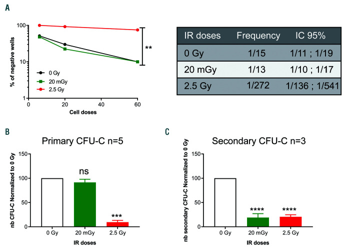
To understand the impact of HSPC exposure to LDIR, we first performed serial long-term culture initiating cell (LTC-IC) and CFU tests as surrogate assays to study human HSPC properties, i.e. self-renewal and differentiation capacities.25–27 Human CD34+CD38lowCD45RA−CD90+ HSPC were purified, irradiated and cultured with the MS5 stromal cell line in LTC medium for five weeks or plated directly in semi-solid methylcellulose cultures. In LTC-IC limiting dilution analyses, every single well was harvested independently at the end of the culture and plated in CFU-C assay. The LTC-IC frequency obtained after a 20 mGy irradiation of HSPC was similar to the LTC-IC frequency of sham-irradiated HSPC (1/15) suggesting that LDIR do not affect LTC-IC frequency. In contrast, a high (2.5 Gy) irradiation dose induced a drastic drop in LTC-IC frequency (1/272) (Figure 1A). In addition, 2.5 Gy-irradiated HSPC directly seeded in CFU-C conditions after IR produced very few colonies compared to control cells whereas 20 mGy-irradiated HSPC generated a similar number of CFU-C to sham-irradiated HSPC (Figure 1B and Online Supplementary Figure S1A). No difference in the quality of CFU-C was observed between sham and 20 mGy conditions (Online Supplementary Figure S1B). These primary LTC-IC and primary CFU-C assays show that 20 mGy LDIR do not alter HSPC frequency and clonogenicity in vitro and do not induce any myelo/erythroid differentiation bias in primary cultures (Online Supplementary Figure S1B). To characterize whether LDIR have deleterious effects on human HSPC self-renewal potential in vitro, serial replatings were performed for both CFU-C and LTC-IC assays.25–27 We observed that 20 mGy and 2.5 Gy-irradiated CD34+CD38lowCD45RA−CD90+ HSPC produced lower numbers of secondary colonies in contrast to sham-irradiated HSPC, showing that 20 mGy alters their serial clonogenic potential (Figure 1C and Online Supplementary Figure S1C). This result was also obtained after picking up and replating individual primary CFU-GM colonies (Online Supplementary Figure S1D). In the case of LTC-IC, secondary/extended cultures were initiated using cells expressing high CD34 surface expression (CD34hi), purified after the initial five weeks of LTC-IC culture (Online Supplementary Figure S1E) and seeded for five additional weeks in LTCIC conditions at limiting dilution. Of note, we were not able to perform such extended LTC-IC with the 2.5 Gy condition due to very low cell quantities in the cultures. Interestingly, there was a 2-fold decrease in the extended LTC-IC frequency of 20 mGy-irradiated HSPC compared to sham-irradiated HSPC (1/275 vs. 1/128) (Online Supplementary Figure S1F) showing a defect in long-term HSPC maintenance. This decrease in secondary LTC-IC frequency induced by 20 mGy LDIR was also found in bulk culture conditions with 20 mGy-irradiated HSPC generating fewer CFU-C than sham-irradiated cells after secondary LTC-IC cultures (Online Supplementary Figure S1G). Taken together, these results suggest that a single exposure to 20 mGy LDIR impairs the in vitro self-renewal potential of human CD34+CD38lowCD45RA−CD90+ HSPC.
Figure 1.
Low doses (LD) of ionizing radiations (IR) exposure of human hematopoietic stem progenitor cells (HSPC) leads to deficient serial colony forming unit-cell assay (CFU-C) and primary and extended long-term culture initiating cell (LTC-IC) potentials. CD34+ CD38low CD45RA− CD90+ HSPC were sorted from pools of independent cord blood (CB) samples by cell sorting and exposed to the indicated IR doses prior to in vitro cultures. (A) LTC-IC assay in limiting dilution (pool of 2 experiments, 120 wells/IR dose). Irradiated CD34+ CD38low CD45RA−CD90+ HSPC were seeded on MS5 stromal cells in limiting dilution for five weeks then plated in methylcellulose for 12 days. LTC-IC frequency was calculated using LCALC software. (B) Primary CFU-C assay (cumulative results from 4 independent experiments with HSPC isolated from 4 independent pools of CB samples). HSPC (500 cells/plate) were plated in CFU-C condition for 12-14 days and the number (nb) of CFU-C was quantified. Results are normalized to the non-irradiated conditions. (C) Primary CFU-C were pooled and replated in methylcellulose for 12-14 days. Shown are the nb of secondary CFU-C. Results are normalized to the sham-irradiated conditions (cumulative results from 3 independent experiments). Results are shown as mean±standard error of mean. **P<0.01, ***P<0.001, ****P<0.0001 (Mann-Whitney statistics).
A 20 mGy dose of irradiation decreases human hematopoietic stem/progenitor cell hematopoietic reconstitution potential
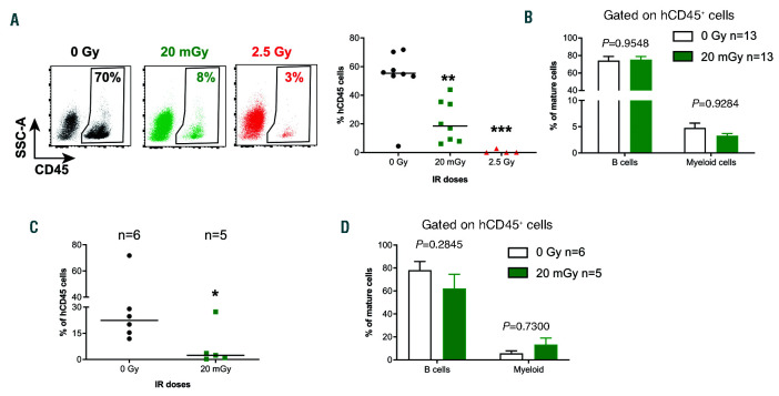
We then studied the effect of 20 mGy LDIR on in vivo HSC functions. To do so, NSG mice were first engrafted with human CD34+ cells. Sixteen weeks later, once human hematopoiesis was stabilized, engrafted mice were exposed to 0, 20 mGy and 2.5 Gy IR doses and sacrificed immediately after irradiation. Bone marrow (BM) cells were harvested and phenotype analysis was performed. The levels of human CD45+ cells and the percentage of LinnegCD34+ cells recovered from NSG BM were similar in the irradiated and non-irradiated groups (Online Supplementary Figure S2A and B). Furthermore, no increase of apoptosis of human cells engrafted in NSG mouse BM was observed in mice irradiated at 20 mGy compared to non-irradiated mice (Online Supplementary Figure S2C). To study the consequences of irradiation on human HSC functions (i.e. reconstitution and differentiation potential), BM cells containing 5.104 human CD45+CD34+CD19−cells were transplanted in secondary NSG mice. Human hematopoietic development in the secondary recipient mice was analyzed 13-16 weeks later. Human engraftment levels in mice receiving 20 mGy and 2.5 Gy-irradiated BM cells were decreased compared to sham-irradiated BM cells (Figure 2A and Online Supplementary Figure S2D) showing that 20 mGy-irradiated BM cells are less efficient than non-irradiated BM cells to reconstitute human hematopoiesis in secondary recipient mice. However, in the 20 mGy condition, some human cells were still detected in the BM of secondary recipient mice. Among them, human B CD19+ lymphocytes and CD14+/CD15+ myeloid cells were produced at the same proportion than in non-irradiated conditions, indicating that the few HSC that survived LDIR had maintained their differentiation capacities (Figure 2B). To exclude a non-cell autonomous effect of LDIR on HSPC mediated by irradiated hematopoietic or non-hematopoietic BM cells during the transplantation, we purified human CD34+CD38low cells from the BM of control and 20 mGy-irradiated mice before transplantation in secondary NSG mice. As in the previous experiment, human cell engraftment in secondary recipients showed a reduced hematopoietic reconstitution capacity when 20 mGy-irradiated CD34+CD38low cells were used (Figure 2C) and again no difference in the differentiation of the engrafted HSC was shown (Figure 2D) showing a cell-autonomous effect of LDIR in HSPC. Taken together these results indicate that 20 mGy LDIR affects the hematopoietic reconstitution capacity of human HSPC.
Figure 2.
Hematopoietic reconstitution capacities of human hematopoietic stem cells (HSC) after in vivo exposure to low doses (LD) of ionizing radiations (IR). NSG mice were transplanted with 5.104 CD34+ CB cells and 13 to 16 weeks post graft mice were irradiated or not with the indicated doses then immediately sacrificed. Bone marrow (BM) cells were recovered and characterized by flow cytometry (Online Supplementary Figure S2A and B). An equivalent of 5.104 CD34+ CD19− BM cells were injected in secondary recipient mice. (A) Dot plots of representative engraftment levels (left: human hCD45+ cells) in secondary recipient mice and human engraftment levels obtained in the BM of 9 secondary NSG mice (right: 2 independent experiments are pooled). Results of a third experiment is shown in Online Supplementary Figure S2D since engraftment levels of control mice were lower. (B) Proportion of engrafted human B cells and myeloid cells based respectively on CD19 and CD14/CD15 expression gated in human CD45+ cells, in secondary recipient mice. (C) Human hematopoietic reconstitution of CD34+ CD38low Linneg BM cells purified by cell sorting from primary mice and transplanted in secondary recipient mice. Shown are human cell reconstitution in the BM of secondary recipient mice 13 weeks later. (D) Proportion of engrafted human B cells and myeloid cells gated within human CD45+ cell compartment in secondary mice after human CD34+ CD38low Linneg BM cell transplants. Results are shown as mean±standard error of mean. *P<0.05; **P<0.01, ***P<0.001 (Mann-Whitney statistics).
Low doses of ionizing radiations do not induce apoptosis nor alter cell cycle of hematopoietic stem/progenitor cells
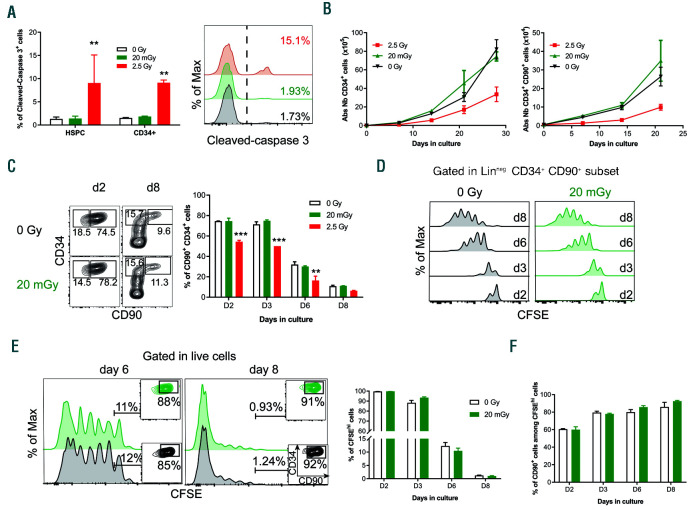
To characterize the mechanisms activated by 20 mGy LDIR in human primary HSPC, we first investigated if LDIR trigger apoptosis. Analysis of caspase 3 cleavage showed that 2.5 Gy irradiation increases CD34+CD38lowCD45RA−CD90+ HSPC apoptosis (Figure 3A, red histograms), whereas 20 mGy LDIR had no significant effect on the percentage of cleaved caspase 3-expressing HSPC compared to sham-irradiated HSPC (Figure 3A, green histograms). As increased HSPC cell cycle can lead to self-renewal defects and HSC exhaustion,28 we also determined if 20 mGy LDIR could alter HSPC proliferation. 20 mGy and sham-irradiated CD34+CD38lowCD45RA−CD90+ HSPC generated the same number of CD34+ and CD34+CD90+ cells in vitro (Figure 3B, left and right panel, respectively) whereas 2.5 Gy-irradiated HSPC did not proliferate efficiently. Moreover, the proportion of CD34+CD90+ cells evolved similarly during culture period after no irradiation or 20 mGy LDIR, strengthening the lack of effect of a 20 mGy irradiation on HSPC differentiation (Figure 3C). We studied the cell division rate of irradiated HSPC after staining with CFSE and during culture in serum free medium with cytokines. The number of cell divisions was the same for sham- or 20 mGy-irradiated HSPC (Figure 3D). After 6 and 8 days of culture, no difference in CFSEhi cell (the most immature cells)28 proportion was observed between 20 mGy and control HSPC (Figure 3E) and 90% of these CFSEhi cells expressed the immature markers CD34 and CD9029 regardless of the culture time and irradiation conditions considered (Figure 3F). These results show that 20 mGy LDRI does not impair either proliferation or differentiation of the most immature HSPC in cytokine-stimulating conditions.
Figure 3.
Low doses (LD) of ionizing radiations (IR) do not induce apoptosis and do not modify the cell cycle in human hematopoietic stem progenitor cells (HSPC). (A) CD34+ cells were irradiated and cultured for 6 hours (h) at 37°C, then stained for cell surface markers and fixed. Cleaved-caspase 3 protein expression was analyzed by FACS. Percentage of cleaved-caspase 3+ cells on CD34+ CD38low CD45RA− CD90+ HSPC and on total CD34+ (left panel) and overlay histograms of cleaved-caspase 3 expression on HSPC (right panel) are represented in function of IR doses. One representative experiment out of two is shown. (B) Sorted CD34+ CD38low CD45RA−CD90+ HSPC were irradiated or not and co-cultured with MS5 stroma cell line for several days. At several time points, cells were numerated and stained for cell surface markers. The numbers of CD34+ cells (left) and LinnegCD34+CD90+ cells (right) were monitored over time. One representative experiment out of two is shown. (C-F) Sorted CD34+ CD38low CD45RA− CD90+ HSPC were first stained with carboxyfluorescein hydroxysuccinimidyl ester (CFSE), irradiated and cultured for several days. One representative experiment out of two is shown. (C) Differentiation of CD34+ CD38low CD45RA− CD90+ HSPC in culture was followed by using expression levels of CD90 and CD34 surface markers. Dot plots (left panel) represent CD90 and CD34 expression after 2 and 8 days of culture for control and 20 mGy-irradiated HSPC. Histogram bars (right panel) represent the percentage of LinnegCD34+CD90+ cells at different days of culture after IR. (D) Levels of carboxyfluorescein succinimidyl ester (CFSE) fluorescence in the LinnegCD34+CD90+ subset at different days of culture in control and 20 mGy conditions. No differences in cell division can be detected between both conditions. (E) Histogram representing CFSE staining in the HSPC-derived bulk cells at days 6 and 8 of culture in control and 20 mGy conditions (left panels). Histogram bars show CFSE labeling loss over culture time in the bulk population (right panel). (F) Percentage of LinnegCD34+CD90+ cells in CFSEhi cells in control and in 20 mGy conditions. Results are shown as mean±standard error of mean. **P<0.01, ***P<0.001 (Mann and Whitney statistics). Abs Nb: absolute numbers.
Low doses of ionizing radiations do not induce DNA double strand breaks nor activate ATM or p53 signaling pathway
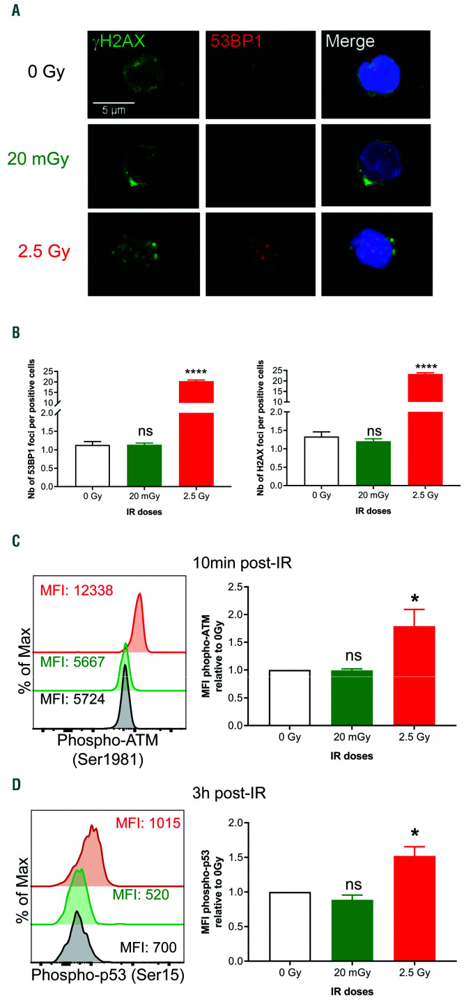
Since irradiation usually induces DNA double strand breaks (DSB), we quantified the number of H2AX and 53BP1 foci 30 min post irradiation (Figure 4A). In contrast to a 2.5 Gy irradiation, a 20 mGy irradiation did not increase the number of H2AX and 53BP1 foci compared to sham-irradiated CD34+CD38lowCD45RA−CD90+ HSPC indicating that 20 mGy LDIR does not induce DNA DSB (Figure 4B). We then studied the DNA damage response (DDR) path way after exposure to LDIR by quantification of ATM and p53 phosphorylation 10 min and 3 h after irradiation. As expected, 2.5 Gy-irradiated HSPC exhibited an increased ATM and p53 phosphorylation compared to control HSPC (Figure 4C and D). In contrast, no increase in ATM or p53 phosphorylation was detected after exposure to 20 mGy (Figure 4C and D). Importantly, the expression of p53 protein was not modified by IR (Online Supplementary Figure S3). Altogether these results indicate that 20 mGy LDIR does not induce DNA DSB nor activate p53 and ATM-dependent DNA damage repair pathway in human HSPC.
Figure 4.
Low doses (LD) of ionizing radiations (IR) do not induce DNA double strand breaks nor activate ATM-dependent/p53-dependent DNA repair pathway in human hematopoietic stem progenitor cells (HSPC). CD34+ CD38low CD45RA− CD90+ HSPC were purified by cell sorting and exposed to different doses of IR as indicated. (A) γH2AX and 53BP1 foci were examined by confocal microscopy 30 minutes (min) post IR (at least 100 cells by condition were analyzed. (B) Number (Nb) of 53BP1 (left panel) and γH2AX (right panel) foci by positive HSPC. (C and D) CD34+ cells were irradiated, cultured 10 min (C) or 3 hours (h) (D) at 37°C, stained for cell surface markers then fixed. (C) Analysis of ATM-phosphorylation on Ser1981 by FACS in CD34+ CD38low CD45RA− CD90+ HSPC (one representative experiment out of 4). (D) Analysis of p53-phosphory-lation on Ser15 (left) and p53 protein expression (right) in CD34+ CD38low CD45RA− CD90+ HSPC by FACS 3 h post IR (one representative experiment out of 5). Results are shown as mean±standard error of mean. *P<0.05, ****P<0.0001 (Mann and Whitney statistics).
Low doses of ionizing radiations increase reactive oxygen species levels, 8-oxo-dG lesions, induce NRF2 translocation into the nucleus, activate p38MAPK pathway and delay mitochondrial activation
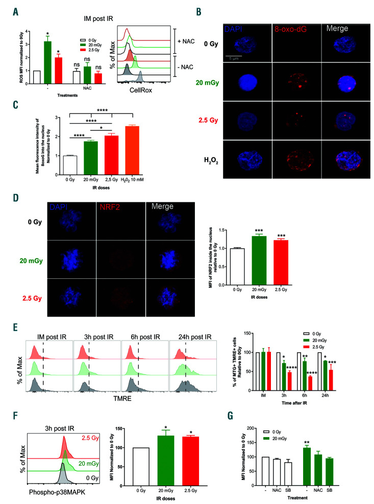
Since irradiation is known to promote ROS production,10,18,30 we next quantified ROS levels after LDIR exposure. ROS levels in CD34+CD38lowCD45RA− CD90+ HSPC after exposure to LDIR were measured immediately or 3 h after irradiation of CD34+ cells. Menadione and NAC treatments were used to respectively induce and inhibit ROS production. Increased ROS levels in HSPC were observed immediately after exposure to 20 mGy LDIR and to a lesser extent after exposure to 2.5 Gy, as compared to no irradiation (Figure 5A and Online Supplementary Figure S4A and B). These ROS increased levels were transient as no further difference in ROS levels could be detected 3 h after irradiation (Online Supplementary Figure S4C). NAC pretreatment of HSPC significantly decreased this early burst of ROS after 20 mGy and 2.5 Gy exposure. As increased ROS levels can lead to 8-oxo-dG lesions, as well as NRF2 translocation into the nucleus, we looked for 8-oxo-dG lesions in DNA of irradiated versus sham-irradiated HSPC30,31 and NRF2 location into HSPC.22,24 As expected sham-irradiated and H2O2-treated (control) cells exhibited respectively no and highly detectable anti-8-oxo-dG nuclear labeling. After exposure to 20 mGy, 8-oxo-dG staining was detected in the HSPC nucleus showing that 20 mGy LDIR can induce 8-oxo-dG lesions in DNA (Figure 5B and C). Similarly, the NRF2 protein was found in the nucleus of 20 mGy- and 2.5 Gy-irradiated HSPC compared to sham-irradiated cells (Figure 5D). As an increase in ROS is also associated with a delay in mitochondrial activation,32 we used mitotracker green (MTG) and TMRE probes to study respectively mitochondrial mass and membrane potential. Of note, CB CD34+ cells and CB HSPC are mainly quiescent, therefore there is very little mitochondrial activation (TMREneg) (Figure 5E, first left panel, and data not shown). HSPC exposure to LDIR did not alter the mitochondrial mass (MTG) of CD34+ cells in short-term culture (Online Supplementary Figure S5A). However, a delay in mitochondrial activation occurred (MTG+ TMRE+ HSPC) as soon as 3 h post IR (Figure 5E and Online Supplementary Figure S5B), suggesting that LDIR affect mitochondrial activity. In line with mitochondria activation, autophagy activation was monitored after IR (Online Supplementary Figure S6). The CytoID probe was used to follow autophagy in HSPC.33,34 As expected, after treatment with chloroquine and rapamycine, autophagy was detected in CD34+ cells (Online Supplementary Figure S6A). Besides, LDIR did not induce autophagy in HSPC after a different culture time (Online Supplementary Figure S6B and C). Finally, we investigated whether the observed increase of ROS can lead to p38MAPK activation as previously documented.19 Thr180/Tyr18 phosphorylation was used as a marker of p38MAPK activation. As a positive control of p38MAPK activation, increased p38MAPK phosphorylation (P-p38MAPK) can be detected in PMA-treated HSPC (Online Supplementary Figure S5C). In irradiated HSPC, we observed an increase of P-p38MAPK after exposure to 20 mGy and 2.5 Gy IR compared to sham-irradiated controls, suggesting that LDIR can activate p38MAPK pathway in HSPC similarly to high irradiation doses (2.5 Gy)35 (Figure 5F). To further confirm that p38MAPK acti vation was due to the early transient increase in ROS levels, HSPC were treated with NAC or SB203580, a p38MAPK inhibitor, prior to 20 mGy irradiation. As expected, SB203580 prevented increased p38MAPK phosphorylation in 20 mGy-irradiated HSPC (Figure 5G). NAC treatment resulted in the same decrease in p38MAPK phosphorylation in 20 mGy-irradiated HSPC (Figure 5G). Altogether, these results show that LDIR increase ROS levels leading to DNA 8-oxo-dG lesions, NRF2 translocation into the nucleus and p38MAPK activation in 20 mGy-irradiated HSPC.
Figure 5.
Low doses (LD) of ionizing radiations (IR) induce transitory reactive oxygen species (ROS) increase, 8-Oxo-dG DNA lesions and p38MAPK activation with altered mitochondrial activity in hematopoietic stem progenitor cells (HSPC). (A) ROS levels were quantified in CD34+ CD38low CD45RA− CD90+ HSPC using CellRox Orange probe immediately after IR. (Left) Pool of CellRox Orange mean of fluorecence relative to 0 Gy condition, right overlay histograms showing CellRox Orange fluorescence. One representative experiment out of four is shown (see also Online Supplementary Figure S4). Results are shown as mean±standard error of mean. (B and C) CD34+ CD38low CD45RA− CD90+ HSPC were purified by cell sorting and exposed to different doses of IR or H2O2 as indicated. Shown are 8-oxo-dG lesions quantified by confocal microscopy 30 minutes post IR (at least 50 cells were screened by condition in 3 independent experiments. Blue: Dapi, Red: 8-oxo-dG). Histograms represent the intensity of fluorescence of 8-oxo-dG staining within HSPC nucleus. To avoid heterogeneity, mean fluorescence intensity (MFI) has been normalized to the sham-irradiated condition. (D) CD34+ CD38low CD45RA− CD90+ HSPC were purified by cell sorting and exposed to different doses of IR as indicated. Shown are NRF2 staining quantified by confocal microscopy 2 hours (h) post IR (at least 50 cells were screened by condition in 2 independent experiments. Blue: Dapi, Red: NFR2). Histograms represent the intensity of fluorescence of NRF2 staining within HSPC nucleus. To avoid heterogeneity, MFI has been normalized to the sham-irradiated condition. (E) Mitochondrial activity was monitored over time by using TMRE (membrane potential, left panel) and MTG (mitochondrial mass) probes in HSPC. Shown is the frequency of TMRE+ cells over time in culture for one representative experiment out of three independent experiments and the mitochondria activation (% of MTG+ TMRE+, right panel) over time in culture (pool of the 3 independent experiments). (F) CD34+ cells were irradiated and cultured 2 h at 37°C followed by cell surface marker staining and then fixed. Phosphorylation of p38MAPK on Thr180/Tyr182 was analyzed by flow cytometry. Overlay histograms of p38MAPK phosphorylation on CD34+ CD38low CD45RA− CD90+ HSPC (left panel) are represented for the three irradiation conditions. Overlay histograms are from one representative experiment out of three. Histogram bars (right panel) show the MFI of phospho-p38MAPK in CD34+ CD38low CD45RA− CD90+ HSPC (n=3 independent experiments). (G) CD34+ cells were treated with NAC, SB203580 (SB) or untreated for 1 h at 37°C then irradiated and cultured 2 h at 37°C. Staining for cell surface markers was performed and then cells were fixed. Phosphorylation of p38MAPK on Thr180/Tyr182 was analyzed by flow cytometry. Histogram bars show mean of fluorescence of phospho-p38MAPK in CD34+ CD38low CD45RA− CD90+ HSPC (n=3 independent experiments). Results are shown as mean±standard error of mean. *P<0.05, **P<0.01, ***P<0.001, ****P<0.0001 (Mann-Whitney statistics).
20 mGy-dependent reactive oxygen species increase and p38MAPK activation lead to defects in the serial clonogenic potential of hematopoietic stem/progenitor cells
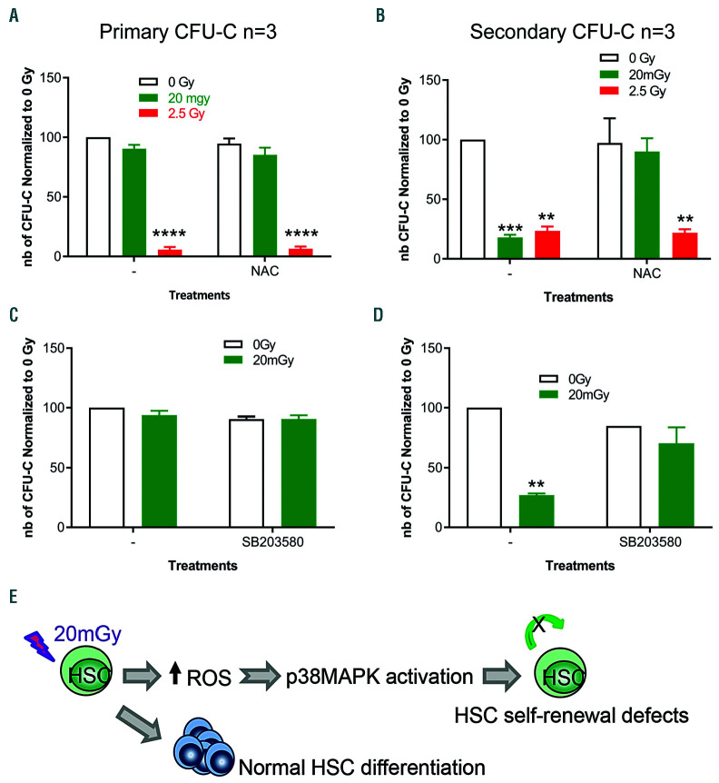
As increased ROS levels can lead to HSC loss of potentials,18 we then asked if ROS-dependent pathways could explain the HSPC functional defects after LDIR exposure. To this end, serial CFU-C assays were performed using sorted CD34+CD38lowCD45RA−CD90+ HSPC pre-treated or not with NAC before exposure to LDIR and cultures. 20 mGy-irradiated HSPC generated the same number of primary CFU-C compared to sham-irradiated HSPC with or without NAC treatment (Figure 6A and Online Supplementary Figure S7A). However, 20 mGy-irradiated HSPC treated with NAC before IR, but not 2.5 Gy-irradiated cells, were capable of generating equivalent numbers of secondary CFU-C compared to sham-irradiated HSPC, showing that NAC treatment prior to exposure to 20 mGy protected HSPC from the loss of in vitro serial clonogenic potential (Figure 6B and Online Supplementary Figure S7B). This result was obtained when the serial plating assays were performed with the whole cell population harvested from primary CFU cultures (Figure 6B), and also after picking up and replating individual primary CFU-GM colonies (Online Supplementary Figure S7C). Rescue of secondary replating properties of HSPC after 20 mGy LDIR was also obtained using HSPC pretreatment with Catalase, another antioxidant enzyme (Online Supplementary Figure S7D). These results show that preventing ROS production with antioxidants before LDIR exposure rescues the in vitro serial clonogenic potentials of HSPC.
Figure 6.
Low doses (LD) of ionizing radiations (IR) induce a transitory increase of ROS in CD34+ CD38low CD45RA−CD90+ hematopoietic stem progenitor cells (HSPC) that alters their serial clonogenic potential. (A) Colony forming unit-cell (CFU-C) assay. Cumulative results from 3 independent experiments with CD34+ CD38low CD45RA−CD90+ HSPC from 3 independent pools of cord blood (CB) samples. Sorted CD34+ CD38low CD45RA− CD90+ HSPC were pre-treated or not with N-acetylcysteine (NAC) prior to IR and plated (500 cells/plate) in CFU-C conditions for 12-14 days. Shown are the number (nb) of CFU-C (primary CFU-C). Results are normalized to the sham-irradiated conditions. (B) Primary CFU-C were pooled and replated in CFU-C conditions for 12-14 days. Shown are the nb of secondary CFU-C, normalized to the sham-irradiated conditions (cumulative results from 3 independent experiments). (C) Sorted CD34+ CD38low CD45RA− CD90+ HSPC were pre-treated or not with SB203580 prior to IR and plated (500 cells/plate) in CFU-C conditions for 12-14 days. Shown are the nb of CFU-C (primary CFU-C). Results are normalized to the sham-irradiated conditions. (D) Primary CFU-C were pooled and replated in CFU-C conditions for 12-14 days. Shown are the nb of secondary CFU-C, normalized to the sham-irradiated conditions (cumulative results from 2 experiments with CD34+ CD38low CD45RA− CD90+ HSPC from two independent pools of CB samples. (E) Model explaining how LDIR can impair HSC self-renewal through ROS-p38MAPK dependent pathway. Results are shown as mean+standard error of mean. **P<0.01, ***P<0.001, ****P<0.0001 (Mann-Whitney statistics).
Finally, we wondered whether ROS-mediated p38MAPK activation was involved in LDIR-induced HSC self-renewal defects. HSPC were pre-treated with SB203580, a specific inhibitor of p38MAPK, prior to 20 mGy irradiation and serial CFU-C assays. No difference in the number of primary CFU-C was detected with SB203580 pretreated HSPC regardless of the irradiation dose used (Figure 6C and Online Supplementary Figure S7E). However, whereas SB203580-untreated 20 mGy-irradiated HSPC generated very few secondary CFU-C, SB203580 treatment of HSPC protected their capacity to generate secondary CFU-C as efficiently as sham-irradiated HSPC (Figure 6D and Online Supplementary Figure S7F), suggesting that p38MAPK pathway activation participates in LDIR-mediated HSPC defects. Based on all these results, we propose a model in which 20 mGy LDIR rapidly increases ROS amounts in HSPC that induce p38MAPK activation altogether leading to a defect in the long-term maintenance of the clonogenic potential of CD34+CD38lowCD45RA−CD90+ HSPC (Figure 6E).
Discussion
Here we show that exposure to a single 20 mGy LDIR alters the functional properties of human HSPC. No defect in HSPC differentiation potential tested in primary cultures was detected after exposure to LDIR. However, HSPC irradiated at 20 mGy in vivo in the NSG mouse BM harbored a defect in human hematopoietic reconstitution potential. This defect was cell-intrinsic since 20 mGy-irradiated CD34+CD38low cells isolated after in vivo irradiation failed to serially reconstitute NSG mice as efficiently as non-irradiated cells; the same was observed with in vivo 20 mGy-irradiated bulk BM cells. This in vivo phenotype was also observed in vitro when using LDIR-exposed CD34+CD38-/lowCD45RA−CD90+ HSPC in serial CFU-C and LTC-IC assays, supporting the fact that these effects are cell-autonomous and not limited to transplantation conditions. Likewise, in vitro, HSPC exposure to 20 mGy induced a loss of secondary CFU-C potential as well as a decrease in secondary LTC-IC frequency. Altogether, based on the use of in vivo assays and in vitro surrogate assays to evaluate the self-renewal potential,25–27 these functional results strongly argue for an effect of 20 mGy LDIR on the long-term HSC functional properties, most likely through a loss of self-renewal potential. Of note, 20 mGy LDIR has been shown to decrease self-renewal capacity in murine HSC as well.22
High-dose ionizing radiations (HDIR) (2.5 Gy) are known to induce DNA DSB in human HSPC, rejoining is delayed, and H2AX foci persist leading to a loss of HSC functions partly related to apoptosis and activation of p53 pathway.23 Despite the publication of several studies over the past few years,36–38 little is known about which pathway is used to repair DNA DSB in human HSPC.16 In the present work, we tested if a single 20 mGy LDIR can alter cell cycle and induce apoptosis, and cause DNA DSB in HSPC. Surprisingly, and in contrast to HDIR, 20 mGy irradiation did not induce obvious cell cycle defects nor promote apoptosis in HSPC, since no increased cleaved caspase 3 protein was detected after exposure to 20 mGy LDIR. Moreover, no significant increase in H2AX and 53BP1 foci numbers was revealed, suggesting that 20 mGy LDIR does not produce DNA DSB. Finally, neither p53 nor ATM pathway was activated after 20 mGy exposure. However, and similarly to HDIR, 20 mGy irradiation led to 8-oxo-dG lesions in HSPC DNA. No such lesion was observed in sham-irradiated cells. Altogether, 20 mGy LDIR does not induce classic DNA damage and repair pathways usually activated by γ-irradiation but rather triggers 8-oxo-dG-dependent DNA damage that can be linked to uncontrolled increase in ROS levels. Moreover, NRF2 protein was found in the nucleus of 20 mGy-irradiated HSPC. Indeed, increased ROS levels were detected immediately after HSPC exposure to 20 mGy and, in line with this, we could observe that 20 mGy-irradiated HSPC had a delay in mitochondrial activation compared to control cells. Our results and those from Romeo’s lab22 suggest that the transient increase in ROS levels is likely to be responsible for HSPC defects after LDIR exposure. We tested this hypothesis using antioxidant treatment of HSPC prior to exposure to LDIR. Importantly, pre-treatment of HSPC with NAC or Catalase prior to LDIR exposure did rescue the loss of in vitro serial clonogenic potential of HSPC. In mouse, irradiation can induce p38MAPK activation through increased levels of ROS.19,35 Prevention of p38MAPK activation leads to decreased IR toxicity in HSC.35 It is also known that dormant HSC have little or no p38MAPK activation, and that p38MAPK activation in HSC is associated with differentiation and loss of HSC self-renewal.17,39 In humans, the function of the p38MAPK pathway is still not fully understand but preventing p38MPAK activation allows HSC maintenance/expansion in vitro.40,41 Interestingly, in our model, we observed a ROS-dependent p38MAPK activation in human HSPC after exposure to both 2.5 Gy and 20 mGy IR. The involvement of the p38MAPK pathway in the LDIR-mediated HSC self-renewal defects was then confirmed in serial replating CFU-C assays. Indeed, pre-treatment of HSPC with a specific inhibitor of p38MAPK prior to LDIR rescued their serial replating capacities. This is in agreement with the fact that HSC treatments either with NAC or p38MAPK inhibitor increase LTC-IC frequency and promote higher hematopoietic reconstitution upon serial transplantation.18,19 It is important to highlight that two other studies on the effect of LDIR have also shown that LDIR did not induce classic DNA damage and repair pathways, but rather an oxidative stress (increase in ROS level and NRF2 nuclear localization).22,42 Therefore, oxidative stress induction seems to be a feature of exposure to LDIR, leading either to a differentiation defect in the case of cycling stem cells42 or a self-renewal defect in the case of quiescent stem cells, as we observed for human HSPC; this is also the case for mouse HSC.22
Increased ROS levels as well as p38MAPK activation in HSC are associated with aging and stress during serial transplantation.17,18,43 The aging phenomenon is clearly a strong driver of differentiation and expansion of myeloid-biased HSPC.44 Here we were not able to detect any bias toward myelopoiesis when analyzing the progeny of the surviving LDIR-treated human HSC after serial transplantation, maybe due to the NSG mouse model, as the NSG BM microenvironment is more supportive of B-cell rather than myeloid-cell differentiation.45 Moreover, all experiments were performed with HSPC from CB, i.e. young HSPC. Thus, although it is tempting to speculate that exposure to LDIR may induce early/accelerated aging of the human HSC, we have no formal proof of that. Since radiation sensitivity and transplantation efficiency are highly dependent on the ontogenic origin of HSPC,46–48 aged HSPC may be more sensitive to LDIR. Another feature of HSC aging is higher risk of leukemic transformation, especially in the presence of an oncogenic-initiating event such as a mutation of the epigenetic modifiers DNMT3a or TET2, as observed in blood from elderly people.49 A very interesting and important question for the future would be to determine if aged HSC exposed to LDIR are more prone to (pre)leukemic transformation, especially when HSC contain primary oncogenic mutations.
To sum up, in contrast to HDIR, 20 mGy does not induce DNA DSB, nor apoptosis and a defect in the cell cycle. However, both 20 mGy and 2.5 Gy IR induce 8-oxo-dG lesions in DNA, increase ROS levels, and activate the p38MAPK pathway leading to HSC self-renewal defects. Nevertheless, only 20 mGy-LDIR effects were counteracted by use of antioxidants prior to irradiation exposure, indicating there are major differences between these two IR doses. These results show for the first time that a dose as low as 20 mGy can have a huge impact on human HSC through both similar and also different molecular mechanisms to those of high IR doses.
Acknowledgments
We acknowledge the midwives from Clinique des Noriets in Vitry-sur-Seine and the Cell therapy department of Hôpital Saint Louis in Paris, France, especially Prof. J. Larghero, for providing cord blood samples free of charge and the families who agreed to donate the samples for research purposes. This work was made possible thanks to multiple platforms (Flow cytometry, Microscopy, Irradiation and animal facilities) of IRCM, Fontenay-aux-Roses, France. We are thus grateful to J Tilliet and N Dechamps for their great technical help during this project. A special thanks to J Bernardino and JB Lahaye for their devoted work at the irradiation platform from IRCM but also to C. Villagrasa, Y. Ristic and M. Razanajotovo-Derichebourg for their precious help at the irradiation platform, laboratory of dosimetry of ionizing radiation, IRSN, Fontenay-aux-Roses, France. Finally, we acknowledge Paul-Henri Roméo, Franck Yates, Christophe Carles, Benjamin Uzan and Rima Haddad for helpful discussions and manuscript reviewing.
Footnotes
Check the online version for the most updated information on this article, online supplements, and information on authorship & disclosures: www.haematologica.org/content/105/8/2044
Funding
This study was supported by grants from INSERM, CEA (Segment Radiobiologie), Ligue Nationale contre le Cancer (équipe labellisée from 2014 to 2017), Région Ile de France/Cancéropole, Electricité de France and the European network RISK-IR. I-SS was supported by the European network RISK-IR and EH is a PhD fellow from the Ligue Nationale Contre le Cancer.
References
- 1.Mendelson A, Frenette PS. Hematopoietic stem cell niche maintenance during homefostasis and regeneration. Nat Med. 2014; 20(8):833–846. [DOI] [PMC free article] [PubMed] [Google Scholar]
- 2.Doulatov S, Notta F, Laurenti E, Dick JE. Hematopoiesis: a human perspective. Cell Stem Cell. 2012;10(2):120–136. [DOI] [PubMed] [Google Scholar]
- 3.Majeti R, Park CY, Weissman IL. Identification of a hierarchy of multipotent hematopoietic progenitors in human cord blood. Cell Stem Cell. 2007;1(6):635–645. [DOI] [PMC free article] [PubMed] [Google Scholar]
- 4.Arcangeli ML, Frontera V, Bardin F, et al. JAM-B regulates maintenance of hematopoietic stem cells in the bone marrow. Blood. 2011;118(17):4609–4619. [DOI] [PubMed] [Google Scholar]
- 5.Yu VW, Scadden DT. Hematopoietic Stem Cell and Its Bone Marrow Niche. Curr Top Dev Biol. 2016;118:21–44. [DOI] [PMC free article] [PubMed] [Google Scholar]
- 6.Benyoucef A, Calvo J, Renou L, et al. The SCL/TAL1 Transcription Factor Represses the Stress Protein DDiT4/REDD1 in Human Hematopoietic Stem/Progenitor Cells. Stem Cells. 2015;33(7):2268–2279. [DOI] [PubMed] [Google Scholar]
- 7.Brunet de la Grange P, Armstrong F, Duval V, et al. Low SCL/TAL1 expression reveals its major role in adult hematopoietic myeloid progenitors and stem cells. Blood. 2006; z108(9):2998–3004. [DOI] [PubMed] [Google Scholar]
- 8.Correia NC, Arcangeli ML, Pflumio F, Barata JT. Stem Cell Leukemia: how a TALented actor can go awry on the hematopoietic stage. Leukemia. 2016; 30(10):1968–1978. [DOI] [PubMed] [Google Scholar]
- 9.Orkin SH. Diversification of haematopoietic stem cells to specific lineages. Nat Rev Genet. 2000;1(1):57–64. [DOI] [PubMed] [Google Scholar]
- 10.Ito K, Hirao A, Arai F, et al. Regulation of oxidative stress by ATM is required for self-renewal of haematopoietic stem cells. Nature. 2004;431(7011):997–1002. [DOI] [PubMed] [Google Scholar]
- 11.Nijnik A, Woodbine L, Marchetti C, et al. DNA repair is limiting for haematopoietic stem cells during ageing. Nature. 2007; 447(7145):686–690. [DOI] [PubMed] [Google Scholar]
- 12.Rossi DJ, Bryder D, Seita J, Nussenzweig A, Hoeijmakers J, Weissman IL. Deficiencies in DNA damage repair limit the function of haematopoietic stem cells with age. Nature. 2007;447(7145):725–729. [DOI] [PubMed] [Google Scholar]
- 13.Cheng T, Rodrigues N, Dombkowski D, Stier S, Scadden DT. Stem cell repopulation efficiency but not pool size is governed by p27(kip1). Nat Med. 2000;6(11):1235–1240. [DOI] [PubMed] [Google Scholar]
- 14.Matsumoto A, Takeishi S, Kanie T, et al. p57 is required for quiescence and mainte nance of adult hematopoietic stem cells. Cell Stem Cell. 2011;9(3):262–271. [DOI] [PubMed] [Google Scholar]
- 15.Zou P, Yoshihara H, Hosokawa K, et al. p57(Kip2) and p27(Kip1) cooperate to maintain hematopoietic stem cell quiescence through interactions with Hsc70. Cell Stem Cell. 2011;9(3):247–261. [DOI] [PubMed] [Google Scholar]
- 16.Biechonski S, Milyavsky M. Differences between human and rodent DNA-damage response in hematopoietic stem cells: at the crossroads of self-renewal, aging and leukemogenesis. Transl Cancer Res. 2013; 2(5):372–383. [Google Scholar]
- 17.Jang YY, Sharkis SJ. A low level of reactive oxygen species selects for primitive hematopoietic stem cells that may reside in the low-oxygenic niche. Blood. 2007;110(8):3056–3063. [DOI] [PMC free article] [PubMed] [Google Scholar]
- 18.Yahata T, Takanashi T, Muguruma Y, et al. Accumulation of oxidative DNA damage restricts the self-renewal capacity of human hematopoietic stem cells. Blood. 2011; 118(11):2941–2950. [DOI] [PubMed] [Google Scholar]
- 19.Ito K, Hirao A, Arai F, et al. Reactive oxygen species act through p38 MAPK to limit the lifespan of hematopoietic stem cells. Nat Med. 2006;12(4):446–451. [DOI] [PubMed] [Google Scholar]
- 20.Pearce MS. Patterns in paediatric CT use: an international and epidemiological perspective. J Med Imaging Radiat Oncol. 2011;55(2):107–109. [DOI] [PubMed] [Google Scholar]
- 21.Pearce MS, Salotti JA, Little MP, et al. Radiation exposure from CT scans in childhood and subsequent risk of leukaemia and brain tumours: a retrospective cohort study. Lancet. 2012;380(9840):499–505. [DOI] [PMC free article] [PubMed] [Google Scholar]
- 22.Rodrigues-Moreira S, Moreno SG, Ghinatti G, et al. Low-dose irradiation promotes persistent oxidative stress and decreases self-renewal in hematopoietic stem cells. Cell Rep. 2017;20(13):3199–3211. [DOI] [PubMed] [Google Scholar]
- 23.Milyavsky M, Gan OI, Trottier M, et al. A distinctive DNA damage response in human hematopoietic stem cells reveals an apoptosis-independent role for p53 in self-renewal. Cell Stem Cell. 2010;7(2):186–197. [DOI] [PubMed] [Google Scholar]
- 24.Picou F, Debeissat C, Bourgeais J, et al. n-3 Polyunsaturated fatty acids induce acute myeloid leukemia cell death associated with mitochondrial glycolytic switch and Nrf2 pathway activation. Pharmacol Res. 2018;136:45–55. [DOI] [PubMed] [Google Scholar]
- 25.Hao QL, Thiemann FT, Petersen D, Smogorzewska EM, Crooks GM. Extended long-term culture reveals a highly quiescent and primitive human hematopoietic progenitor population. Blood. 1996;88(9):3306–3313. [PubMed] [Google Scholar]
- 26.Petzer AL, Hogge DE, Landsdorp PM, Reid DS, Eaves CJ. Self-renewal of primitive human hematopoietic cells (long-term-culture-initiating cells) in vitro and their expansion in defined medium. Proc Natl Acad Sci U S A. 1996;93(4):1470–1474. [DOI] [PMC free article] [PubMed] [Google Scholar]
- 27.Rizo A, Dontje B, Vellenga E, de Haan G, Schuringa JJ. Long-term maintenance of human hematopoietic stem/progenitor cells by expression of BMI1. Blood. 2008;111(5):2621–2630. [DOI] [PubMed] [Google Scholar]
- 28.Nakamura-Ishizu A, Takizawa H, Suda T. The analysis, roles and regulation of quiescence in hematopoietic stem cells. Development. 2014;141(24):4656–4666. [DOI] [PubMed] [Google Scholar]
- 29.Notta F, Doulatov S, Laurenti E, Poeppl A, Jurisica I, Dick JE. Isolation of single human hematopoietic stem cells capable of long-term multilineage engraftment. Science. 2011;333(6039):218–221. [DOI] [PubMed] [Google Scholar]
- 30.Walter D, Lier A, Geiselhart A, et al. Exit from dormancy provokes DNA-damage-induced attrition in haematopoietic stem cells. Nature. 2015;520(7548):549–552. [DOI] [PubMed] [Google Scholar]
- 31.Haghdoost S, Czene S, Naslund I, Skog S, Harms-Ringdahl M. Extracellular 8-oxo-dG as a sensitive parameter for oxidative stress in vivo and in vitro. Free Radic Res. 2005;39(2):153–162. [DOI] [PubMed] [Google Scholar]
- 32.Cabon L, Bertaux A, Brunelle-Navas MN, et al. AIF loss deregulates hematopoiesis and reveals different adaptive metabolic responses in bone marrow cells and thymocytes. Cell Death Differ. 2018;25(5):983–1001. [DOI] [PMC free article] [PubMed] [Google Scholar]
- 33.Chabi S, Uzan B, Naguibneva I, et al. Hypoxia regulates lymphoid development of human hematopoietic progenitors. Cell Rep. 2019;29(8):2307–2320.e6. [DOI] [PubMed] [Google Scholar]
- 34.Gomez-Puerto MC, Folkerts H, Wierenga AT, et al. Autophagy Proteins ATG5 and ATG7 Are Essential for the Maintenance of Human CD34(+) Hematopoietic Stem-Progenitor Cells. Stem Cells. 2016; 34(6):1651–1663. [DOI] [PubMed] [Google Scholar]
- 35.Wang Y, Liu L, Zhou D. Inhibition of p38 MAPK attenuates ionizing radiation-induced hematopoietic cell senescence and residual bone marrow injury. Radiat Res. 2011;176(6):743–752. [DOI] [PMC free article] [PubMed] [Google Scholar]
- 36.de Laval B, Pawlikowska P, Petit-Cocault L, et al. Thrombopoietin-increased DNA-PK-dependent DNA repair limits hematopoietic stem and progenitor cell mutagenesis in response to DNA damage. Cell Stem Cell. 2013;12(1):37–48. [DOI] [PubMed] [Google Scholar]
- 37.Kraft D, Rall M, Volcic M, et al. NF-kappaB-dependent DNA damage-signaling differ entially regulates DNA double-strand break repair mechanisms in immature and mature human hematopoietic cells. Leukemia. 2015;29(7):1543–1554. [DOI] [PubMed] [Google Scholar]
- 38.Shao L, Feng W, Lee KJ, Chen BP, Zhou D. A sensitive and quantitative polymerase chain reaction-based cell free in vitro non-homologous end joining assay for hematopoietic stem cells. PLoS One. 2012;7(3):e33499. [DOI] [PMC free article] [PubMed] [Google Scholar]
- 39.Tesio M, Tang Y, Mudder K, et al. Hematopoietic stem cell quiescence and function are controlled by the CYLD-TRAF2-p38MAPK pathway. J Exp Med. 2015;212(4):525–538. [DOI] [PMC free article] [PubMed] [Google Scholar]
- 40.Baudet A, Karlsson C, Safaee Talkhoncheh M, Galeev R, Magnusson M, Larsson J. RNAi screen identifies MAPK14 as a drug-gable suppressor of human hematopoietic stem cell expansion. Blood. 2012; 119(26):6255–6258. [DOI] [PubMed] [Google Scholar]
- 41.Zou J, Zou P, Wang J, et al. Inhibition of p38 MAPK activity promotes ex vivo expansion of human cord blood hematopoietic stem cells. Ann Hematol. 2012;91(6):813–823. [DOI] [PMC free article] [PubMed] [Google Scholar]
- 42.Fernandez-Antoran D, Piedrafita G, Murai K, et al. Outcompeting p53-Mutant Cells in the Normal Esophagus by Redox Manipulation. Cell Stem Cell. 2019;25(3):329–341.e6. [DOI] [PMC free article] [PubMed] [Google Scholar]
- 43.Rube CE, Fricke A, Widmann TA, et al. Accumulation of DNA damage in hematopoietic stem and progenitor cells during human aging. PLoS One. 2011;6(3):e17487. [DOI] [PMC free article] [PubMed] [Google Scholar]
- 44.Akunuru S, Geiger H. Aging, Clonality, and Rejuvenation of Hematopoietic Stem Cells. Trends Mol Med. 2016;22(8):701–712. [DOI] [PMC free article] [PubMed] [Google Scholar]
- 45.Ito R, Takahashi T, Katano I, Ito M. Current advances in humanized mouse models. Cell Mol Immunol. 2012;9(3):208–214. [DOI] [PMC free article] [PubMed] [Google Scholar]
- 46.Hernandez L, Terradas M, Camps J, Martin M, Tusell L, Genesca A. Aging and radiation: bad companions. Aging Cell. 2015;14(2):153–161. [DOI] [PMC free article] [PubMed] [Google Scholar]
- 47.Holyoake TL, Nicolini FE, Eaves CJ. Functional differences between transplantable human hematopoietic stem cells from fetal liver, cord blood, and adult marrow. Exp Hematol. 1999;27(9):1418–1427. [DOI] [PubMed] [Google Scholar]
- 48.Kollman C, Howe CW, Anasetti C, et al. Donor characteristics as risk factors in recipients after transplantation of bone marrow from unrelated donors: the effect of donor age. Blood. 2001;98(7):2043–2051. [DOI] [PubMed] [Google Scholar]
- 49.Xie M, Lu C, Wang J, et al. Age-related mutations associated with clonal hematopoietic expansion and malignancies. Nat Med. 2014;20(12):1472–1478. [DOI] [PMC free article] [PubMed] [Google Scholar]


