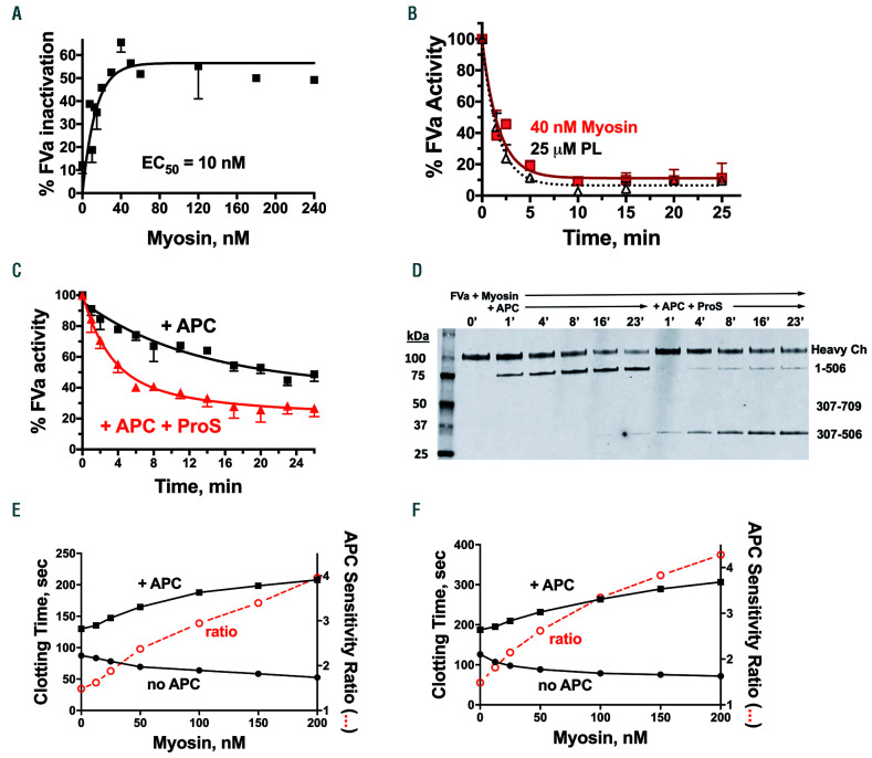
Figure 1.
Myosin supports inactivation of activated factor V by activated protein C in purified human coagulation factor reaction mixtures and in human plasma coagulation assays. (A) Skeletal muscle myosin (myosin) dose-response curve for inactivation of 1.25 nM activated factor V (FVa) by 0.4 nM activated protein C (APC) over 4 min. Data shown are the composite from three experiments performed on different days. (B) Time course for inactivation of 1 nM FVa by 0.4 nM APC in the presence of 40 nM myosin, or 25 μM phospholipids (PL). Residual FVa activity was measured in a second-stage standard prothrombinase assay with PL, activated factor X (FXa) and prothrombin. One hundred percent FVa activity corresponded to generation of ~1 nM thrombin/min from the sub-aliquot used to test for prothrombinase activity in the absence of APC in the second stage of the assay. Data shown are the composite from three or four experiments performed on different days. (C) Inactivation of FVa (3.7 nM final concentration) by APC (0.6 nM final concentration) with or without protein S (100 nM final concentration), as indicated, in the presence of 120 nM myosin. Data shown are combined from experiments performed on three different days. (D) Immunoblot analysis of reaction mixtures from FVa inactivation studies. Inactivation of FVa (3.7 nM final concentration) by APC (1 nM final concentration) with or without protein S (100 nM final concentration) in the presence of 120 nM myosin was monitored at the indicated times. Aliquots were taken over time into 80°C LDS Li-Cor sample buffer containing 10 mM EDTA for 10 min for immunoblotting in sodium dodecylsulfate polyacrylamide gel electrophoresis. Immunoblotting of FVa was performed using a monoclonal anti-FVa heavy chain antibody with an epitope in the region of residues 307-506. (E) Factor Xa one-stage assay in FX-depleted plasma with FXa added shows the effects of myosin on clotting times in the presence of 17 nM APC (+ APC) (■) and in the absence of added APC (no APC) (●).The APC sensitivity ratio (○) was obtained by dividing the clotting times in the presence of APC by the clotting times in the absence of APC at each myosin concentration. (F) Kaolin clotting time assays in FV-deficient plasma with FVa added show the effects of myosin on clotting times in the presence of 17 nM APC (+ APC) (■) and in the absence of added APC (no APC) (●). The APC sensitivity ratio was calculated as in (E).

