

Abstract
Cold environments can trigger a variety of conditions, which, in their acute phase often present to the Emergency Department. Primary acrocyanosis is a distinct, rare condition which may be missed resulting in misdiagnosis and mismanagement. Primary acrocyanosis is a peripheral vascular disorder defined by painless, symmetrical discoloration of the distal appendages and uniquely characterized by persistence of the skin color changes after cold exposure. We present a case of a 24-year-old female who presented to the Emergency Department with peripheral cyanosis after cold exposure and was eventually diagnosed with primary acrocyanosis by Rheumatology. The prognosis for primary acrocyanosis is quite good in comparison to other acrosyndromes and once secondary causes of acrocyanosis have been ruled, out can be managed conservatively with lifestyle modifications and potential follow-up with Rheumatology.
Keywords: Acrocyanosis, Cyanosis, Peripheral vascular disorder
1. Introduction
Cold weather can bring a variety of environmentally related issues to the Emergency Department, from critical hypothermia to less urgent presentations such as Raynaud's phenomenon. While the recognition and management of these conditions are familiar to the Emergency Physician, primary acrocyanosis, a distinct, rare condition may be missed, resulting in misdiagnosis and mismanagement. Acrocyanosis is a functional peripheral vascular disorder defined by painless, symmetrical discoloration of the distal appendages and uniquely characterized by persistence of the skin color changes after cold exposure [1,2]. Primary acrocyanosis is a distinct cause of peripheral cyanosis which must be recognized and differentiated from both secondary causes of acrocyanosis as well as similarly presenting conditions including Raynaud's phenomena and the pernio-like lesions of COVID-19 due to the varied management [4]. Key differences in recognition and management of primary acrocyanosis will be illustrated in the following case presentation.
2. Case report
A 24-year-old female presented to the Emergency Department with discoloration of her hands after an outdoor run. She reported that after 90 min of running in –19 °C weather she began experiencing pruritus in her fingers associated with new onset bluish discoloration. She reported that the discomfort had resolved however the discoloration had worsened despite rewarming. She was otherwise well including being a non-smoker and taking only the oral contraceptive pill as medication.
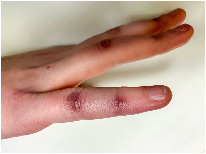
On physical examination, there was notable peripheral cyanosis presenting as painless discoloration at the DIP and PIP joints involving all digits bilaterally (Figs. 1 and 2 ). Digits were cool to the touch with the remainder of the physical examination being unremarkable including normal motor, power and sensation with a capillary refill <3 s and a negative Allen's test. Additionally, oxygen saturation was 100% with no sign of central cyanosis. Routine blood work and subsequent inflammatory markers to further investigate alternate diagnoses on the differential for peripheral cyanosis as well as assess for secondary causes of acrocyanosis were negative. Rheumatology was consulted in the Emergency Department who diagnosed her clinically with primary acrocyanosis and outpatient follow-up was arranged. At discharge we advised her to avoid running in the cold climate to prevent re-exacerbation.
Fig. 1.

Clinical photograph highlighting the bilateral, symmetrical nature of acrocyanosis at the PIP and DIP joins.
Fig. 2.
Clinical photograph highlighting the degree of cyanosis at the PIP and DIP joints.
3. Discussion
Cold environments can trigger a variety of conditions, which, in their acute phase may present to the Emergency Department. Primary acrocyanosis is a rare, functional peripheral vascular disorder caused by chronic vasospasm of the small cutaneous arterioles with compensatory dilatation in the capillary venules causing cyanosis in the setting of cold exposure [1,2]. While the prevalence of primary acrocyanosis is uncertain due to the rare nature of the disease, it is largely a disorder affecting young adults in cool climates with a female predominance [1]. Primary acrocyanosis presents as a symmetric, painless, discoloration of the distal appendages which can be precipitated by cold exposure and must be differentiated from secondary causes of acrocyanosis as well as more commonly presenting acrosyndromes such as Raynaud's phenomenon, erythromelalgia and more novel pernio-like lesions of COVID-19 [1,2,4]. Clinical criteria to diagnose acrocyanosis in general include painless cyanosis of extremities, local hypothermia, sweatiness and elastic infiltration of the skin [2]. This criterion differentiates acrocyanosis from the more commonly presenting Raynaud's phenomenon, by the relative persistence of skin color changes, symmetrical presentation and lack of paroxysmal pallor [1]. Pernio-like lesions of COVID-19, also known as chilblains, a novel cause of the blue digit, can be characterized by rapid onset, itching, pain and tenderness and quick improvement with re-warming, very similar to primary acrocyanosis [1]. Chilblains have been reported in the setting of cold weather as well as in the setting of COVID-19 infection but unlike primary acrocyanosis are associated with pain and improvement with Nifedipine [1,5]. Primary acrocyanosis is a diagnosis of exclusion, however once its possibility has been raised, secondary causes of acrocyanosis including connective tissue diseases, hypoxemia and drug exposures among other conditions should be ruled out [1,3]. Careful history, physical examination and ancillary testing including capillary oximetry, CBC, ANA, ESR and CRP will help guide the clinical decisions [1,3]. Additionally, we propose that a nasopharyngeal swab be sent to differentiate primary acrocyanosis from the novel COVID-19 associated chilblains, given the similarity in presentation [1,4]. As a precaution, all patients with suspected COVID-19 infection should be instructed to self-isolate [5]. Once the diagnosis of primary acrocyanosis has been made conservative management including counselling on lifestyle modifications including smoking cessation, avoiding cold weather and using insulated clothing to improve local circulation and decrease arteriospasm should be initiated [1]. In the Emergency Department follow-up may be arranged with outpatient Rheumatology to ensure adequate control of symptoms with conservative management and monitor the need for vasoactive drugs [1].
Declarations of Competing Interest
None.
Financial disclosures
This research did not receive any specific grant from funding agencies in the public, commercial, or not-for-profit sectors.
References
- 1.Kurklinsky A.K., Miller V.M., Rooke T.W. Acrocyanosis: the flying dutchman. Vasc Med. 2011;16:288–301. doi: 10.1177/1358863X11398519. [DOI] [PMC free article] [PubMed] [Google Scholar]
- 2.Das S., Maiti A. Acrocyanosis: an overview. Indian J Dermatol. 2013;58:417–420. doi: 10.4103/0019-5154.119946. [DOI] [PMC free article] [PubMed] [Google Scholar]
- 3.Nousari H.C., Kimyai-Asadi A., Anhalt G.J. Chronic idiopathic acrocyanosis. J Am Acad Dermatol. 2001;45:207–208. doi: 10.1067/mjd.2001.103266. [DOI] [PubMed] [Google Scholar]
- 4.Freeman E.E., McMahon D.E., Lipoff J.B., Rosenbach M., Kovarik C., Takeshita J. Pernio-like skin lesions associated with COVID-19: a case series of 318 patients from 8 countries. J Am Acad Dermatol. 2020;83:486–492. doi: 10.1016/j.jaad.2020.05.109. [DOI] [PMC free article] [PubMed] [Google Scholar]
- 5.Ladha M.A., Dupuis E.C. SARS-CoV-2-related chilblains. CMAJ. 2020;192:804. doi: 10.1503/cmaj.201348. [DOI] [PMC free article] [PubMed] [Google Scholar]