

Abstract
Subcutaneous flexor carpi radialis (FCR) tendon ruptures secondary to osteoarthritis of the scapho-trapezio-trapezoidal (STT) joint are very rare. A 53-year-old female suffered a subcutaneous FCR tendon rupture after noticing pain in her right wrist. Because of continuing pain and decreasing strength in her right hand, surgery of the STT joint arthrodesis and an FCR tendon reconstruction with free tendon graft was performed. Her left wrist gradually showed the same symptoms 9 years later. The STT fusion for her left wrist was then performed. The FCR tendon was partially worn out on the spur of the trapezial ridge as well. Only 10 cases of FCR tendon ruptures have been reported due to definite STT osteoarthritis in six English papers. We believe STT fusion should be done without hesitation before tendon rupture occurs, and when motion pain around the STT joint and decreasing grip strength exists. However, it is controversial whether the FCR tendon reconstruction should be done in addition to STT fusion.
Keywords: FCR tendon, injury, STT fusion, reconstruction
Subcutaneous flexor carpi radialis (FCR) tendon ruptures are very rare. The most common causes of the ruptures are bone spurs due to scapho-trapezio-trapezoidal (STT) osteoarthritis. Our particular case is a bilateral FCR tendon injury with STT osteoarthritis. Whether the ruptured FCR tendon should be reconstructed or not in addition to STT fusion is still controversial. Now that we are facing an aging society, such cases are expected to increase. Therefore, we think it is beneficial to report this.
Case Report
In April 2007, a right-handed 53-year-old female visited our clinic with pain around the radiovolar side of her right wrist joint. She had no past history of rheumatoid arthritis or trauma and had a normal blood test. Physical examination revealed tenderness on the FCR tendon and mild tenderness on the STT joint that was provoked by a resistive active motion. X-rays showed osteoarthritis in both the STT joints, including osteosclerotic change and narrow joint space ( Fig. 1 ).
Fig. 1.

Bilateral radiograph of the wrist joints. The posteroanterior radiograph shows the scapho-trapezio-trapezoidal joint narrowing and sclerotic changes of the subchondral bone (arrow).
We diagnosed osteoarthritis in both of her hands with FCR tendinitis in her right wrist. Conservative treatments were initiated including the use of nonsteroidal anti-inflammatory drugs and an intra-articular steroid injection (twice). Seven months later, when the patient lifted up her bed, acute pain suddenly occurred in her right wrist with a snapping sound. Upon examination there was a palpable defect measuring 7 cm in line with the FCR tendon's distal palpable end, which also had tenderness. The motion pain and tenderness along the distal FCR tendon continued for 4 months after this episode. Moreover, grip strength on the right hand decreased to 17 kg (22 kg left hand). Surgery of the STT joint arthrodesis and FCR tendon reconstruction was performed. The distal end of the ruptured FCR tendon was on the ridge of the trapezium. The proximal end of the ruptured FCR tendon was located 7 cm from the distal end. The STT joint showed remarkable synovial proliferation, and the formation of osteophytes by which the FCR tendon had been torn. The joint cartilage was highly degenerated, entirely, and showed partial defect. In addition, we found that the tendon-like synovial tissue was newly formed between the proximal and distal tendon ends. The myostatic contracture of the FCR remained minimal and the muscle was still functional ( Fig. 2 ). The sclerotic bones and the osteophytes of the STT joint were removed. The STT joint was fixed with five criss-cross Kirschner wires that were buried under soft tissues. In addition, the ruptured FCR tendon was reconstructed with a free tendon graft using the ipsilateral palmaris longus tendon (PL) by means of interlacing suture ( Fig. 3 ). Cast immobilization from the arm to the palm was applied for 3 weeks, after which exercise of wrist motion was permitted. The wires were removed 8 weeks after surgery. Eight years after surgery the patient still has no pain or complaints regarding the right wrist in daily activities. The range of motion of her right wrist is 60 degrees in flexion and 50 degrees in extension. The grip strength showed 19.5 kg on the right hand (left 22.5 kg,) 2 years after the surgery, and increased to 25 kg (left 19 kg) 9 years after the surgery. We also measured the flexion strength of the wrist joints ( Fig. 4 ). It showed 11 kg (left 10 kg) 2 years after surgery, and increased to 14.5 kg (left 12 kg) 9 years after the surgery.
Fig. 2.

The distal end was located on the ridge of the scapho-trapezio-trapezoidal joint (arrow A ) and the proximal end was retracted 7 cm from the distal end (arrow B ). Both ends were connected by a tendon-like synovial tissue (dot arrow). Palmaris longus tendon (star).
Fig. 3.

The flexor carpi radialis tendon was reconstructed with free tendon graft by interlacing suture using the palmaris longus tendon (arrow).
Fig. 4.
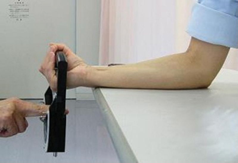
Measurement of the flexion strength of the wrist joint. The forearm is placed on the table with the elbow flexed at 90 degrees and the wrist is placed off the table. The grip dynamometer grasped in her hand is pulled down vertically by an examiner, then the maximum flexion strength is measured.
Her left wrist gradually showed the same symptoms such as motion pain, tenderness along the distal FCR tendon, and decreasing of the grip strength. The STT fusion surgery for her left wrist was then performed 8 years after the right hand. We found the FCR tendon was worn out partially on the spur of the trapezial ridge ( Fig. 5 ).
Fig. 5.

Flexor carpi radialis tendon (star); partial rupture (solid arrow); the ridge of the trapezial spur (left dotted arrow); and wide scapho-trapezio-trapezoidal joint space due to ligament loosening (right dotted arrow).
Two years after left-hand surgery, X-rays showed the fusion of her left STT joint had been completed and the patient had no complaints.
Discussion
In our case, the bony spurs of the trapezial ridge of STT osteoarthritis caused the frictional tendinitis of the FCR tendon, eventually leading to a complete attritional rupture on the right wrist, and an incomplete rupture on the left wrist. Fitton et al 1 was the first to describe such a case. Six cases were treated surgically with only osteophyte resection or synovectomy, but none of them were repaired. Irwin et al 2 recommended the excision of the tendon stump. Tonkin and Stern 3 reported that their three cases showed minimal disability, and were treated without reconstruction. None of these cases featured repairs or reconstruction. On the contrary, Allred and Rayan 4 recommended surgical repair for the FCR tendon rupture, as it can be distinctly disabling in young and active patients if left unrepaired. This is still a controversial issue.
According to Brand et al, 5 the flexion force of the wrist joint is comprised of the FCR, 4.1, the PL, 1.2, and the flexor carpi ulnaris, 6.7. As the force of the PL tendon is the least of these three tendons, the FCR tendon reconstruction by the PL tendon graft was deemed to be a suitable treatment for recovering sufficient wrist strength.
As for the problem of myostatic contracture, we think that it should not easily become symptomatic after a 4-month delay of reconstruction. In our case, the FCR muscle had maintained mobility. Currently, the patient's range of wrist motion is sufficient for daily life, which is supposed to be more than 40 degrees of flexion and extension. 6 7 The grip strength and wrist flexion strength have shown an increase 9 years after the surgery.
In conclusion, we recommend STT fusion surgery should be performed quickly when painful STT osteoarthritis is accompanied with pain along the FCR tendon and decreasing grip strength so that it does not rupture.
Moreover, we believe FCR tendon reconstruction using the PL tendon graft at the same time with STT fusion could result in better functional ability of the wrist joint. The vast majority of the cases in the literature call for no reconstruction with STT fusion. They also only discuss osteoarthritis as the primary cause.
Footnotes
Conflict of Interest None declared.
References
- 1.Fitton J, Shea F W, Goldie W. Lesions of the flexor carpi radialis tendon and sheath causing pain at the wrist. J Bone Joint Surg Br. 1968;50(02):359–363. [PubMed] [Google Scholar]
- 2.Irwin L R, Outhwaite J, Burge P D. Rupture of the flexor carpi radialis tendon associated with scapho-trapezial osteoarthritis. J Hand Surg [Br] 1992;17(03):343–345. doi: 10.1016/0266-7681(92)90126-m. [DOI] [PubMed] [Google Scholar]
- 3.Tonkin M A, Stern H S. Spontaneous rupture of the flexor carpi radialis tendon. J Hand Surg [Br] 1991;16(01):72–74. doi: 10.1016/0266-7681(91)90133-9. [DOI] [PubMed] [Google Scholar]
- 4.Allred D W, Rayan G M. Flexor carpi radialis tendon rupture following chronic wrist osteoarthritis: a case report. J Okla State Med Assoc. 2003;96(05):211–212. [PubMed] [Google Scholar]
- 5.Brand P W, Beach R B, Thompson D E. Relative tension and potential excursion of muscles in the forearm and hand. J Hand Surg Am. 1981;6(03):209–219. doi: 10.1016/s0363-5023(81)80072-x. [DOI] [PubMed] [Google Scholar]
- 6.Brumfield R H, Champoux J A.A biomechanical study of normal functional wrist motion Clin Orthop Relat Res 1984;(187):23–25. [PubMed] [Google Scholar]
- 7.Palmer A K, Werner F W, Murphy D, Glisson R. Functional wrist motion: a biomechanical study. J Hand Surg Am. 1985;10(01):39–46. doi: 10.1016/s0363-5023(85)80246-x. [DOI] [PubMed] [Google Scholar]