To the Editor:
Conventional radial artery (RA) cannulation at the wrist interferes with electrode placement for intraoperative neurophysiological monitoring (IONM) and is often troubled by dampened arterial waveforms after postural changes. We report the use of distal radial artery (DRA) cannulation in the anatomical snuffbox for arterial blood pressure (ABP) monitoring during a spine surgery requiring IONM and postural changes.
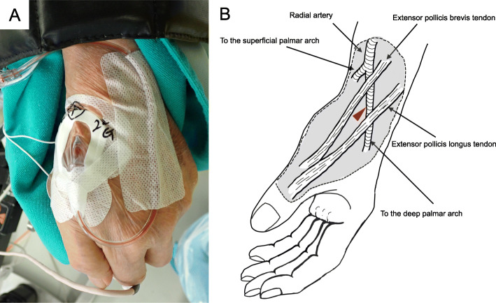
An 89-year-old man underwent extreme lateral interbody fusion in the right lateral decubitus and prone positions with motor evoked potential (MEP) monitoring. After inducing anesthesia with propofol and remifentanil, his wrist was adducted on a roll with the point of cannulation facing upwards. We successfully inserted a 22-gauge cannula (Surflo; TERUMO, Tokyo, Japan) in the DRA in the anatomical snuffbox on the first attempt under ultrasound guidance (Fig. 1a). This technique of DRA cannulation provided good ABP monitoring throughout surgery.
Fig. 1.
a Distal radial artery cannulation in the anatomical snuffbox. An electrode for recording the motor evoked potential from the abductor pollicis brevis is attached to the thenar area. The left forearm is pronated and fixed on a padded armrest in the right lateral decubitus position. b Anatomical scheme of the entry point for cannulation of the dorsal radial artery of the left hand. The entry point is indicated by the red arrowhead
In recent times, the DRA has been increasingly cannulated for cardiac and neurosurgical interventions [1, 2]. Based on our experience, the advantages of using the DRA for ABP monitoring in neurosurgery are as follows: (1) the stimulating electrodes for the assessment of somatosensory evoked potentials and the recording electrodes for MEP are placed above the median nerve at the wrist and the abductor pollicis brevis, respectively. Thus, RA cannulation at the wrist interferes with the proper placement of these electrodes. DRA cannulation avoids this interference and enables the adequate stimulation and recording that is necessary for reliable IONM. (2) The arterial waveform is stable in the prone and lateral decubitus positions because DRA cannulation in the anatomical snuffbox permits pronation of the patient’s forearm. In the prone and lateral decubitus positions, the palmar aspects of the pronated arms face the padded armrests. This frequently dampens the arterial waveform monitored by the conventional RA cannulation method. Additionally, the arterial wave forms obtained by cannulation of the DRA are less affected by wrist flexion during postural changes than those obtained by conventional RA cannulation [3].
The DRA is distal to the division of the superficial palmar branch of the RA (Fig. 1b). It continues as the deep palmar arch and receives collateral flow from the superficial palmar branch and the ulnar artery [2, 4]. Therefore, DRA cannulation confers lesser risk of ischemia compared to the conventional technique of RA cannulation [2, 4]. It has also been reported that the occurrence of other complications, including hematomas and neuropathy, did not significantly differ between these two approaches [1, 3]. However, the possibility of nerve injury should be considered because the superficial branch of the radial nerve is near the entry point of DRA cannulation [3]. Further well-designed studies are needed for ensuring the safety of DRA cannulation during the perioperative period. In addition, the DRA may be more difficult to cannulate than the RA due to its smaller diameter [1], which is a limitation of DRA cannulation.
In conclusion, the DRA in the anatomical snuffbox can be a useful site for ABP monitoring in neurosurgery and enables adequate electrode positioning for neurophysiological assessments.
Acknowledgements
The authors would like to thank Kengo Fujita M.D. (Ina Central Hospital, Ina City, Nagano, Japan) for illustrating Fig 1b.
Abbreviations
- ABP
Arterial blood pressure
- DRA
Distal radial artery
- IONM
Intraoperative neurophysiological monitoring
- MEP
Motor evoked potential
- RA
Radial artery
Authors’ contributions
RT and TS participated in the anesthetic management and drafted the manuscript. CK and JS revised the manuscript. All authors have read and approved the final manuscript.
Funding
Not applicable
Availability of data and materials
Not applicable
Ethics approval and consent to participate
Not applicable
Consent for publication
Written informed consent was obtained from the patient for the publication of this report and accompanying images.
Competing interests
The authors declare that they have no competing of interests.
Footnotes
Publisher’s Note
Springer Nature remains neutral with regard to jurisdictional claims in published maps and institutional affiliations.
References
- 1.Coomes EA, Haghbayan H, Cheema AN. Distal transradial access for cardiac catheterization: a systematic scoping review. Catheter Cardiovasc Interv. 2019. 10.1002/ccd.28623 [Epub ahead of print]. [DOI] [PubMed]
- 2.McCarthy DJ, Chen SH, Brunet MC, Shah S, Peterson E, Starke RM. Distal radial artery access in the anatomical snuffbox for neurointerventions: Case Report. World Neurosurg. 2019;122:355–359. doi: 10.1016/j.wneu.2018.11.030. [DOI] [PubMed] [Google Scholar]
- 3.Kimura Y, Kimura S, Inoue H, Yamauchi M, Sumita S. Comparison of usefulness of the dorsal branch of the radial artery with the radial artery for arterial cannulation (in Japanese with English abstract) Masui. 2012;61:728–732. [PubMed] [Google Scholar]
- 4.Deepika K, Palaniappan D, Fuhrman T, Saltzmanm B. Anatomic snuffbox radial artery cannulation. Anesth Analg. 2010;111:1078–1079. doi: 10.1213/ANE.0b013e3181ef343a. [DOI] [PubMed] [Google Scholar]
Associated Data
This section collects any data citations, data availability statements, or supplementary materials included in this article.
Data Availability Statement
Not applicable


