Abstract
Background and Purpose
The traditional Chinese medicine, diosgenin (Dio), has attracted increasing attention because it possesses various therapeutic effects, including anti-tumor, anti-infective and anti-allergic properties. However, the commercial application of Dio is limited by its extremely low aqueous solubility and inferior bioavailability in vivo. Soluplus, a novel excipient, has great solubilization and capacity of crystallization inhibition. The purpose of this study was to prepare Soluplus-mediated Dio amorphous solid dispersions (ASDs) to improve its solubility, bioavailability and stability.
Methods
The crystallization inhibition studies were firstly carried out to select excipients using a solvent shift method. According to solubility and dissolution results, the preparation methods and the ratios of drug to excipient were further optimized. The interaction between Dio and Soluplus was characterized by differential scanning calorimetry (DSC), fourier transform infrared (FT-IR) spectroscopy, scanning electron microscopy (SEM), powder X-ray diffraction (PXRD) and molecular docking. The pharmacokinetic study was conducted to explore the potential of Dio ASDs for oral administration. Furthermore, the long-term stability of Dio ASDs was also investigated.
Results
Soluplus was preliminarily selected from various excipients because of its potential to improve solubility and stability. The optimized ASDs significantly improved the aqueous solubility of Dio due to its amorphization and the molecular interactions between Dio and Soluplus, as evidenced by dissolution test in vitro, DSC, FT-IR spectroscopy, SEM, PXRD and molecular docking technique. Furthermore, pharmacokinetic studies in rats revealed that the bioavailability of Dio from ASDs was improved about 5 times. In addition, Dio ASDs were stable when stored at 40°C and 75% humidity for 6 months.
Conclusion
These results indicated that Dio ASDs, with its high solubility, high bioavailability and high stability, would open a promising way in pharmaceutical applications.
Keywords: diosgenin, amorphous solid dispersions, solubility, bioavailability, stability
Introduction
Dioscin, a typical natural saponin compound, is abundant in dioscorea, such as Dioscorea nipponica Makino, Dioscorea zingiberensis C. H. Wright and Dioscorea panthaica Prain et Burkill.1 It is the main active ingredient of some Chinese patent medicines, such as Liuwei Dihuang Wan, Weiaoxin Tablets and Di’ao Xinxuekang Capsules.2–4 Diosgenin (Dio, the structural formula is shown in Figure 1) is the main metabolite from oral administration of dioscin.5 The Dio shows extensive pharmacological activities, including anti-tumor effect,6 cardiovascular protection,7 blood lipid regulation,8 anti-nervous system disorders,9 immune modulation and inflammation10 and other pharmacological activities.11,12 Moreover, Dio is also used as an intermediate in the pharmaceutical industry to synthesize steroid hormones and steroid contraceptives.13 Meanwhile, it has been reported that Dio has a good safety for long-term traditional use.14 Therefore, Dio is considered to be a potential compound with wide clinical application. However, like many new compounds with therapeutic properties, Dio is characterized by poor aqueous solubility.15,16 The strong hydrophobicity (logP 5.7) of Dio leads to the low bioavailability.17 The results of pharmacokinetics show that the absolute bioavailability of Dio in rats is only about 7%.18
Figure 1.
The structural formulas of dioscin and Dio.
Lots of Dio formulations strategies have been used to overcome the above-mentioned issues, including liquid crystalline,18 cyclodextrin complexes19 and nanocrystal.20 However, the preparation process of liquid crystalline technology was complicated, and cyclodextrin had the safe issues to some extent. Moreover, nanosuspension could only increase the bioavailability of Dio for about 2 times compared with Dio solution. Therefore, it is necessary to develop a simple and effective method to improve the bioavailability of Dio. Recently, studies have focused on amorphous solid dispersion (ASD).21 ASD is one of the most effective and simple methods to enhance the solubility and dissolution for insoluble drugs.22 The increased dissolution rate of amorphous form can provide a drug concentration to supersaturation in the solution.23 Such an increase in gastrointestinal concentration can improve the overall absorption of drugs.24 However, due to their thermodynamic instability, it will tend to recrystallize and lose its improved solubility. Dispersion of hydrophobic drugs in hydrophilic carriers can improve the dynamic stability of amorphous.25 Based on their chemical versatility, various excipients such as polyvinyl pyrrolidone (PVP), Eudragit, polyethylene glycol (PEG), polyvinyl alcohol, gelucire, hydroxypropyl cellulose, Soluplus and Kollidone26 are used to encapsulate and protect bioactive drugs in ASDs. In addition, ASDs can be prepared in many ways, such as rapid cooling of the melt, precipitation of drug-carrier solution or by direct solid conversion. These methods can be classified into heat-based method, solvent-based method and mechanochemistry-based method. Because different preparation methods change the performance of the final product, it is necessary to reasonably select the manufacturing technology.27
Soluplus is an amphiphilic polyvinyl caprolactam-polyvinyl acetate-polyethylene glycol graft co-polymer with great solubilizing nature for water-insoluble substances.28 From a biopharmaceutical perspective, it is attractive because of its advantageous properties, including minimum toxicity, low hygroscopicity, low glass transition temperature, and good thermal stability.29 Moreover, it can greatly increase the wettability of drug highly dispersed in ASD and inhibit the precipitation or crystallization during dissolution. Soluplus also prevents the aging of ASD during storage to a certain extent by forming hydrogen bond with the drug.30 So far, Soluplus has been used as an ASD carrier for a variety of drugs including curcumin,31 quercetin,32 andrographolide33 and other traditional Chinese medicine ingredients.34
In the present study, we aimed to explore a new Dio ASD formulation to alleviate the physical and chemical constraints. A solvent shift method was used to select excipients. According to solubility and dissolution, the preparation method and the ratio of drug to excipient for Dio ASD were further optimized. The interactions between Dio and Soluplus were studied by differential scanning calorimetry (DSC), fourier transform infrared (FT-IR) spectroscopy, scanning electron microscopy (SEM), powder X-ray diffraction (PXRD) and molecular docking. The bioavailability was explored the potential of Dio ASDs for oral administration. In addition, the long stability of Dio ASDs was also studied.
Materials and Methods
Materials
Dio with purity above 98% was obtained from Nanjing Spring & Autumn Biological Engineering Co., Ltd. (Nanjing, China). The chemical standards of Dio and internal standard (IS) tanshinone ⅡA (purity >98%) were purchased from the National Institute for Food and Drug Control (Beijing, China). Sodium dodecyl sulfate (SDS) was supplied by Tianjin Bodi Chemical Holding Co., Ltd. (Tianjin, China). Soluplus, polyvinylpyrrolidone K30 (PVPK30), polyethylene glycol 4000 (PEG4000), hydroxypropylmethyl cellulose E30 (HPMC-E30) and Pluronic F68 were provided by BASF Co., Ltd. (Shanghai, China). Methanol, acetonitrile and formic acid of MS grade were purchased from Thermal Fisher Scientific (Tustin, CA, USA). Acetonitrile and methanol of chromatographic grade were supplied by Beijing MREDA Technology Co. Ltd. (Beijing, China). Deionized distilled water was prepared in the laboratory and used throughout the research process.
Animals
Healthy male Sprague-Dawley rats were obtained from the Beijing Vital River Laboratory Animal Technology Co., Ltd. (Beijing, China) with license number SCXK2016-0006. All animals were exposed to the environment with a temperature of 22±1°C, a relative humidity of 50±1% and a light/dark cycle of 12/12 h. The rats were given free access to standard chow and sterile water and then fasted for 12 h prior to the experiment. All animal-related protocols were approved by Animal Ethical Experimentation Committee of Chengde Medical University (Ethical approval NO.CDMULAC-20,180,410,015, Chengde, China) according to the requirements of the National Act on the Use of Experimental Animals (People’s Republic of China).
HPLC Analysis of Dio
The content of Dio in the preparation was determined by high-performance liquid chromatography (HPLC) to evaluate the solubility and dissolution. Agilent 1260 Infinity liquid chromatography (Agilent Technologies, Inc., Santa Clara, CA, USA) equipped with quaternary solvent delivery system, online degasser, autosampler, column temperature controller and diode-array detection system was used, and UV spectra within the range of 190–400 nm were recorded. Chromatography was carried out on a Diamonsil® Plus C18 column (150 × 4.6 mm, 5 μm; Dikma Technologies Co., Ltd., Beijing, China). The mobile phase was composed of acetonitrile-deionized water (84:16, v/v) and set at a flow rate of 1 mL/min. The detection wavelength was set at 203 nm, and the injection volume was 10 µL. The column oven temperature was maintained at 30°C. The standard curve was linear (r2 > 0.999) with a mass range of 0.1235–24.7 μg/mL. The detection limit was the concentration at S/N=3. The inter-day and intra-day accuracies were less than 2.0%. The average recovery was over 99%, with an RSD of 1.53%.
UPLC-MS/MS Method
A Waters Acquity UPLC liquid chromatography included diode array detector, injection manager, binary pump and ACQUITY Console software. Chromatographic separations were carried out on an ACQUITY UPLC® BEH C18 column (2.1 × 50 mm, 1.7 μm; Waters Corporation, Ireland) equipped with a guard column. The Dio was eluted with an isocratic mobile phase system (80:20, v/v) consisting of acetonitrile and 0.1% formic acid. The experimental conditions were set as follows: the column temperature was kept at 35°C, the flow rate was 0.3 mL/min, the injection volume was 2 μL, and the total running time was 5 min.
The mass spectrometer was equipped with a Q-Trap® 5500 triple quadruple mass spectrometer (AB SCIEX, USA) using turbo ionspray electrospray ionization (ESI) source for sample analysis. The positive MRM mode was used for Dio and the IS tanshinone ⅡA. MS/MS parameters were set as follows: ion spray voltage, +5500 V; curtain gas, 35 psi; temperature, 500°C; ion source gas 1, 50 psi; ion source gas 2, 50 psi; the interface heater was on, and the collision gas was medium. The mass transitions were: Dio, m/z 415.2 → 271.2, tanshinone ⅡA, m/z 295.2 → 277.1; the declustering voltage of Dio was 71.96 V, the collision voltage was 23.03 eV; the declustering voltage of tanshinone ⅡA was 120 V, and the collision voltage was 25.92 eV.
The standard curve of Dio was linear (r2 > 0.99) within the concentration range of 1.33–1358 ng/mL and the lower limit of quantification was 1.33 ng/mL. The detection limit was the concentration at S/N=3. The inter-day and intra-day accuracy were less than 7.0%, and the accuracy (relative error) ranged from 1.41% to 3.40%. The extraction recovery and matrix effect of Dio were 103.89% and 92.88%, respectively, which were 103.22% and 95.56% for IS, respectively.
Evaluation on Crystallization Inhibition of Excipients
Solvent-shift method was used to study the crystallization inhibition of Dio by different excipients. Refering to Shi’s experiment35 for specific experimental steps, 27 mg of Dio was dissolved in 5 mL 95% ethanol to make a uniformly supersaturated solution simulating a supersaturated drug system. Moreover, 81 and 162 mg of excipient were dissolved in 900 mL of water, respectively. After equilibration for a period of time, the above-mentioned supersaturated drug solution was quickly added to the excipient solution. The solution without excipient was used as a control, the medium was maintained at 37°C, and the rotation speed was 50 r/min. Next, 5 mL sample was taken at 5, 10, 15, 20, 30, 60, 90, 120, 150, 180, 210 and 240 min, respectively, and 5 mL solution without drug was replenished quickly. The sample was passed through a 0.22-µm microporous membrane, and the filtrate was taken for HPLC determination. The experiment was repeated for three times.
Preparation of Dio ASDs and the Physical Mixture (PM)
Co-Precipitation Method
Mixtures of Dio and Soluplus at varying w/w ratios 1:2, 1:4, 1:6, 1:8, 1:10 and 1:12 were dissolved in anhydrous ethanol. The organic solvent was dried by rotating steam. Then, they were dried in the DZ-2BCIV vacuum drying oven (Tianjin Tester Instrument Co., Ltd., China). The ASDs were ground and sieved (60 mesh) to obtain uniform particles named as Dio-C-ASDs. Every experiment was repeated for three times.
Freeze-Drying Method
Mixtures of Dio and Soluplus at varying w/w ratios 1:2, 1:4, 1:6, 1:8, 1:10 and 1:12 were dissolved in 2% ethanol solution. The solution was frozen at −40°C for 8 h, and then lyophilized at −45°C for 24 h using LGL-22D freeze dryer (Shanghai Hongxin Biological Technology Co., Ltd., China). The ASDs were ground and sieved (60 mesh) to obtain uniform particles named as Dio-F-ASDs. Every experiment was repeated for three times.
Microwave Quenching Method
Mixtures of Dio and Soluplus at varying w/w ratios 1:2, 1:4, 1:6, 1:8, 1:10 and 1:12 were placed in crucibles. The mixtures were placed in a Galanz P70D2OTL-D4 microwave oven (Guangdong Galanz Group Co. Ltd., China) at high temperature of microwave (power 700 w) for 10 min. Then, they were removed to quench with liquid nitrogen at −196°C and dried in a DZ-2BCIV vacuum drying oven (Tianjin Taisite Instrument Co., Ltd.). The ASDs were ground with a mortar and pestle, and then sieved (60 mesh) to obtain uniform particles named as Dio-M-ASDs. Every experiment was repeated for three times.
PM of Dio and Soluplus
PM was gently prepared by mixing Dio and Soluplus using simple tumbling method for 15 min. Furthermore, the prepared homogeneous PM was stored in a desiccator.
Determination of Saturated Solubility of ASDs
An excess amount of Dio ASDs was added to 5 mL distilled water, and the suspension was shaken in an HZQ-C thermostatically controlled gas bath (Tianjin Experimental Instrument Factory, Tiantan, China) at 37±0.5°C for 48 h. The samples were then filtered through a 0.45-μm membrane filter, and the concentration of the solution was determined by HPLC.
Dissolution Testing of ASDs
Dissolution testing of Dio ASDs was tested in a sink condition dissolution study (ie the dissolution medium can dissolve >3 times of the total drug dose in the dissolution study)36 according to the USP Apparatus 2 setup using RC806D dissolution tester (Tianjin Tianda Tianfa Technology Co., Ltd., Tianjin, China). Briefly, 55 mg of sample powder (containing 5 mg Dio) was accurately weighed, filled into #0 gelatin capsules and then placed in a settling basket. The dissolution tests were performed at 37°C in 900 mL 0.1% SDS with a paddle speed of 100 rpm. Subsequently, 10 mL sample was withdrawn at 5, 15, 30, 60, 90 and 120 min and immediately passed through 0.45-μm filters. About 1 mL initial filtrate was discarded, and the remaining filtrate was then analyzed by HPLC.
Characterization of Dio ASDs
DSC Studies
Thermograms for Dio, Soluplus, Dio ASDs and PM were obtained. The samples were sealed in aluminum pans and analyzed using a TA DSC-250 (TA Instruments, USA). The samples were heated in a nitrogen atmosphere at a constant heating rate of 10°C/min within the range of 25–300°C to obtain thermograms.
FT-IR Spectroscopy
FT-IR spectra were obtained by Bruker Tensor27 FT-IR spectrometer (Bruker, Germany) to characterize the possible interactions between the drug and the carrier in solid state. About 2 mg sample was gently ground, mixed with IR-grade dry potassium bromide, and then compressed in a 10-ton hydraulic press for 5 min to form discs. The spectra of Dio, Soluplus, Dio ASDs and PM were scanned over a frequency range of 4000–400 cm−1 with a resolution of 4 cm−1.
SEM Analysis
The surface morphology of Dio, Soluplus, Dio ASDs and PM was evaluated by SEM (Jeol-JSM-7500F scanning microscope, Tokyo, Japan) operating at 5 kV. All samples were simply mixed to avoid surface changes. Before observation, the samples were fixed on the glass stump with double-sided adhesive tape, and vacuum argon plating was used. Micrographs were acquired at different magnifications to study the morphological and surface characteristics of the ASDs.
PXRD
Diffraction patterns of Dio, Soluplus, Dio ASDs and PM were determined in a Scintag X-ray diffractometer (PW3040/60 PANalytical, Netherlands) using Cu Kα radiation with a nickel filter. Samples were scanned in a series of 3–35°of 2θ with a step width of 0.03°/min and the generator settings were 40 kV and 100 mA.
Molecular Docking
The Dio structure was created using ChemDraw software. The 3D structures of Dio, Soluplus, HPMC-E30, PVP-K30, Pluronic-F68 and PEG-4000 were built by optimizing with the Sybyl 6.9.1 software package (Tripos Associates: St. Louis, MO, 2003). The optimized parameters of energy change were 0.005 (kcal/mol), and the maximum number of iterations was 10,000 times. Powell method (Tripos force field) was used to reduce the distributed charge to an energy change of 0.005 kcal/(mol × A). All other parameters remained at default. The molecular docking of Dio and excipients was carried by AutoDock 4.0 software. Optimized AutoDocking parameters were as follows: the maximum number of energy evaluations per time was 25,000,000; the local search of Solis and Wets was 3000 times; the number of generations was 100, and the individual number in population was 300. As a result, differences of the position root-mean-square deviation less than 2 Å were clustered together.
Pharmacokinetic Study
Pathogen-free male Sprague-Dawley rats, weighing 220±20 g, were kept in an environmentally controlled breeding room. The Dio ASDs were freshly prepared before the pharmacokinetic test. All samples were suspended in saline containing 0.05% carboxymethylcellulose sodium (CMC-Na) as the coarse suspension. A total of 30 rats were randomly and evenly divided into five groups. The animals were fasted overnight before the experiment. The samples were taken orally at a single intragastric dose of Dio (100 mg/kg body weight). The dosage was determined according to literature20 and through preliminary experiments. Heparinized plasma samples (0.4 mL) were collected from the ophthalmic veins using sterile capillary tube before administration and at 0.083, 0.25, 0.5, 0.75, 1, 2, 3, 4, 6, 8, 12, 24, 36, 48 and 72 h post-administration. Rats were given a standard meal at 12 h after administration. All blood samples were centrifuged immediately at 13,000 rpm for 15 min. The supernatants were aspirated and stored at −80°C until further analysis. All plasma samples were thawed at room temperature prior to analysis. Briefly, the IS solution (50 μL, 41.85 ng/mL) was mixed with 50 μL of the plasma sample in a 0.5-mL Eppendorf tube. The solution was vortexed for 30 s, additionally mixed with 250 μL methanol-acetonitrile (1:1), consequently vortexed for 2 min and centrifuged at 15,000 rpm for 15 min. The supernatant was injected into the UPLC-MS/MS system for analysis.
The maximum plasma concentration (Cmax) and the time to reach Cmax (Tmax) of Dio were obtained directly from the plasma concentration profiles. Other pharmacokinetic parameters were calculated using the DAS 3.0 pharmacokinetic software.
Storage Stability Experiment
Stability testing was carried out for Dio ASD. The PXRD was first utilized to show the morphological changes with time. Moreover, the in vitro release after different storage durations was compared. Samples were stored in an LHH-150GSP stability chamber (Shanghai Yiheng Scientific Instrument Co., Ltd., Shanghai, China) at 40°C and 75% RH. Stability studies were conducted at 0, 3 and 6 months, the crystal content of samples was tested by PXRD, and in vitro release of the samples was determined by HPLC.
Statistical Analysis
Results were expressed as the mean±standard deviation (SD). Statistical analysis was performed in the aqueous solubility determinations, dissolution tests and pharmacokinetic study using one-way ANOVA. P<0.05 was considered as statistically significant.
Results and Discussion
Supersaturation of Dio in the Presence of Excipients
In the field of oral administration, increasing intraluminal concentration through supersaturation is expected to enhance the intestinal absorption.37,38 It has been proved that the addition of precipitation inhibitors can stabilize and prolong supersaturation. The selection of different polymer materials as excipients has different effects on the dissolution rate and recrystallization ability of pharmaceutical preparations. Herein, a high-throughput precipitation screening was conducted to select the excipients and assess the supersaturation potential during the lead selection/optimization process.39,40 The solvent shift method, an effective technique for rapid screening excipients, can inhibit drug crystallization in supersaturated solution.40,41 Many studies have utilized this method to assess the significance of ASD systems in achieving dissolution and oral bioavailability improvement.42 In this study, we investigated the effect of five different water-soluble excipients on Dio crystal inhibition using the solvent shift method.
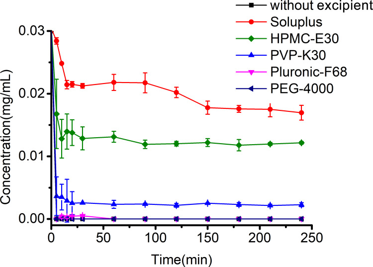
The Dio-95% ethanol solution was injected into the medium containing amorphous excipients Soluplus, HPMC-E30 and PVP-K30, semicrystalline excipients Pluronic-F68 and PEG-4000, respectively. The ratio of drug to excipients was 1:6. The main properties of the five excipients are shown in Table 1. Figure 2 shows that Dio was rapidly precipitated in the dissolution medium without excipient, and the concentration was lower than the detection limit after 5 min. Semicrystalline excipients can delay, promote or not affect the crystallization kinetics of different active pharmaceutical ingredients. However, the Pluronic-F 68 and PEG-4000 could not inhibit crystallization of Dio in supersaturated solution during 240 min. PVP-K30 had very weak crystallization inhibitory effect. HPMC-E30 had a certain crystallization inhibitory effect. Although Dio was rapidly precipitated in the first 15 min in the medium containing HPMC-E30, and it could maintain a high concentration later. Soluplus had the strongest inhibitory effect on crystallization. The gradual sedimentation and high concentration of Dio in the Soluplus-containing supersaturated solution persisted for 4 h. The results showed that the crystallization inhibition capability in the supersaturated solution could be ranked as follows: Soluplus > HPMC-E30 > PVP-K30 > Pluronic-F 68 and PEG-4000, and amorphous excipients were more effective at inhibiting the crystallization of Dio than semicrystalline excipients over the time.
Table 1.
The Physical and Chemical Properties of Different Excipients
| Excipients | Chemical Structure | Molecular Weight(g/mol) | Solubility in Water | Glass Transition Temperature (°C) |
|---|---|---|---|---|
| Soluplus | 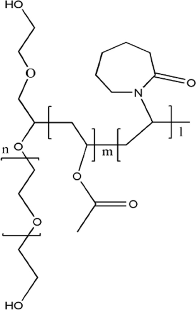 |
90,000–140,000 | Soluble | Around 70 |
| HPMC-E30 | 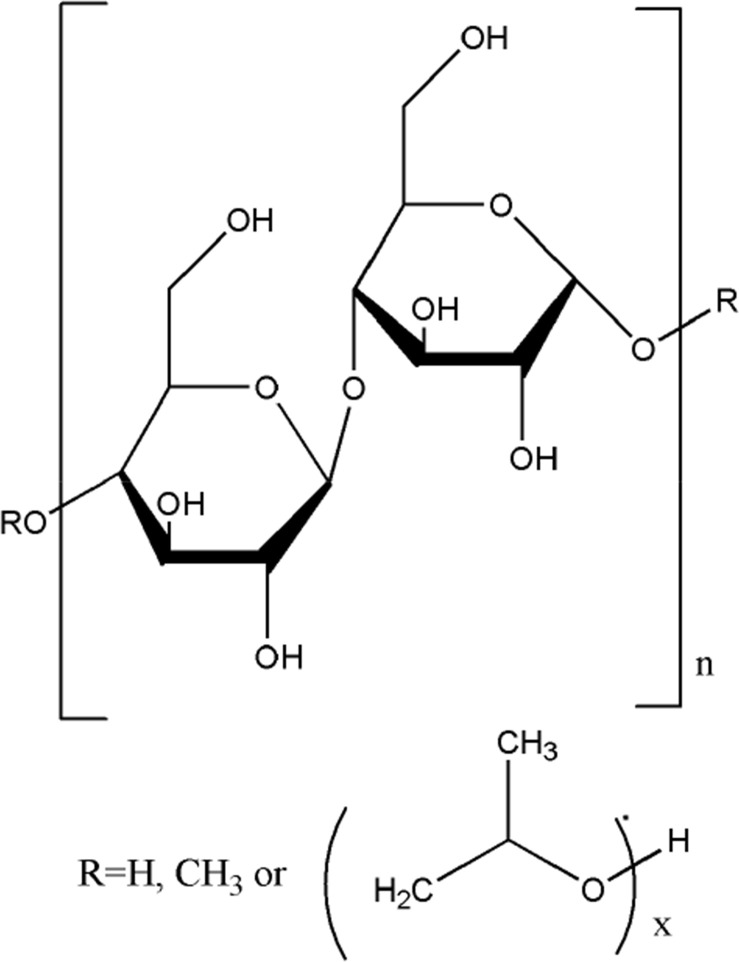 |
9000–12,000 | Soluble | Around 180 |
| PVP-K30 | 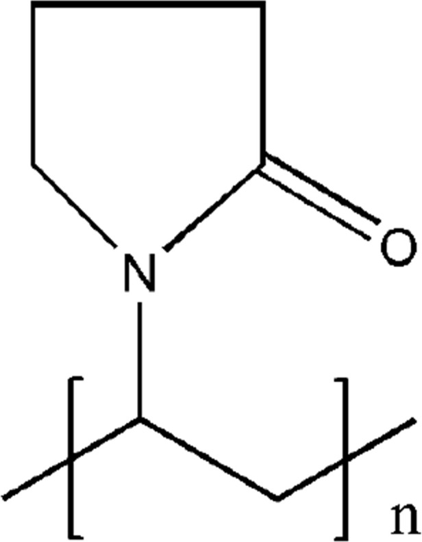 |
25,000–40,000 | Soluble | Around 180 |
| Pluronic-F 68 |  |
7680–9510 | Soluble | – |
| PEG-4000 |  |
3600–4400 | Soluble | Around 55 |
Figure 2.
Concentration-time profiles of Dio in the medium of 900 mL solution in the presence and absence of excipients (drug: excipient = 1:6). Data are presented as the mean ± SD (n=3).
We also observed the inhibitory effect of various excipients on Dio when the ratio of drug to excipient was 1:3. It was found that except for Soluplus which had a weak crystal inhibition effect (the concentration of Dio could be maintained at 0.003 mg/mL for 2.5 h), all other excipients had no crystal inhibition effect. This showed that the increase in excipient concentration had a beneficial effect on the inhibition of Dio crystallization.
Aqueous Solubility and Dissolution of Dio-ASD Formulations
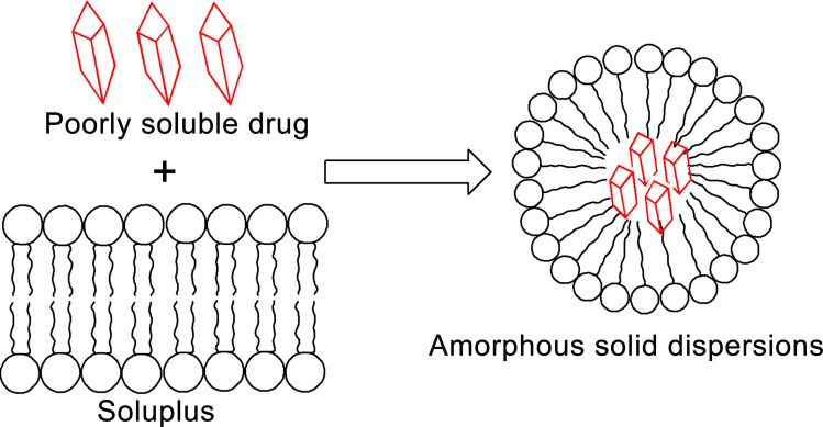
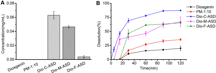
Figure 3 depicts the possible scheme of Dio-Soluplus ASD synthesis. The ASDs appeared as a pale yellow, free-flowing powder mass. The suitable mass ratios of drug to Soluplus were optimized based on solubility and dissolution studies. As shown in Figure 4, the co-precipitation, microwave quenching and freeze-drying of ASDs with a mass ratio of 1:10 w/w showed the best dissolution profile. These batches of ASDs were selected for further studies. Figure 5 shows the solubility and dissolution results.
Figure 3.
A schematic representation of drug-Soluplus ASDs.
Figure 4.
The dissolution of different Dio/Soluplus ratios in ASDs. Each point represents the mean ± SD (n = 3).
Figure 5.
The solubility (A) and dissolution (B) of different Dio ASDs with a mass ratio of 1:10 w/w for drug and excipient. Each point represents the mean ± SD (n = 3).
ASD basically contains storage of potential energy, similar to a spring-compressed mechanical system. In the desired medium, the potential energy is released, and the dispersion “bounces” the molecular entity into a supersaturated state. Since supersaturation is thermodynamically unstable, the formulation must supply a “parachute” to maintain a high concentration of drugs to achieve maximum absorption amounts. Previous studies have used various types of excipients to stabilize supersaturated system dynamics against recrystallization.43 For supersaturated systems, the ideal carrier should dissolve quickly to produce a sufficiently high drug concentration and effectively maintain the supersaturated concentration.44 As shown in Figure 5A, the solubility of Dio and PM was lower than the detection limit, so the value was recorded as 0. The solubility of Dio was significantly improved in ASDs as follows: Dio-C-ASD> Dio-M-ASD ≫ Dio-F-ASD. Figure 5B shows the dissolution of Dio in ASDs. In 0.1% SDS solution, the dissolution of all ASDs was significantly higher compared with the drug and PM. More dissolution of Dio from ASDs might be attributed to improved wettability of the drug in the dissolution medium and the transition of the drug from a crystalline state to an amorphous state.45 Interestingly, there was a “spring” phenomenon in the ASDs, while no “parachute” effect was observed in the 120-min dissolution test. The drug concentration was always increased over time. Moreover, all ASDs were dissolved slowly, which might be attributed to the presence of Soluplus with a high viscosity at 37°C in the solution. Shi et al46 have found that Soluplus could solubilize the drugs and inhibit the crystallization of drugs in supersaturated solution by self-micellization. The special grafted structure provides amphiphilicity to Soluplus, which makes it easy to form micelles, and the critical micelle concentration (CMC) is only 7.6 mg/L.47 In this study, the concentration of Soluplus in Dio ASDs was 61 mg/L, which was higher than the CMC of Soluplus, and thus the solubility of Dio was steadily improved through the formation of micelles.
Characterization of Dio ASDs
DSC Analysis
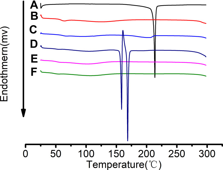
Figure 6 shows the thermal behaviors of pure Dio, Soluplus, PM and ASDs. Dio showed a sharp endothermic peak at 213.67°C, indicating that Dio existed in crystal structure. The endothermic peak of Dio disappeared in the Dio-C-ASD and Dio-M-ASD, so it is confirmed that the drug was miscible with excipients and the formation of amorphous state was achieved. Miscibility of Dio and Soluplus improved the physical stability of amorphous Dio in ASDs by reducing the molecular fluidity of Dio.48 Dio is a water-insoluble compound that has the potential tendency to form hydrates and solvates. When formed from different solvents, drug compounds can crystallize into polymorphs, hydrates and solvates.49 In Ningbo Gonga’s experiment, four solvents were used to prepare Dio solvates. DSC curves showed that there were more than two endothermic peaks in the solvate, and Dio solvate lost the solvent through endothermic process to achieve the stable form.50 In the Dio-F-ASD, Dio still existed in crystal form. It could be explained in this way as follows: In the Dio-F-ASD preparation, Dio was dissolved in water with 2% ethanol, and recrystallization might occur in the freeze-drying process. Therefore, Dio showed a melting-mediated polymorphism with a melting endotherm at 158.6°C, followed by a small exotherm at about 160.8°C corresponding to the recrystallization of Dio into a more stable form. While a second endotherm at 168.7°C corresponded to the melting of the stable form. Theoretically, the endothermic peak of Dio should exist at around 213°C in PM during the crystal melting process. However, no obvious endothermic peak was found in this experiment. It might be attributed to that when the PM was tested in the DSC experiment, heating of DSC program made the first molten carrier a good solvent of Dio, which was gradually dissolved in the molten carrier before reaching the melting point. This melting process was similar to that used to prepare the ASD of Dio, which was dissolved in molecular state in Soluplus. Alternatively, it might be explained by that the crystallization of the drug was inhibited by Soluplus, which reduced the characteristic endothermic peak of Dio. Therefore, there was no apparent endothermic peak of Dio in PM. This result was consistent with the research results of Yang et al on sulfadiazine solid dispersion.51
Figure 6.
DSC curves of Dio (A), Soluplus (B), PM-1:10 (C), Dio-F-ASD (D), Dio-C-ASD (E) and Dio-M-ASD (F).
FT-IR Analysis
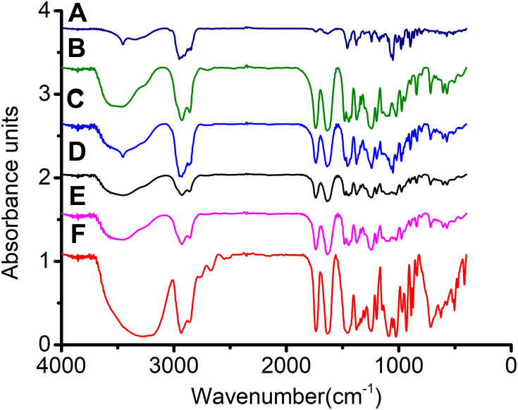
FT-IR spectra of Dio, Soluplus, PM and ASDs were recorded to evaluate any solid–solid interactions that may exist between the drug and excipients, and Figure 7 shows the results. Dio had the following characteristic peaks (cm−1): 3452.2 (-OH), 1241.5, 1053.1 (3β-OH, ∆5), 980.4, 918.7<897.4, 866.3 (25R spirane). The Soluplus spectra showed an O=C-S- carbonyl peak at 1739.2 cm−1, an O=C-C=C- carbonyl peak at 1637.7 cm−1, and an -OH peak at 3465.3 cm−1. The FT-IR spectra of Dio in PM were similar to pure Dio, indicating that there was no obvious interaction between the drug and the excipient. In contrast, the ASDs FT-IR spectra showed the shift of the hydroxyl peak and the peak associated with the -OH group of Dio, indicating that there was a hydrogen bond between the drug and the excipient. In the ASDs, the characteristic peak of Dio 3452.2 (-OH) was weakened and widened toward the higher wavenumber end. It was merged into a wide and dull absorption peak with the hydroxyl peak of Soluplus, which was located at 3459.8 cm−1. The broadening of the -OH peak might indicate an intermolecular hydrogen bond between the hydroxyl group of the Dio and the carbonyl group (hydrogen bond acceptor) of Soluplus. It was worth noting that the interaction between the drug and the excipient was an additional benefit for the ASDs, because they could not only inhibit the crystallization of the drug but also enhance the solid solubility of the drug into the hydrophilic excipient.52 The results were consistent with the dissolution results.
Figure 7.
FT-IR spectra of Dio (A), Soluplus (B), PM-1:10 (C), Dio-C-ASD (D), Dio-M-ASD (E) and Dio-F-ASD (F).
SEM Analysis
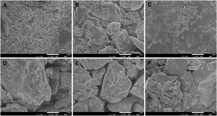
The morphology of Dio, Soluplus, PM and ASDs was examined using SEM, and Figure 8 illustrates the representative photographs. Dio powder appeared as rod-shaped or granular crystalline structure. Soluplus existed in an amorphous form. Dio in PM was still dispersed in the excipient by crystal structure. However, there was no Dio crystal structure in ASDs. This result further confirmed that Dio existed in the ASD system in a molecular or amorphous form and had a small specific surface area, thereby achieving a change in the dissolution rate.
Figure 8.
SEM photomicrographs of Dio (A), Soluplus (B), PM-1:10 (C), Dio-C-ASD (D), Dio-M-ASD (E) and Dio-F-ASD (F).
PXRD Measurements
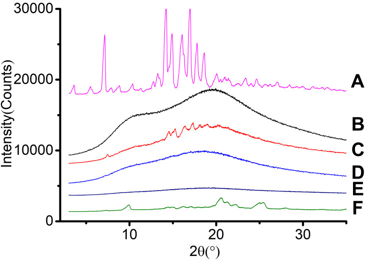
Figure 9 shows the PXRD images of Dio, Soluplus, PM and ASDs. Pure Dio showed sharp diffraction peaks within the range of 5°-20° at 2θ angles, at 7.14°, 14.22°, 14.90°, 16.08°, 16.98°, 17.78° and 18.64°, suggesting that Dio existed in a crystal form. When mixed with Soluplus, the interaction between Dio and excipient could cover the drug peak, thus reducing the intensity of the drug peak. However, all the major characteristic crystal peaks of Dio could be clearly observed in the PM diffractogram. The situation was changed when Dio was loaded into Soluplus using ASD strategy. The characteristic peaks of crystal Dio disappeared in Dio-C-ASD and Dio-M-ASD, indicating that the interaction between the excipient and Dio could effectively transform the crystal state of the drug into an amorphous state. Compared with the crystal Dio, the amorphous state of Dio with higher energy could be better wetted and released from the solid dispersion excipient. In addition, compared with the PM of Dio and Soluplus, ASD technology was more effective in enhancing the molecular interaction and performing function.33 However, some typical crystal peaks of Dio were clearly observed in the Dio-F-ASD diffractogram, indicating that some Dio did not form an amorphous state. This was confirmed by DSC test results. Combination of DSC, FT-IR and SEM results indicated that the Dio in the Dio-C-ASD and Dio-M-ASD was present in an amorphous form. Although Dio was amorphous in the Dio-C-ASD and Dio-M-ASD, the peak halos of the two methods were different from the X-ray. This indicated that the molecular structure of the whole system may be different in the different preparation processes.
Figure 9.
PXRD spectra of Dio (A), Soluplus (B), PM-1:10 (C), Dio-C-ASD (D), Dio-M-ASD (E) and Dio-F-ASD (F).
Molecular Docking
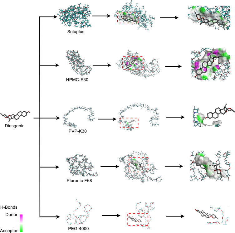
The molecular interactions between drug and excipients were studied by computer simulation. The optimization results of the complexes represented the most stable excipient conformation, which was the lowest total energy. Figure 10 describes the molecular docking results of Dio with excipients by showing intermolecular interaction or the hydrogen bond distribution at the interface. Obviously, due to the hydrogen bond forces (green-dotted line) and hydrophobic forces (pink-dotted line), a strong binding energy was formed between Dio and Soluplus. Only hydrogen bond force and no hydrophobic force were formed between Dio and HPMC-E30, resulting in its lower binding energy compared with Soluplus. No hydrogen bond force and hydrophobic force were formed between Dio and PVP-K30, Pluronic-F68 and PEG-4000, resulting in their extremely low binding energy. There was excellent agreement between these computation results and FT-IR.
Figure 10.
Molecular docking of Dio with excipients.
Generally speaking, excipients that form strong molecular interactions with Dio were very effective crystallization inhibitors, while weaker hydrogen bonding interactions were crystallization inhibitors with poor physical stability. In computer simulations, the binding energy (ΔG) can be used to assess the strength of interactions between Dio and excipients. Negative free energy (ΔG<0) indicates that the system is stable, whereas positive ΔG indicates the phase separation. The calculated binding energy values were Soluplus (−6.83 kcal/mol)>HPMC-E30 (−6.58 kcal/mol)>PVP-K30 (−6.54 kcal/mol)>Pluronic-F68 (−5.75 kcal/mol)>PEG-4000 (−5.62 kcal/mol). Therefore, Soluplus could form strong hydrogen bond interactions with Dio, which was consistent with the experimental results of crystal inhibition. The trend of binding affinity was consistent with the above-mentioned increase in stability.
In vivo Pharmacokinetic Results
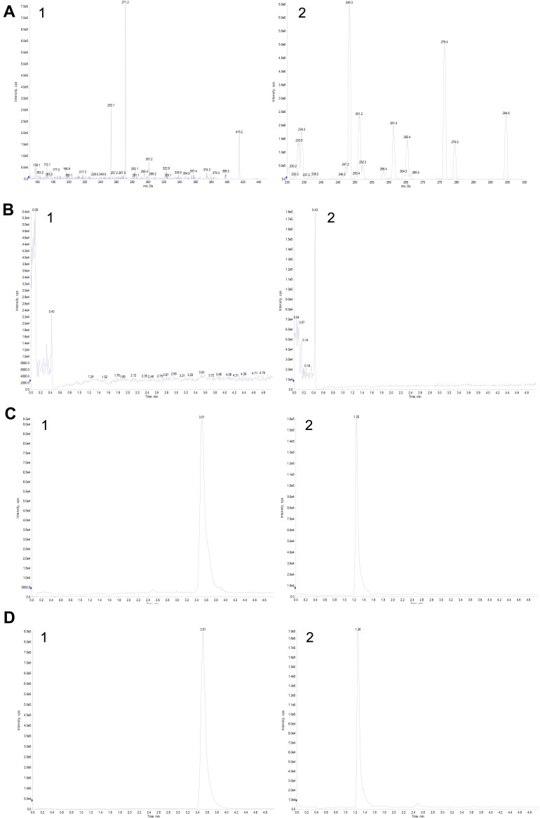
The blank plasma samples were analyzed to check their selectivity. Under LC-MS/MS conditions, no significant interference peaks of endogenous substances were observed in the Dio and IS retention regions. The results showed that the method had good selectivity. A typical chromatogram is shown in Figure 11.
Figure 11.
Mass spectra and representative chromatograms of Dio (1) and IS (2): (A) Mass spectra; (B) blank plasma; (C) blank plasma spiked with Dio and IS; and (D) a representative plasma sample 8 h after drug administration.
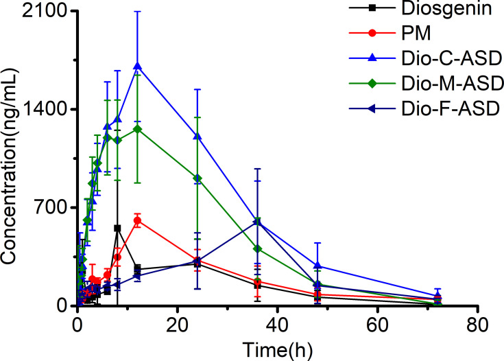
Figure 12 shows the mean plasma concentration–time curves following oral administration of ASDs and Dio. Table 2 lists the pharmacokinetic parameters. Significant enhancement in the in vivo performance was observed for Dio-C-ASD and Dio-M-ASD. The AUC0–t and Cmax of the Dio-C-ASD were 52,239.26±18,464.56 μg/L*h and 1742.65±491.46 μg/L, which were 5.69- and 3.15-fold higher compared with the Dio, respectively, yielding a relative bioavailability of 576.69%. Dio-M-ASD had a lower relative bioavailability than Dio-C-ASD with AUC0–t and Cmax of 40,727.418±1561.36 μg/L*h and 1443.50±511.96 μg/L, respectively. The Tmax of the Dio-C-ASD (11.00±2.45 h) and Dio-M-ASD (9.00±3.28 h) was little longer than that of Dio (8.50±5.44 h), while there was no significant difference. Soluplus might delay the absorption of Dio, while the effect was not obvious. The apparent volume of Dio-C-ASD and Dio-M-ASD distribution Vz/F and clearance rate CLz/F were significantly lower compared with Dio, with a 6.25-fold and 5.15-fold decrease in Vz/F and a 4.56-fold and 4.34-fold decrease in CLz/F, indicating that the retention time of Dio-C-ASD and Dio-M-ASD in vivo was significantly longer compared with the Dio. The result indicated that the bioavailability of Dio was greatly improved when it was prepared into ASD by co-precipitation and microwave quenching method. The dissolution rate, stability, morphology and particle size of ASDs prepared by different methods are often different.53,54 Karmwar et al55 found that although the drugs in the ASD prepared by the two methods were both amorphous, the molecular structure of the whole system was different, so the dissolution rate of the two methods was also different. Similar results were observed in this experiment. Although Dio existed in an amorphous state in both Dio-C-ASD and Dio-M-ASD, the dissolution and bioavailability of Dio were different, which may be caused by the different molecular structure of the system due to the different preparation processes. Compared with Dio, Dio-F-ASD had little improvement in bioavailability. According to DSC and PXRD results, Dio recrystallized during freeze-drying, and eventually still partially existed in the crystalline state, which may be the reason for its low bioavailability. At last, the formulation Dio-C-ASD was selected as it was found to be better than other ASD formulations for in vivo evaluation.
Figure 12.
Plasma concentration-time profiles following oral administration of ASDs and Dio n = 6.
Table 2.
Pharmacokinetic Parameters Following Oral Administration of ASDs and Dio (n = 6)
| Parameter | Dio | PM | Dio-C-ASD | Dio-M-ASD | Dio-F-ASD |
|---|---|---|---|---|---|
| AUC0-t(μg/L*h) | 9167.87±4168.42 | 11,037.87±7350.23 | 52,239.26±18,464.56** | 40,727.418±1561.36* | 14,631.44±4848.35 |
| AUC0-∞(μg/L*h) | 9275.51±4225.99 | 11,333.60±7529.67 | 53,490.51±19,648.62** | 41,535.50±1607.51* | 14,967.91±5146.81 |
| MRT0-t(h) | 21.67±4.66 | 22.68±4.32 | 21.83±3.30 | 20.22±2.68 | 22.22±7.24 |
| t1/2z(h) | 12.33±3.02 | 9.08±2.37* | 10.61±1.99 | 11.43±1.99* | 9.15±5.33* |
| Tmax(h) | 8.50±5.44 | 12.50±6.68 | 11.00±2.45 | 9.00±3.28 | 12.00±0.45 |
| CLz/F(L/h/kg) | 12.11±6.52 | 12.51±4.79 | 2.18±1.11** | 2.79±1.21* | 7.28±2.18* |
| Vz/F(L/kg) | 231.16±161.21 | 162.03±72.32 | 31.88±12.94** | 44.88±17.46* | 91.59±51.89* |
| Cmax(μg/L) | 552.96±294.54 | 581.03±83.30 | 1742.65±491.46** | 1443.50±511.96* | 689.776±140.79 |
Notes: Statistically significant compared with the Dio: *P<0.05, **P<0.01.
Stability Study
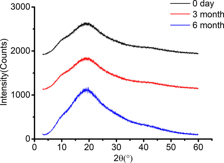
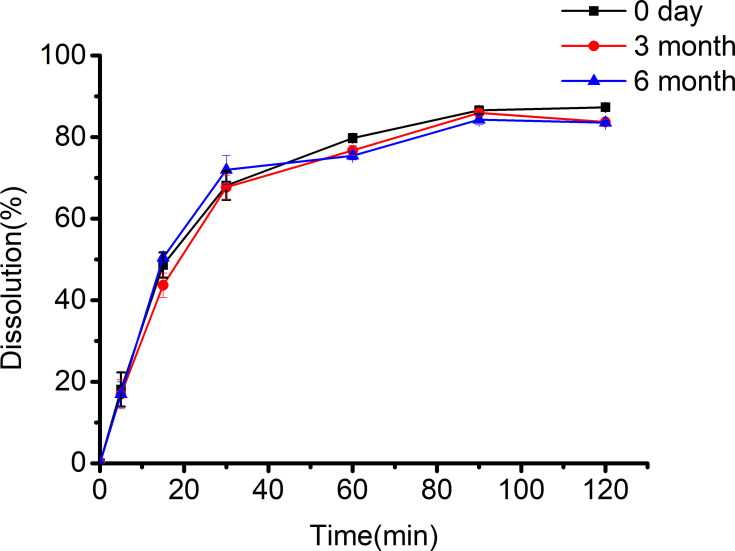
One problem with ASD is the possibility of crystallization of amorphous drugs during storage. Another problem is the effect of water on storage stability, because the presence of water may increase the fluidity of drugs and promote the drug crystallization.56 In this experiment, the stability of ASD samples was studied for 6 months. As shown in the PXRD diffraction peak diagram (Figure 13) that Dio-C-ASD after storage was similar to the initial systems and did not show any diffraction peak of Dio. The dissolution profile (Figure 14) of Dio-C-ASD at 40°C and 75% humidity for 6 months had no substantial differences compared with the fresh ASD. It was further confirmed that the amorphous drug did not crystallize in the ASD and had a good physical stability. Generally, excipients used at high concentrations stabilize the amorphous form of drugs by increasing the glass transition temperature (Tg) of the system, thereby reducing the molecular mobility of amorphous drugs in solid dispersion systems. Another important stabilization mechanism is that the crystallization inhabitation of amorphous drugs is attributed to the drug-excipient molecular interaction in the ASD.33,57 The strong intermolecular interactions, especially hydrogen bonding between Soluplus and Dio, might further lower the mobility of Dio molecular, and delay the recrystallization upon storage.
Figure 13.
PXRD spectra for Dio-C-ASD stored for 0 days, 3 months and 6 months.
Figure 14.
Dissolution profile of Dio in Dio-C-ASD stored for 0 days, 3 months and 6 months (n = 3).
Conclusions
In conclusion, among all the candidate excipients, Soluplus provided Dio with superior solubilization and stability in the test solution. With the ratio 1:10 Dio to Soluplus, co-precipitation, microwave quenching and freeze-drying methods of preparation, Dio-ASDs demonstrated significant improvement in solubility, which could be attributed to amorphization according to DSC, PXRD and SEM. FT-IR study indicated that hydrogen bond formed between the Dio and Soluplus. The use of molecular docking technique provided unique insights, revealing the drug-excipient interaction at a molecular level. In addition, pharmacokinetic studies in rats revealed that the bioavailability of Dio was greatly improved when it was prepared into ASDs. Besides, Dio-C-ASD could be stored at 40°C and 75% humidity for 6 months without changes, which was confirmed by the stability study. These above-mentioned results showed that ASD technology was an effective and promising strategy to enhance the oral bioavailability of Dio along with other poorly water-soluble and insoluble drugs.
Funding Statement
This study was financially supported by the Key Discipline Construction Projects of Higher School, Science and Technology Research Youth Fund Project of National Natural Science Foundation of China (No.81703736), Hebei Province Natural Science Foundation of China (No. H2018406038), Science and Technology Research Youth Fund Project of Higher School in Hebei Province (No. QN2015127), and Program for Innovative Talents of Higher Education of Liaoning (2012520005).
Disclosure
The authors declare that they have no conflicts of interest.
References
- 1.Tang YN, Pang YX, He XC, et al. UPLC-QTOF-MS identification of metabolites in rat biosamples after oral administration of Dioscorea saponins: a comparative study. J Ethnopharmacol. 2015;165:127–140. doi: 10.1016/j.jep.2015.02.017 [DOI] [PubMed] [Google Scholar]
- 2.Cheng X, Su X, Chen X, et al. Biological ingredient analysis of traditional Chinese medicine preparation based on high-throughput sequencing: the story for Liuwei Dihuang Wan. Sci Rep. 2014;4:5147. doi: 10.1038/srep05147 [DOI] [PMC free article] [PubMed] [Google Scholar]
- 3.Zhou W, Cheng X, Zhang Y. Effect of liuwei dihuang decoction, a traditional Chinese medicinal prescription, on the neuroendocrine immunomodulation network. Pharmacol Ther. 2016;162:170–178. doi: 10.1016/j.pharmthera.2016.02.004 [DOI] [PubMed] [Google Scholar]
- 4.Jia Y, Chen C, Ng CS, et al. Meta-analysis of randomized controlled trials on the efficacy of di’ao xinxuekang capsule and isosorbide dinitrate in treating angina pectoris. Evid Based Complement Alternat Med. 2012;904147. [DOI] [PMC free article] [PubMed] [Google Scholar]
- 5.Feng JF, Tang YN, Ji H, et al. Biotransformation of Dioscorea nipponica by rat intestinal microflora and cardioprotective effects of diosgenin. Oxid Med Cell Longev. 2017;4176518. [DOI] [PMC free article] [PubMed] [Google Scholar]
- 6.Wang WC, Liu SF, Chang WT, et al. The effects of diosgenin in the regulation of renal proximal tubular fibrosis. Exp Cell Res. 2014;323(2):255–262. doi: 10.1016/j.yexcr.2014.01.028 [DOI] [PubMed] [Google Scholar]
- 7.Manivannan J, Balamurugan E, Silambarasan T, et al. Diosgenin improves vascular function by increasing aortic eNOS expression, normalize dyslipidemia and ACE activity in chronic renal failure rats. Mol Cell Biochem. 2013;384(1–2):113–120. doi: 10.1007/s11010-013-1788-2 [DOI] [PubMed] [Google Scholar]
- 8.Frohnert BI, Bernlohr DA. Protein carbonylation, mitochondrial dysfunction, and insulin resistance. Adv Nutr. 2013;4(2):157–163. doi: 10.3945/an.112.003319 [DOI] [PMC free article] [PubMed] [Google Scholar]
- 9.Xiao L, Guo D, Hu C, et al. Diosgenin promotes oligodendrocyte progenitor cell differentiation through estrogen receptor-mediated ERK1/2 activation to accelerate remyelination. Glia. 2012;60(7):1037–1052. doi: 10.1002/glia.22333 [DOI] [PubMed] [Google Scholar]
- 10.Khosravi Z, Sedaghat R, Baluchnejadmojarad T, et al. Diosgenin ameliorates testicular damage in streptozotocin-diabetic rats through attenuation of apoptosis, oxidative stress, and inflammation. Int Immunopharmacol. 2019;70:37–46. doi: 10.1016/j.intimp.2019.01.047 [DOI] [PubMed] [Google Scholar]
- 11.Sultana N. Microbial biotransformation of bioactive and clinically useful steroids and some salient features of steroids and biotransformation. Steroids. 2018;136:76–92. doi: 10.1016/j.steroids.2018.01.007 [DOI] [PubMed] [Google Scholar]
- 12.Haratake A, Watase D, Setoguchi S, et al. Effect of orally ingested diosgenin into diet on skin collagen content in a low collagen skin mouse model and its mechanism of action. Life Sci. 2017;174:77–82. doi: 10.1016/j.lfs.2017.02.013 [DOI] [PubMed] [Google Scholar]
- 13.Quiñones JP, Iturmendi A, Henke H, et al. Polyphosphazene-based nanocarriers for the release of agrochemicals and potential anticancer drugs. J Mater Chem B. 2019;7(48):7783–7794. doi: 10.1039/C9TB01985E [DOI] [PubMed] [Google Scholar]
- 14.Qin Y, Wu X, Huang W, et al. Acute toxicity and sub-chronic toxicity of steroidal saponins from Dioscorea zingiberensis C.H. Wright in rodents. J Ethnopharmacol. 2009;126(3):543–550. doi: 10.1016/j.jep.2009.08.047 [DOI] [PubMed] [Google Scholar]
- 15.Lei G, Liu G, Ma J, et al. Application of drug nanocrystal technologies for oral drug delivery of poorly soluble drugs. Pharm Res. 2012;30(2):307–324. doi: 10.1007/s11095-012-0889-z [DOI] [PubMed] [Google Scholar]
- 16.Sugano K, Kansy M, Artursson P, et al. Coexistence of passive and carrier-mediated processes in drug transport. Nat Rev Drug Discov. 2010;9(8):597–614. doi: 10.1038/nrd3187 [DOI] [PubMed] [Google Scholar]
- 17.Rytting E, Lentz KA, Chen X, et al. Aqueous and cosolvent solubility data for drug-like organic compounds. AAPS J. 2005;7(1):E78–E105. doi: 10.1208/aapsj070110 [DOI] [PMC free article] [PubMed] [Google Scholar]
- 18.Okawara M, Hashimoto F, Todo H, Sugibayashi K, Tokudome Y. Effect of liquid crystals with cyclodextrin on the bioavailability of a poorly water-soluble compound, diosgenin, after its oral administration to rats. Int J Pharm. 2014;472(1–2):257–261. doi: 10.1016/j.ijpharm.2014.06.032 [DOI] [PubMed] [Google Scholar]
- 19.Okawara M, Tokudome Y, Todo H, et al. Effect of β-cyclodextrin derivatives on the diosgenin absorption in caco-2 cell monolayer and rats. Biol Pharm Bull. 2014;37(1):54–59. doi: 10.1248/bpb.b13-00560 [DOI] [PubMed] [Google Scholar]
- 20.Liu CZ, Chang J, Zhang L, et al. Preparation and evaluation of diosgenin nanocrystals to improve oral bioavailability. AAPS PharmSciTech. 2017;18(6):2067–2076. doi: 10.1208/s12249-016-0684-y [DOI] [PubMed] [Google Scholar]
- 21.Li B, Konecke S, Wegiel LA, et al. Both solubility and chemical stability of curcumin are enhanced by solid dispersion in cellulose derivative matrices. Carbohydr Polym. 2013;98(1):1108–1116. doi: 10.1016/j.carbpol.2013.07.017 [DOI] [PubMed] [Google Scholar]
- 22.Xiong XN, Zhang M, Hou Q, et al. Solid dispersions of telaprevir with improved solubility prepared by co-milling: formulation, physicochemical characterization, and cytotoxicity evaluation. Mater Sci Eng C Mater Biol Appl. 2019;105:0928–4931. doi: 10.1016/j.msec.2019.110012 [DOI] [PubMed] [Google Scholar]
- 23.França MT, O’Reilly Beringhs A, Nicolay Pereira R, et al. The role of sodium alginate on the supersaturation state of the poorly soluble drug chlorthalidone. Carbohydr Polym. 2019;209:207–214. doi: 10.1016/j.carbpol.2019.01.007 [DOI] [PubMed] [Google Scholar]
- 24.Price DJ, Ditzinger F, Koehl NJ, et al. Approaches to increase mechanistic understanding and aid in the selection of precipitation inhibitors for supersaturating formulations-a PEARRL review. J Pharm Pharmacol. 2019;71(4):483–509. doi: 10.1111/jphp.12927 [DOI] [PubMed] [Google Scholar]
- 25.França MT, Nicolay RP, Riekes MK, et al. Investigation of novel supersaturating drug delivery systems of chlorthalidone: the use of polymer-surfactant complex as an effective carrier in solid dispersions. Eur J Pharm Sci. 2018;533(1):266. [DOI] [PubMed] [Google Scholar]
- 26.Andreas S, Jorg Hand Maxim P. Mechanisms of increased bioavailability through amorphous solid dispersions: a review. Drug Deliv. 2020;27(1):110–127. doi: 10.1080/10717544.2019.1704940 [DOI] [PMC free article] [PubMed] [Google Scholar]
- 27.Boel E, Smeets A, Vergaelen M, et al. Comparative study of the potential of poly (2- ethyl −2- oxazoline) as carrier in the formulation of amorphous solid dispersions of poorly soluble drugs. Eur J Pharm Biopharm. 2019;144:79–90. doi: 10.1016/j.ejpb.2019.09.005 [DOI] [PubMed] [Google Scholar]
- 28.Shamma RN, Basha M. Soluplus: a novel polymeric solubilizer for optimization of carvedilol solid dispersions: formulation design and effect of method of preparation. Powder Technol. 2013;237:406–414. doi: 10.1016/j.powtec.2012.12.038 [DOI] [Google Scholar]
- 29.Punčochová K, Vukosavljevic B, Hanuš J, et al. Non-invasive insight into the release mechanisms of a poorly soluble drug from amorphous solid dispersions by confocal Raman microscopy. Eur J Pharm Biopharm. 2016;101:119–125. doi: 10.1016/j.ejpb.2016.02.001 [DOI] [PubMed] [Google Scholar]
- 30.Shi X, Xu T, Huang W, et al. Stability and bioavailability enhancement of telmisartan ternary solid dispersions: the synergistic effect of polymers and drug-polymer(s) interactions. AAPS PharmSciTech. 2019;20(4):143. doi: 10.1208/s12249-019-1358-3 [DOI] [PubMed] [Google Scholar]
- 31.Rani S, Mishra S, Sharma M, et al. Solubility and stability enhancement of curcumin in Soluplus ® polymeric micelles: a spectroscopic study. J Disper Sci Technol. 2020;41(4):523–536. doi: 10.1080/01932691.2019.1592687 [DOI] [Google Scholar]
- 32.Singh J, Mittal P, Vasnt B, et al. Design, optimization, characterization and in-vivo evaluation of quercetin enveloped soluplus®/P407 micelles in diabetes treatment. Artif Cell Nanomed B. 2018;46(3):546–555. doi: 10.1080/21691401.2018.1501379 [DOI] [PubMed] [Google Scholar]
- 33.Song B, Wang J, Lu SJ, et al. Andrographolide solid dispersions formulated by soluplus to enhance interface wetting, dissolution, and absorption. J Appl Polym Sci. 2020;137(6):48354. doi: 10.1002/app.48354 [DOI] [Google Scholar]
- 34.Abhijeet DK, Veena SB. Influence of novel carrier soluplus® on aqueous stability, oral bioavailability, and anticancer activity of Morin hydrate. Dry Technol. 2019;37(9):1143–1161. doi: 10.1080/07373937.2018.1488261 [DOI] [Google Scholar]
- 35.Shi NQ, Lei YS, Song LM, et al. Impact of amorphous and semicrystalline polymers on the dissolution and crystallization inhibition of pioglitazone solid dispersions. Powder Technol. 2013;247:211–221. doi: 10.1016/j.powtec.2013.06.039 [DOI] [Google Scholar]
- 36.Qian F, Wang J, Hartley R, et al. Solution behavior of PVP-VA and HPMC-AS-based amorphous solid dispersions and their bioavailability implications. Pharm Res. 2012;29(10):2765–2776. doi: 10.1007/s11095-012-0695-7 [DOI] [PubMed] [Google Scholar]
- 37.Pas T, Bergonzi A, Lescrinier E, et al. Drug-carrier binding and enzymatic carrier digestion in amorphous solid dispersions containing proteins as carrier. Int J Pharm. 2019;563:358–372. doi: 10.1016/j.ijpharm.2019.03.062 [DOI] [PubMed] [Google Scholar]
- 38.Sun DD, Lee PI. Crosslinked hydrogels-a promising class of insoluble solid molecular dispersion carriers for enhancing the delivery of poorly soluble drugs. Acta Pharm Sin B. 2014;4(1):26–36. doi: 10.1016/j.apsb.2013.12.002 [DOI] [PMC free article] [PubMed] [Google Scholar]
- 39.Chavan RB, Rathi S, Shastri NR. Cellulose based polymers in development of amorphous solid dispersions. Asian J Pharm Sci. 2019;3:248–264. doi: 10.1016/j.ajps.2018.09.003 [DOI] [PMC free article] [PubMed] [Google Scholar]
- 40.Bevernage J, Forier T, Brouwers J, et al. Excipient-mediated supersaturation stabilization in human intestinal fluids. Mol Pharm. 2011;8(2):564–570. doi: 10.1021/mp100377m [DOI] [PubMed] [Google Scholar]
- 41.Yamashita T, Ozaki S, Kushida I. Solvent shift method for anti-precipitant screening of poorly soluble drugs using biorelevant medium and dimethyl sulfoxide. Int J Pharm. 2011;419(1–2):170–174. doi: 10.1016/j.ijpharm.2011.07.045 [DOI] [PubMed] [Google Scholar]
- 42.Sun M, Wu C, Fu Q, et al. Solvent-shift strategy to identify suitable polymers to inhibit humidity-induced solid-state crystallization of lacidipine amorphous solid dispersions. Int J Pharm. 2016;503(1–2):238–246. doi: 10.1016/j.ijpharm.2016.01.062 [DOI] [PubMed] [Google Scholar]
- 43.Everaerts M, Van den Mooter G. Complex amorphous solid dispersions based on poly (2 - hydroxyethyl methacrylate): study of drug release from a hydrophilic insoluble polymeric carrier in the presence and absence of a porosity increasing agent. Int J Pharm. 2019;566:77–88. doi: 10.1016/j.ijpharm.2019.05.040 [DOI] [PubMed] [Google Scholar]
- 44.Peng R, Huang J, He L, et al. Polymer/lipid interplay in altering in vitro supersaturation and plasma concentration of a model poorly soluble drug. Eur J Pharm Sci. 2020;146:105262. doi: 10.1016/j.ejps.2020.105262 [DOI] [PubMed] [Google Scholar]
- 45.Vasconcelos T, Marques S, Neves J, Sarmento B. Amorphous solid dispersions: rational selection of a manufacturing process. Adv Drug Deliv Rev. 2016;100:85–101. doi: 10.1016/j.addr.2016.01.012 [DOI] [PubMed] [Google Scholar]
- 46.Shi NQ, Wang SR, Zhang Y, et al. Hot melt extrusion technology for improved dissolution, solubility and “spring-parachute” processes of amorphous self-micellizing solid dispersions containing BCS II drugs indomethacin and fenofibrate: profiles and mechanisms. Eur J Pharm Sci. 2019;130:78–90. doi: 10.1016/j.ejps.2019.01.019 [DOI] [PubMed] [Google Scholar]
- 47.Zi P, Zhang C, Ju C, et al. Solubility and bioavailability enhancement study of lopinavir solid dispersion matrixed with a polymeric surfactant - soluplus. Eur J Pharm Sci. 2019;134:233–245. doi: 10.1016/j.ejps.2019.04.022 [DOI] [PubMed] [Google Scholar]
- 48.Trivino A, Gumireddy A, Meng F, et al. Drug–polymer miscibility, interactions, and precipitation inhibition studies for the development of amorphous solid dispersions for the poorly soluble anticancer drug flutamide. Drug Dev Ind Pharm. 2019;45(8):1277–1291. doi: 10.1080/03639045.2019.1606822 [DOI] [PubMed] [Google Scholar]
- 49.Llinàs A, Goodman JM. Polymorph control: past, present and future. Drug Discov Today. 2008;13(5–6):198–210. [DOI] [PubMed] [Google Scholar]
- 50.Gong N, Wang Y, Zhang B, et al. Screening, preparation and characterization of diosgenin versatile solvates. Steroids. 2019;143:18–24. doi: 10.1016/j.steroids.2018.11.016 [DOI] [PubMed] [Google Scholar]
- 51.Yang HY, Zhou ZL, Zhou XP, et al. Preparation and properties of sulfadiazine solid dispersion. Chin Pharm J. 2017;52(9):750–754. [Google Scholar]
- 52.Riekes MK, Kuminek G, Rauber GS, et al. HPMC as a potential enhancer of nimodipine biopharmaceutical properties via ball-milled solid dispersions. Carbohydr Polym. 2014;99:474–482. doi: 10.1016/j.carbpol.2013.08.046 [DOI] [PubMed] [Google Scholar]
- 53.Agrawal AM, Dudhedia MS, Patel AD, et al. Characterization and performance assessment of solid dispersions prepared by hot melt extrusion and spray drying process. Int J Pharm. 2013;457(1):71–81. doi: 10.1016/j.ijpharm.2013.08.081 [DOI] [PubMed] [Google Scholar]
- 54.Josimar OE, Juliana MM. Solid dispersions containing ursolic acid in poloxamer 407 and PEG 6000: a comparative study of fusion and solvent methods. Powder Technol. 2014;253:98–106. doi: 10.1016/j.powtec.2013.11.017 [DOI] [Google Scholar]
- 55.Karmwar P, Graeser K, Gordon KC. Effect of different preparation methods on the dissolution behaviour of amorphous indomethacin. Eur J Pharm Biopharm. 2012;80(2):459–464. doi: 10.1016/j.ejpb.2011.10.006 [DOI] [PubMed] [Google Scholar]
- 56.Vasanthavada M, Tong W, Joshi Y, et al. Phase behavior of amorphous molecular dispersions i: determination of the degree and mechanism of solid solubility. Pharm Res. 2005;21(9):1598–1606. doi: 10.1023/B:PHAM.0000041454.76342.0e [DOI] [PubMed] [Google Scholar]
- 57.Ahmad N, Ahmad R, Alam MA, et al. Daunorubicin oral bioavailability enhancement by surface coated natural biodegradable macromolecule chitosan based polymeric nanoparticles. Int J Biol Macromol. 2019;128:825–838. doi: 10.1016/j.ijbiomac.2019.01.142 [DOI] [PubMed] [Google Scholar]


