

We read with interest the letter by Garcia and colleagues, as well as their recent publication assessing MET amplification in the anaplastic thyroid cancer (ATC)-derived TTA-1 cell line (1). In their correspondence, the authors asked whether MET amplification was observed in TTA-1 cells in our study (2). We retrieved the specific data for MET in TTA-1 and compared it with other cell lines. As shown in Fig. 1A, TTA-1 was the only cell line displaying a focal MET amplification, which correlated with the highest mRNA expression levels (Fig. 1B). Furthermore, we looked at our unpublished data assessing phospho-protein levels in these specimens, showing that phospho-MET is significantly elevated in TTA-1 (Fig. 1C). We studied the earliest TTA-1 passage available in our laboratory and observed a MET copy number fold change = 11.1, whereas Garcia and colleagues reported 20 copies. These differences in copy number magnitude are likely attributable to the methods used for copy number calling (targeted sequencing vs. FISH) and, as the authors pointed out, to some degree of clonal evolution during passaging of cultured cells. Although we did not have access to the original tumor from which this cell line was derived 25 years ago (3), the nonoverlapping pattern of occurrence of MET amplification with other known thyroid cancer driver genes, the signaling consequences of this genetic event, along with our recent observation of Met amplification as a mechanism of resistance to Braf inhibition in murine ATCs (4), strongly suggests that MET activation is a genetic driver in the TTA-1 cell line, and perhaps in a subset of ATCs. To our knowledge, this is the first example of a thyroid cancer cell line driven by an oncogenic amplification event.
Figure 1.
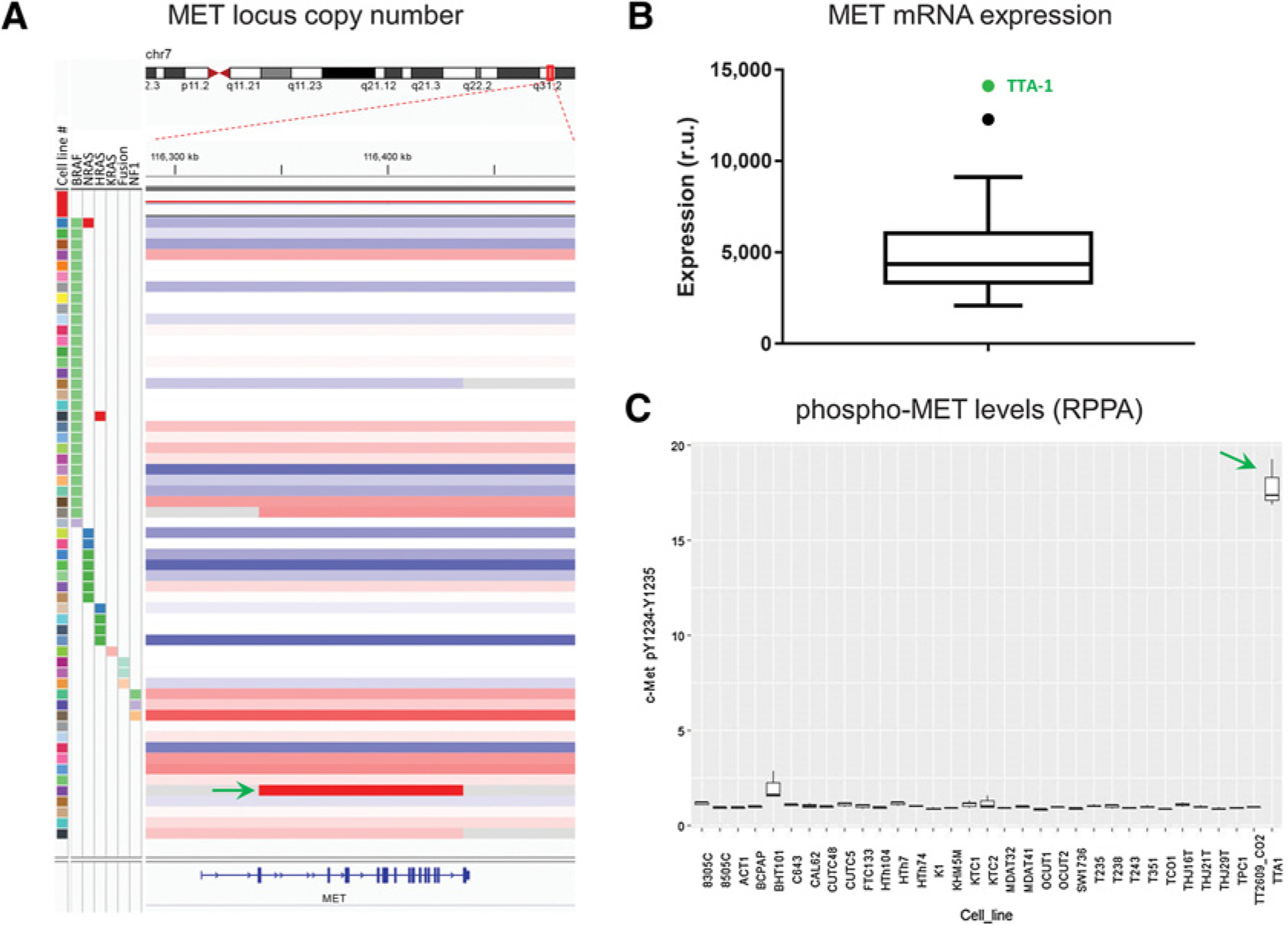
MET status in 58 thyroid cancer cell lines. A, Copy number alteration around MET locus for all cell lines, sorted by genetic driver (left). Copy number changes are expressed as shades of red (gain) or blue (loss). TTA-1 cell line (green arrow) displays a focal copy number amplification (log ratio = 3.5; fold change = 11.1; P < 0.001). B, Tukey boxplot showing relative levels of MET mRNA expression. TTA-1 cell line (green dot) is an outlier and displays the highest expression of this dataset. C, Levels of phospho-MET (pY1234-Y1235) evaluated by reverse phase protein array (RPPA), with TTA-1 showing the highest values.
Footnotes
Disclosure of Potential Conflicts of Interest
No potential conflicts of interest were disclosed.
References
- 1.Garcia C, Buffet C, El Khattabi L, Rizk-Rabin M, Perlemoine K, Ragazzon B, et al. MET overexpression and activation favors invasiveness in a model of anaplastic thyroid cancer. Oncotarget 2019;10:2320–34. [DOI] [PMC free article] [PubMed] [Google Scholar]
- 2.Landa I, Pozdeyev N, Korch C, Marlow LA, Smallridge RC, Copland JA, et al. Comprehensive genetic characterization of human thyroid cancer cell lines: a validated panel for preclinical studies. Clin Cancer Res 2019;25:3141–51. [DOI] [PMC free article] [PubMed] [Google Scholar]
- 3.Yoshida A, Asaga T, Masuzawa C, Kawahara S, Yanoma S, Harada M, et al. Production of cytokines by thyroid carcinoma cell lines. J Surg Oncol 1994; 55:104–7. [DOI] [PubMed] [Google Scholar]
- 4.Knauf JA, Luckett KA, Chen KY, Voza F, Socci ND, Ghossein R, et al. Hgf/Met activation mediates resistance to BRAF inhibition in murine anaplastic thyroid cancers. J Clin Invest 2018;128:4086–97. [DOI] [PMC free article] [PubMed] [Google Scholar]