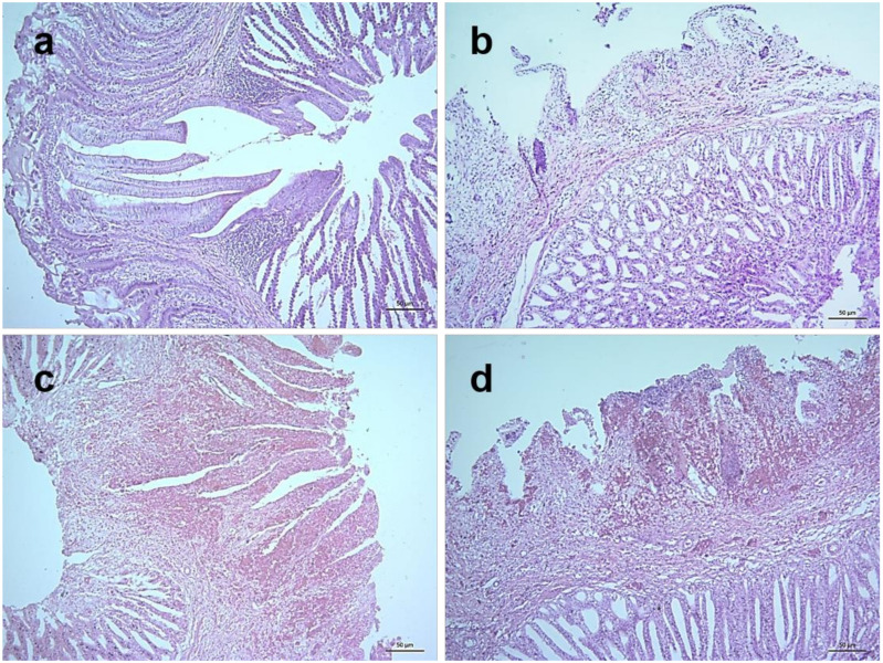
Figure 7.
Histopathological changes in the proventriculus of NDV infected chickens. Section of the proventriculus of control chickens showing normal histology (a). Sections of the proventriculus of infected chickens showing slight congestion and edema at 3 dpi (b), severe hemorrhages in the finger-like mucosal folds (plicae), edema and mild infiltration of inflammatory cells at 4 dpi (c), and severe hemorrhages and necrosis in plicae, loss of lining epithelium, fusion and shortening of plicae and infiltration of large number of inflammatory cells at 5 dpi (d). H&E stain, bar indicates magnification.

