

ABSTRACT
Background:
Since the introduction of digitization in cephalometrics, orthodontics has experienced a new horizon. Technological advancement is usually followed by comparisons between the methods.
Aims:
The aim of this study was to compare values of cephalometric analysis performed by CephNinja and NemoCeph for Downs’s analysis.
Settings and Design:
This prospective study was conducted using 100 diagnostic digital lateral cephalograms taken from the same machine. The samples were collected by non-probability convenience sampling procedures.
Materials and Methods:
The diagnostic images were cropped to standard lateral cephalogram film dimension; a scale image was placed on the top for calibration, numbered 1–100 for identification and was saved in Joint Photographic Experts Group (JPEG) format. A laptop with mouse-controlled cursor was used for NemoCeph and an android phone controlled with finger touch screen was used for CephNinja. Landmark identification for cephalometric analysis was carried out as demanded by the software.
Statistical Analysis Used:
One-way analysis of variance (ANOVA) was used for comparison between the variables, and one-way ANOVA followed by post hoc test was carried out to check the level of significance using Statistical Package for the Social Sciences software program, version 11.0.
Results:
The result showed that the difference of mean values obtained using the two software showed no statistical significance for 70% variables. Y-axis, incisor occlusal plane angle, and the upper incisor to A-Pog showed a statistically significant difference.
Conclusion:
CephNinja presented a satisfactory result with NemoCeph, and can be used interchangeably with confidence.
KEYWORDS: Cephalometry, digital image, orthodontics, software
INTRODUCTION
Radiograph originally developed as research laboratory tool has become a diagnostic necessity in orthodontics.[1] Cephalometric radiographs made a paradigm shift in clinical orthodontics and research.[2,3] William Downs (1948) is credited with developing the first cephalometric analysis. With time, several cephalometric analyses and population-specific cephalometric norms were provided for clinical use.[4]
The standards of facial profile vary with races, places, and time. The morphological features are ethnic characteristics too.[4] This variation leads to the need for different cephalometric norms for individual population group.[5,6,7,8]
Traditional cephalometric analysis technique has many inherent disadvantages such as time-consuming, tedious, large inventory, archiving records, communication of data, and associated chemical hazard.[4] Recently, computer technology has enabled digital processing and on-screen cephalometric tracing.[3] Computerized cephalometrics has gained popularity because of simplicity, quick, precise, and easy archiving. Facility of resolution enhancement adds to the accuracy of digitization.[9] The accuracy of NemoCeph comparable to hand-tracing has already been established.
The time-consuming nature of conventional tracing and the high license cost of cephalometric software with overall bulk of armamentarium refrain the orthodontists from instant cephalometric reading, especially for outstation consultation. A cost-effective and portable alternative for daily use was much awaited to serve instantaneous cephalometric reading.
Android phone is the most common electronic gadget globally and recently CephNinja cephalometric analysis software for android is gaining popularity because of its time-saving nature and free license. The evolution of technology demands its comparison with the existing. Therefore, the aim of this study was to compare and evaluate the measurement obtained using CephNinja for android and NemoCeph for computers for Downs’s cephalometric analysis. So that, if found reliable CephNinja could be used as a user-friendly and portable alternative for cephalometric analysis with the result in no time.
MATERIALS AND METHODS
STUDY DESIGN
This prospective study was conducted over a period of 2 years at Pacific Dental College, Udaipur and Vardhman Institute of Medical Sciences, Pawapuri. The study was approved by university research committee of Pacific Academy of Higher Education and Research. The routine pretreatment diagnostic lateral cephalograms of 100 (male = 45 and female = 55) [Table 1] prospective orthodontic patients who reported for orthodontic treatment from January 2017 to December 2018 were evaluated for cephalometric analysis. The radiographs were taken by the same operator and the same digital OPG (orthopantomogram) machine (Model- OPTON 2004, Tonisha Electronic Company, New Delhi, India) was used to capture all the images. This study sample used routine diagnostic pretreatment radiographs of the subjects, where no radiographs were taken without a reason and no subjects’ personal data were disclosed. Therefore, ethical committee clearance was not required.
Table 1.
Gender distribution
| Gender wise distribution of radiographs | Male | Female | Total |
|---|---|---|---|
| 45 | 55 | 100 |
SAMPLING CRITERIA
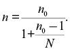
The samples were selected randomly through a non-probability convenience sampling procedures. The sample size was calculated using modified Cochran’s formula as follows:
The samples were selected based on quality of image by expert orthodontists who were blinded for the study. This study evaluated the accuracy of two software for cephalometric analysis. As this was not a follow-up study, so no dropout was noted.
Head position, age gender, and type of malocclusion did not affect the selection criteria.
INCLUSION CRITERIA
The inclusion criteria of the study were as follows:
Radiograph of subjects with permanent set of dentition
Good-quality radiographs with clarity of images
Radiographs with easy landmark identification
Cephalograms with good contrast and sharp-edge images
EXCLUSION CRITERIA
The exclusion criteria of the study were as follows:
Radiographs of subjects with missing upper and/or lower first molars and incisors
Radiographs with poor-quality image
Radiographs with distorted image
Radiographs with artefact
Subjects with craniofacial anomalies
STUDY METHOD
All the pretreatment diagnostic radiographs evaluated in this prospective study were taken using the same digital OPG machine (Model-OPTON 2004, Tonisha Electronic Company) at 65–70 kVp and automatic Milliampere setting. The selected digital lateral cephalograms were standardized and calibrated for uniformity before the commencement of study. All the radiographs were resized to 8 × 10 inch (i.e., equivalent to lateral head film size) for standardization, and a scale image was placed on top of this image for calibration. The digital images were further numbered for identification. Adobe Photoshop software, Version 7.0 (Adobe Systems Inc., CA, USA) was used for this purpose and the final image was saved in Joint Photographic Experts Group (JPEG) format.
OBSERVATIONAL PARAMETERS
The same computer was installed with NemoCeph NX 2009 for Windows (Nemotec, Madrid, Spain). The saved images were evaluated for Downs’s cephalometric analysis using this software. Mouse was used for marking the landmarks. The final cephalometric analysis was saved in the same computer.
One set of these pre-standardized and pre-calibrated cephalometric images were transferred to the android mobile phone using the data cable. CephNinja software (free) was downloaded and installed in the same android mobile phone. The radiographs were opened in the software for Downs’s analysis and the landmarks were identified and marked on screen using finger by the same operator as prompted by the software.
All the radiographs were evaluated by the same examiner and only 5 radiographs were evaluated per session to minimize error. An interval of 24h was set between the two sessions to avoid operator fatigue. The total duration of this prospective study was 2 years including sample collection, cephalometric analysis, and data interpretation. The images were calibrated using the ruler scale image placed on the top of each image during pre-standardization and pre-calibration. Contrast and image enhancement key was used in both the software when required for ease of landmark identification.
STATISTICAL ANALYSIS
The data were tabulated, mean values were calculated for all the variables, and were subjected to statistical test. One-way analysis of variance (ANOVA) was used for comparison between the variables, and one-way ANOVA followed by post hoc test was carried out to check the level of significance using Statistical Package for the Social Sciences software program, version 11.0 (SPSS, Chicago, Illinois).
RESULTS
This study evaluated 100 diagnostic lateral cephalograms of prospective orthodontic patients. Downs advocated the first cephalometric analysis and is still considered gold standard for clinical use owing to its precise and complete reflection of skeletal and dental pattern. The initial studies have established the reliability of NemoCeph for cephalometric analysis with the values comparable to hand-tracing. Therefore, the pre-standardized and pre-calibrated digital lateral cephalometric radiographs evaluated for parameters of Downs’s analysis using the two software, that is, CephNinja and NemoCeph . The values obtained using the CephNinja software for android were compared with the values of NemoCeph software for computers.
The difference of mean values obtained using the two software was comparable clinically in majority and showed no statistical significance for facial angle, angle of convexity, A–B plane angle, mandibular plane angle, cant of occlusal plane, inter incisal angle, and incisor mandibular plane angle at P ≤ 0.5 significant.
The remaining 30% variables, that is, Y-axis, incisor occlusal plane angle, and the upper incisor to A-Pog, showed a significant difference for the readings observed using the two software [Table 2].
Table 2.
Comparison of Downs’s analysis values between groups (one-way analysis of variance)
| Variable | Group | N | Mean ± SD | F-statistics | P value | 95% Confidence interval | ||
|---|---|---|---|---|---|---|---|---|
| Lower | Upper | |||||||
| Facial angle | CephNinja | 100 | 86.14 ± 6.20 | 0.10 | 0.90 | No significance | 84.18 | 88.10 |
| NemoCeph | 100 | 85.78 ± 6.11 | 83.85 | 87.71 | ||||
| Angle of convexity | CephNinja | 100 | 9.99 ± 7.00 | 0.90 | 0.41 | No significance | 7.78 | 12.20 |
| NemoCeph | 100 | 10.20 ± 7.17 | 7.93 | 12.46 | ||||
| A–B plane angle | CephNinja | 100 | 8.71 ± 5.14 | 0.69 | 0.50 | No significance | 7.09 | 10.34 |
| NemoCeph | 100 | 8.93 ± 4.78 | 7.42 | 10.44 | ||||
| Mandibular plane angle | CephNinja | 100 | 24.73 ± 8.63 | 0.58 | 0.56 | No significance | 22.00 | 27.45 |
| NemoCeph | 100 | 23.17 ± 8.78 | 20.40 | 25.94 | ||||
| Y-axis | CephNinja | 100 | 55.10 ± 11.36 | 255.23 | 0.0001 | Significance | 51.52 | 58.69 |
| NemoCeph | 100 | 92.00 ± 6.25 | 90.03 | 93.97 | ||||
| Cant of occlusal plane | CephNinja | 100 | 7.98 ± 5.58 | 2.26 | 0.11 | No significance | 6.22 | 9.74 |
| NemoCeph | 100 | 9.59 ± 5.96 | 7.70 | 11.47 | ||||
| Inter incisal angle | CephNinja | 100 | 118.73 ± 12.16 | 0.32 | 0.72 | No significance | 114.89 | 122.57 |
| NemoCeph | 100 | 120.44 ± 11.58 | 116.78 | 124.09 | ||||
| Incisor occlusal plane angle | CephNinja | 100 | 30.95 ± 7.83 | 321.31 | 0.0001 | Significance | 28.48 | 33.42 |
| NemoCeph | 100 | 25.78 ± 7.40 | 23.44 | 28.12 | ||||
| Incisor mandibular plane angle | CephNinja | 100 | 102.94 ± 6.46 | 0.41 | 0.67 | No significance | 100.90 | 104.98 |
| NemoCeph | 100 | 101.37 ± 8.73 | 98.61 | 104.12 | ||||
| U1 to A-Pog (linear) | CephNinja | 100 | 3.46 ± 1.35 | 175.32 | 0.0001 | Significance | 3.04 | 3.89 |
| NemoCeph | 100 | 0.36 ± 0.13 | 0.32 | 0.40 | ||||
SD = standard deviation
*P ≤ 0.5 significant
A further post hoc test showed the same result [Table 3].
Table 3.
Comparison of Downs’s analysis values between groups (post hoc test)
| Dependent variable | Group | Group | Mean difference | 95% Confidence interval | P value | |
|---|---|---|---|---|---|---|
| Lower limit | Upper limit | |||||
| Facial angle | NemoCeph | CephNinja | –0.36 | –3.92 | 3.20 | 0.97 |
| Angle of convexity | NemoCeph | CephNinja | 0.20 | –3.37 | 3.78 | 0.99 |
| A–B plane angle | NemoCeph | CephNinja | 0.21 | –2.28 | 2.71 | 0.98 |
| Mandibular plane angle | NemoCeph | CephNinja | –1.55 | –6.07 | 2.96 | 0.69 |
| Y-axis | NemoCeph | CephNinja | 36.90 | 32.55 | 41.24 | 0.0001 |
| Cant of occlusal plane | NemoCeph | CephNinja | 1.61 | –1.29 | 4.51 | 0.39 |
| Inter incisal angle | NemoCeph | CephNinja | 1.71 | –4.36 | 7.77 | 0.78 |
| Incisor occlusal plane angle | NemoCeph | CephNinja | –5.17 | –8.89 | –1.45 | 0.0001 |
| Incisor mandibular plane angle | NemoCeph | CephNinja | –1.58 | –5.81 | 2.66 | 0.65 |
| U1 to A-Pog (linear) | NemoCeph | CephNinja | –3.10 | –4.29 | –1.92 | 0.0001 |
| Incisor mandibular plane angle | NemoCeph | CephNinja | –1.55 | –5.81 | 2.71 | 0.66 |
*P ≤ 0.5 significant
DISCUSSION
Cephalometry characterizes the craniofacial skeleton metrically and geometrically. A complete cephalometric analysis is the key for orthodontic diagnosis, treatment planning, and postoperative follow-up. Broadbent, Brodie, and Hofrath provided a new platform for researchers, which subsequently helped in designing a number of different analyses and norms for clinical application.[3,4]
Traditional cephalometric analysis posed many inherent limitations and therefore causes hindrance to its regular practice.[9,10] Digitization of orthodontic office has overcome the limitations of conventional method.
Literature search for this study revealed that digital image of conventional radiography has proved to be a boon. The initial studies focused on testing the accuracy of scanned image of conventional film. The study result of Ganna et al.[11] has proved that scanned lateral cephalogram is preferred alternative to digital image.
It is evident from the subsequent studies that the computerized software programs are less time-consuming compared to hand-tracing.[12,13]
Computer programs are also proven to be reliable and an excellent alternative to conventional method.[14] The study of Iacob et al.[15] reported values comparable to manual and clinically acceptable. In contrast, Eslamian et al.[16] suggested that manual technique is still efficacious. Similarly, Agrawal et al.[10] in their study using CADCAS software for landmark identification reported these software to be unreliable for bilateral structures.
More recently, researchers have compared and evaluated different software programs specific for cephalometric analysis. A comparative study of three software (Planmeca Romexis, Orthalis and AxCeph) by Rusu et al.[17] reported a reliable result with no statistical significant difference.
Although the cephalometric analysis software has proven to be reliable with accuracy comparable to manual technique in majority, the availability and affordability of these commercially available software remained a problem for routine clinical use in the developing country and for the budding orthodontist. The requirement of some instant cephalometric values especially in consultation practice is among the other necessity that remains unfulfilled.
It is the time demand which calls for a cephalometric analysis software that is affordable, quick, and available to the orthodontist anytime and anyplace without an additional monetary and inventory burden. Therefore, this study compared and evaluated the CephNinja for android and NemoCeph for computers for Downs’s cephalometric analysis. The hypothesis to be tested was the readings available using the two software will be comparable clinically and there will be no statistically significant difference between the two.
This study analyzed 10 parameters of the Downs’s analysis by using the two software. The difference of mean values of the observed data using the NemoCeph and CephNinja software was 1°–2° for 70% of the variables and was comparable clinically with no statistically significant difference between the two. This was in consonance with the previous results of Rusa et al.,[17] Nouri et al., [18] and Kumar et al.[19] On the contrary, the mean differences of Y-axis and incisor occlusal plane angle using the two software were 37° and 6°, respectively. This mean difference showed a statistically significant difference. This could be due to difficulty in locating midface structures. The upper incisor to A-Pog (linear) again showed a statistically significant difference between the mean values obtained using the software. A similar difference for Nasion - Point A (Linear) was reported by Correia et al.[20] On the contrary, Tikku et al.[21] in their study comparing NemoCeph with manual also noted a statistically significant difference for L1-NB, but was comparable clinically.
The result of this study showed comparable results for the parameters of Downs’s cephalometric analysis using the two software in majority. Considering the 70% reliability, CephNinja could be considered as a good alternative to the commercially available cephalometric analysis software (NemoCeph). Therefore, CephNinja software can be used alternatively with equivalent confidence for cephalometric analysis.
Considering the 70% reliability and the availability of CephNinja at android play store without the license fee, it could be considered as a good alternative to the commercially available cephalometric NemoCeph software. Therefore, this software can be used alternatively with equivalent confidence for cephalometric analysis.
CONCLUSION
Cephalometric is the backbone of orthodontics. The commercially available cephalometric analysis software has overcome the limitations of traditional method. But these computer programs have not proven to be pocket friendly and available for instant use all the time.
This study found the accuracy of free download CephNinja software for android comparable to commercially available NemoCeph for computers both clinically and statistically in majority. Therefore, CephNinja can be used with reliability as an alternative to commercially available cephalometric analysis software.
The CephNinja is user-friendly and provides cephalometric analysis instantly at one click, anywhere and anytime without a license cost.
FINANCIAL DISCLOSURE
None.
CONFLICT OF INTEREST
There are no conflicts of interest.
AUTHOR CONTRIBUTIONS
Mukesh Kumar: study conception and design, data collection, data analysis and writing. Sommya Kumari: study conception and design, data collection, data analysis and writing. Ambuj Chandna: data acquisition and interpretation, and manuscript editing. Konark: data acquisition and interpretation, and manuscript editing. Anju Singh: data acquisition and interpretation, and manuscript editing. Harsh Kumar: data analysis and interpretation, and manuscript editing. Punita: data analysis and interpretation, and manuscript editing. The seven authors have approved this final version of the manuscript for publication.
ETHICAL POLICY AND INSTITUTIONAL REVIEW BOARD STATEMENT
Study was approved by University Research Committee for Ph.D. Programme, Pacific Academy of Higher Education and Research, Udaipur, Rajasthan, approval number – 31818.
DECLARATION OF PATIENT CONSENT
Not applicable.
DATA AVAILABILITY STATEMENT
Conception and design by Mukesh Kumar and Sommya Kumari; acquisition of data, data analysis, interpretation of data, drafting the article, article revision and final approval was done by all the authors of this research work. Available on request from Mukesh Kumar (mukkukishnaut@yahoo.com).
ACKNOWLEDGEMENT
None.
REFERENCES
- 1.Agarwal D. Cephalometric analysis for diagnosis and treatment of orthodontic patients. J Oral Health Community Dent. 2013;7:75–9. [Google Scholar]
- 2.Gupta G, Tripathi AA, Khandekar S. Cephalometric VTO: A blueprint. Heal Talk. 2013;05:47–50. [Google Scholar]
- 3.Da Silva MBG, Sant’Anna EF. The evolution of cephalometric diagnosis in orthodontics. Dent Press J Orthod. 2013;18:63–71. doi: 10.1590/s2176-94512013000300011. [DOI] [PubMed] [Google Scholar]
- 4.Naragond A, Kenganal S, Sagarkar R, Kumar NS, Sugaradday Diagnostic limitations of cephalometrics in orthodontics: A review. J Dent Med Sci. 2012;3:30–5. [Google Scholar]
- 5.Kommi PB, Venkatesan R, Keerthi N, Kumar AN, Kumar S, Gopinath V. A cephalometric assessment of ideal nasolabial angle range for south Indian population. J Int Oral Health. 2016;8:205–7. [Google Scholar]
- 6.Anh TT, Dang TV, Hong An NP, Ngoc VTN, Phuong NTT, Anh LQ. Cephalometric norms for the Vietnamese population. Trends Orthod. 2016;6:200–4. [Google Scholar]
- 7.Salama EI, Abuaffan AH. Cephalometric hard and soft tissue norms for Sudanese adults. Orthod J Nepal. 2015;5: 28–32. [Google Scholar]
- 8.Alekajbaf I, Zafarmand AH. Posteroanterior cephalometric analysis: The norms for Iranian population. Res Rev: J Dent Sci. 2015;3:49–56. [Google Scholar]
- 9.Nikneshan S, Mohseni S, Nouri M, Hadian H, Kharazifard MJ. The effect of emboss enhancement on reliability of landmark identification in digital lateral cephalometric images. Iran J Radiol. 2015;12:e19302. doi: 10.5812/iranjradiol.19302. [DOI] [PMC free article] [PubMed] [Google Scholar]
- 10.Agrawal MS, Agrawal JSM, Patni V, Nanjannawar L. An evaluation of the reproducibility of landmark identification in traditional versus computer-assisted digital cephalometric analysis system. Trends Orthod. 2015;5:103–10. [Google Scholar]
- 11.Ganna PS, Shetty SK, Yethadka MK, Ansari A. An evaluation of the errors in cephalometric measurements on scanned lateral cephalometric images using computerized cephalometric program and conventional tracings. J Ind Orthod Soc. 2014;48:388–92. [Google Scholar]
- 12.Segura FJE, Valverde AS, Ocampo AM, Angelares PRC. Comparative study between digital and manual cephalometry with digital radiographs. Rev Mex Ortod. 2014;2:93–6. [Google Scholar]
- 13.Kumar D, Solanki R, Sharma R, Khangwal M, Berwal V, Gupta S. Cephalometric landmark identification by conventional and digital direct radiography in skeletal class II individuals. Int J Res Edu Sci Methods. 2015;3:6–10. [Google Scholar]
- 14.Noush MP, Esmaily M. Comparison between digital and ordinary method of analyzing cephalometric radiographic image. Iran J Ortho. 2015;10:e4862. [Google Scholar]
- 15.Iacob M, Rosu S, Zetu I. Accuracy of computer-assisted cephalometric measurements: A comparative study. Int J Med Dent. 2014;4:35–8. [Google Scholar]
- 16.Eslamian L, Hejazi M, Aslani F. Accuracy and characteristics of cephalometric software programs for outcome prediction of orthognathic treatments: A review. J Dent School. 2017;35:215–28. [Google Scholar]
- 17.Rusu O, Petcu AE, Drăgan E, Haba D, Moscalu M, Zetu IN. Reliability and accuracy of three different computerized cephalometric analysis software. Rev Med Chir Soc Med Nat Iasi. 2015;119:248–56. [PubMed] [Google Scholar]
- 18.Nouri M, Hamidiaval S, Baghban AA, Basafa M, Fahim M. Efficacy of a newly designed cephalometric analysis software for McNamara analysis in comparison with dolphin softwahre. J Dent. 2015;12:60–9. [PMC free article] [PubMed] [Google Scholar]
- 19.Kumar M, Kumari S, Shetty P, Kumar RR, Singh DP, Shetty P. Comparative evaluation of NemoCeph and Foxit PDF reader for Steiner’s cephalometric analysis. J Contemp Dent Pract. 2019;20:1051–5. [PubMed] [Google Scholar]
- 20.Correia TRGS, De Sena LM, Da Silva JSP, Pereira HSG. Cephalometric analysis: Concordance between software. Rev Gauch Odontol. 2017;65:321–5. [Google Scholar]
- 21.Tikku T, Khanna R, Maurya RP, Srivastava K, Bhushan R. Comparative evaluation of cephalometric measurements of monitor-displayed images by Nemoceph software and its hard copy by manual tracing. J Oral Biol Craniofac Res. 2014;4:35–41. doi: 10.1016/j.jobcr.2013.11.002. [DOI] [PMC free article] [PubMed] [Google Scholar]
Associated Data
This section collects any data citations, data availability statements, or supplementary materials included in this article.
Data Availability Statement
Conception and design by Mukesh Kumar and Sommya Kumari; acquisition of data, data analysis, interpretation of data, drafting the article, article revision and final approval was done by all the authors of this research work. Available on request from Mukesh Kumar (mukkukishnaut@yahoo.com).