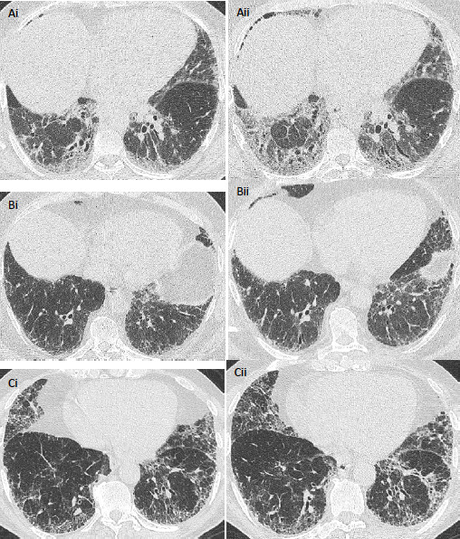
Figure 1.

Serial axial CT images in patients with idiopathic pulmonary fibrosis. In a 50-year-old male patient who did not receive antifibrotic medication and who demonstrated a >10% annualised FVC decline, images acquired 6 months apart (Ai, ii) show change in traction bronchiectasis categorised as markedly worsened (score=5) by scoring radiologists. In a 62-year-old male patient who received antifibrotic medication (Bi, ii), images acquired 13 months apart show annualised FVC decline between 5.0% and 9.9%, and change in traction bronchiectasis was categorised as mildly worsened (score=4). In a 77-year-old man who did not receive antifibrotic medication (Ci, ii) and who had CTs acquired 15 months apart, change in traction bronchiectasis severity (Score=3) and annualised FVC decline (−5.0% to 4.9%) were both considered stable. Parenchymal changes visible on the CT may reflect disease maturation rather than disease progression. FVC, forced vital capacity.
