Abstract
Background:
Parkinson disease (PD) is a common neurodegenerative disorder. Elevations of neurofilament light chain (NfL) concentrations in the cerebrospinal fluid (CSF) and blood are a marker of neuronal/axonal injury and degeneration. However, CSF and blood NfL alterations in patients with PD from existing studies remain inconclusive. To better understand these conflicting data, we will conduct a meta-analysis.
Methods:
We will comprehensively search PubMed, Embase, and Web of Science databases from each database's inception to 7th June, 2020. This protocol will conform to the Preferred Reporting Items for Systematic review and Meta-Analysis Protocols. We will only include original studies published in English that evaluated differences of NfL concentrations in the CSF or blood between idiopathic PD patients and healthy controls. The Newcastle-Ottawa Scale will be used to evaluate the quality of the included studies. Meta-analyses will be carried out using the STATA software version 13.0. Between-group difference of NfL concentrations in the CSF and blood will be expressed as the weighted standardized mean difference. A random-effects model will be used. Supplementary analyses, such as heterogeneity analysis, sensitivity analysis, publication bias, subgroup analysis, and meta-regression analysis will be performed.
Results:
The meta-analysis will provide the differences of NfL concentrations in the CSF and blood between patients with PD and healthy controls and will show the magnitudes of their effect sizes.
Conclusions:
This meta-analysis will provide the evidence of NfL concentrations in the CSF and blood in PD and we hope that our study has an important impact on clinical practice.
Registration number:
INPLASY202060025
Keywords: blood, cerebrospinal fluid, meta-analysis, neurofilament light chain, Parkinson disease
1. Introduction
Parkinson disease (PD) is a common and progressive neurodegenerative disorder.[1] PD affects over 6 million people worldwide in 2016 [1] and is the fastest growing in prevalence, disability, and deaths among the neurological disorders.[2] Pathologically, PD is characterized by a loss of dopaminergic neurons in the substantia nigra and abnormal intracellular α-synuclein accumulation in the form of Lewy neurites and Lewy bodies in the brain.[3] Clinically, PD is characterized by motor triad of resting tremor, bradykinesia, and rigidity and accompanying various nonmotor symptoms.[4,5] However, the neurodegenerative process has started for years in the premotor phase before a diagnosis can be made.[3,6] Sustained efforts have been made to develop reliable biomarkers for early detection, accurate diagnosis, and prognostic assessment.[6]
As a subunit of neurofilament, neurofilament light chain (NfL) is one of the major cytoskeletal components in mature neurons.[7] Elevation of NfL concentrations in the cerebrospinal fluid (CSF) or blood is an index of neuronal/axonal injury and degeneration.[7] While not disease-specific, NfL has been recognized as a promising diagnostic and prognostic biomarker in many neurological diseases, such as multiple sclerosis, Alzheimer disease, and amyotrophic lateral sclerosis.[7–15] However, CSF and blood (ie, plasma and serum) NfL alterations in patients with PD from existing studies remain conflicting. Most of the studies reported increased NfL concentrations in the CSF and blood in patients with PD relative to healthy controls. [16–22] While some other studies found no significant difference in the CSF[23–26] or blood[25,27] NfL levels between patients with PD without dementia and healthy controls. To better understand these conflicting data, Wang et al conducted a meta-analysis that showed no significant difference in CSF NfL level between PD patients and controls.[28] This result is interesting considering that PD is a neurodegenerative disease. However, it should be noted that their meta-analysis in PD included only 5 CSF studies. More recent studies that assessed NfL levels in the CSF and blood in PD have been published.[19–22,25,27,29,30]
In the present study, we will compile the recent evidence and perform 2 meta-analyses to quantitatively examine NfL levels in the CSF and blood separately in patients with PD compared to healthy controls.
2. Methods
2.1. Search strategies
We will comprehensively search PubMed, Embase, and Web of Science databases from each database's inception to 7th June, 2020 with no language or publication restrictions. The following search terms will be used: ((neurofilament light chain) OR nfl) AND ((Parkinson disease) OR Parkinson∗). Additional eligible studies will be obtained through cross-checking cited references. This protocol will conform to the Preferred Reporting Items for Systematic review and Meta-Analysis Protocols (PRISMA-P).[31]
2.2. Eligibility criteria
2.2.1. Inclusion criteria
Studies will be included if they meet the following criteria:
-
(1)
they were original and peer-reviewed articles published in English;
-
(2)
they enrolled patients according to the established diagnostic criteria for idiopathic PD;[5,32,33]
-
(3)
they were case-control studies that evaluated differences of NfL concentrations in the CSF or blood between idiopathic PD patients and healthy controls.
2.2.2. Exclusion criteria
Publications will be excluded if:
-
(1)
they lacked a healthy control comparison group;
-
(2)
they lacked sufficient data to estimate the mean levels and standard deviation of CSF or blood NfL concentrations;
-
(3)
they were nonhuman studies;
-
(4)
the patient sample in one study was overlapped with those with a larger sample size in another study;
-
(5)
they were not an original type, such as review, letter, case report, protocol, editorial, commentary, or conference abstract.
-
(6)
In case of longitudinal studies, only baseline comparison results will be included.
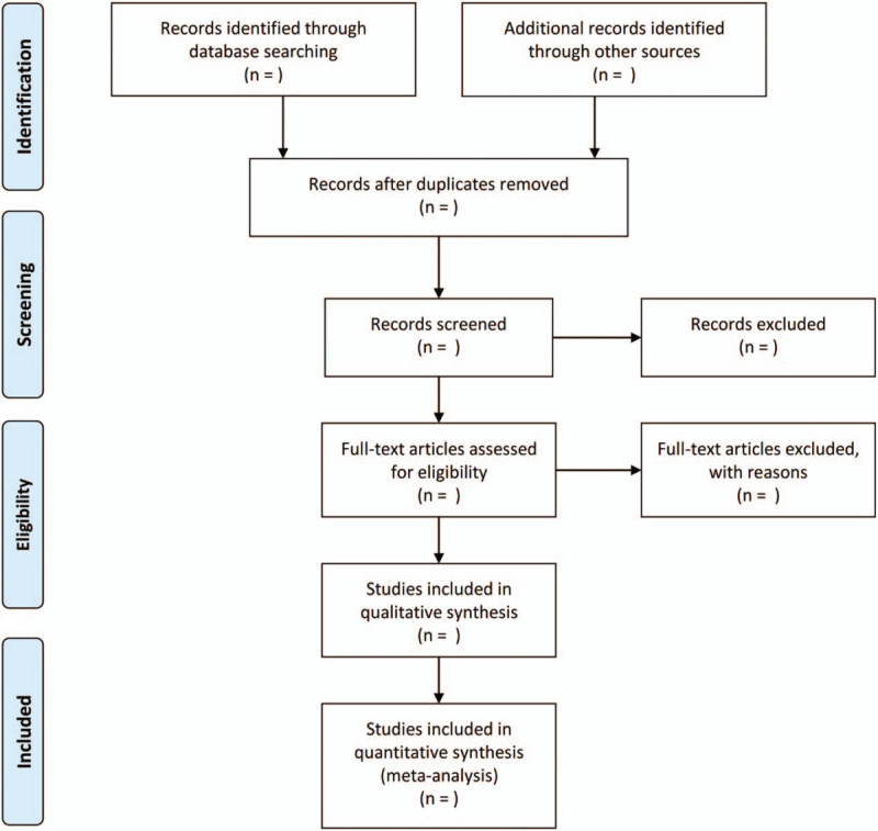
Figure 1 presents the process of selecting eligible articles according to the PRISMA statement.[34] Literature search and study selection will be independently performed by 2 authors.
Figure 1.

Study selection process following the preferred reporting items for systematic review and meta-analysis flowchart.
2.3. Data extraction
Data will be extracted from all eligible studies by two independent investigators using a standard form including the following information: The author's surname, year of publication, age, sex (male percentage), PD severity (Unified Parkinson Disease Rating Scale, part III score), H&Y stage, Mini-Mental State Examination (MMSE) score, disease duration (years), L-dopa equivalent daily dose (mg/day), and the mean and standard deviation of CSF NfL and blood NfL concentrations in PD patients and healthy controls, and NfL analysis methods.
2.4. Quality assessment
The quality of included studies will be assessed using the Newcastle Ottawa Scale (NOS).[35] Using this tool, each study is judged on an 8-item scale with maximum score 9/9 categorized into dimensions (sample selection, comparability of groups, and the assessment of outcome). Quality assessment will be independently performed by 2 authors.
2.5. Data synthesis and statistics
Meta-analyses will be carried out using the STATA software version 13.0 (StataCorp, College Station, TX). Weighted and 95% confidence intervals for results of NfL concentration differences between the PD groups and the healthy control groups will be computed using a random-effects model. Heterogeneity will be assessed using the I2 statistic. Sensitivity analyses will be conducted to test the replicability of the results by repeating the same analyses by consecutively removing 1 study at a time. Potential publication bias will be assessed using Egger linear regression and funnel plot. Subgroup analyses will be conducted in patients with PD with and without dementia. Meta-regression analyses will be conducted to investigate whether NfL concentrations in the CSF and blood were confounded by the moderators, including age, gender, UPDRS-III, H&Y stage, L-dopa equivalent daily dose, and disease duration. The significance level will be set at P < .05.
2.6. Ethics and dissemination
This meta-analysis does not need Ethics approval because it will be performed using the data based on published studies. This meta-analysis will be published in a peer-reviewed scientific journal.
3. Discussion
There is an urgent need to develop a reliable diagnostic and prognostic biomarker in the management of PD. [6] In the last decade, a growing body of evidence supports the NfL as a biomarker of brain injury or neurodegeneration in CSF and blood in a variety of neurological disorders, which may have clinical promise.[7] However, previous studies on NfL concentration in PD were inconsistent. The present meta-analysis will quantitatively examine whether NfL concentrations in the CSF and blood are elevated in patients with PD compared to healthy controls and examine what the magnitudes of their effect sizes are. This meta-analysis will provide the evidence of NfL concentrations in the CSF and blood in PD and we hope that our study has an important impact on clinical practice.
Author contributions
Conceptualization: HongZhou Wang, LiJian Han, PingLei Pan
Data curation: HongZhou Wang, WanHua Wang
Formal analysis: HongZhou Wang
Funding acquisition: PingLei Pan
Investigation: HongZhou Wang, WanHua Wang, HaiCun Shi
Methodology: HongZhou Wang, HaiCun Shi
Project administration: LiJian Han, PingLei Pan
Resources: HongZhou Wang, WanHua Wang, HaiCun Shi
Software: HongZhou Wang
Supervision: LiJian Han
Validation: PingLei Pan
Visualization: HongZhou Wang
Writing – original draft: HongZhou Wang
Writing – review & editing: LiJian Han, PingLei Pan
Footnotes
Abbreviations: CSF = cerebrospinal fluid, NfL= neurofilament light chain, PD = Parkinson disease, PRISMA-P = Preferred Reporting Items for Systematic review and Meta-Analysis Protocols.
How to cite this article: Wang HZ, Wang WH, Shi HC, Han LJ, Pan PL. Cerebrospinal fluid and blood levels of neurofilament light chain in Parkinson disease: a protocol for systematic review and meta-analysis. Medicine. 2020;99:31(e21458).
LH and PP contributed equally to this work.
This work was supported by the National Natural Science Foundation of China (81601161).
The authors report no conflicts of interest.
The datasets generated during and/or analyzed during the current study are available from the corresponding author on reasonable request.
References
- [1].GBD 2016 Neurology Collaborators. Global, regional, and national burden of Parkinson's disease, 1990-2016: a systematic analysis for the Global Burden of Disease Study 2016. Lancet Neurol 2018;17:939–53. [DOI] [PMC free article] [PubMed] [Google Scholar]
- [2].GBD 2015 Neurological Disorders Collaborator Group. Global, regional, and national burden of neurological disorders during 1990-2015: a systematic analysis for the Global Burden of Disease Study 2015. Lancet Neurol 2017;16:877–97. [DOI] [PMC free article] [PubMed] [Google Scholar]
- [3].Braak H, Del Tredici K, Rüb U, et al. Staging of brain pathology related to sporadic Parkinson's disease. Neurobiol Aging 2003;24:197–211. [DOI] [PubMed] [Google Scholar]
- [4].Zis P, Erro R, Walton CC, et al. The range and nature of non-motor symptoms in drug-naive Parkinson's disease patients: a state-of-the-art systematic review. NPJ Parkinsons Dis 2015;1:15013:1-8. [DOI] [PMC free article] [PubMed] [Google Scholar]
- [5].Postuma RB, Berg D, Stern M, et al. MDS clinical diagnostic criteria for Parkinson's disease. Mov Disord 2015;30:1591–601. [DOI] [PubMed] [Google Scholar]
- [6].Parnetti L, Gaetani L, Eusebi P, et al. CSF and blood biomarkers for Parkinson's disease. Lancet Neurol 2019;18:573–86. [DOI] [PubMed] [Google Scholar]
- [7].Gafson AR, Barthelemy NR, Bomont P, et al. Neurofilaments: neurobiological foundations for biomarker applications. Brain 2020;(In press). [DOI] [PMC free article] [PubMed] [Google Scholar]
- [8].Cai L, Huang J. Neurofilament light chain as a biological marker for multiple sclerosis: a meta-analysis study. Neuropsychiatr Dis Treat 2018;14:2241–54. [DOI] [PMC free article] [PubMed] [Google Scholar]
- [9].Forgrave LM, Ma M, Best JR, et al. The diagnostic performance of neurofilament light chain in CSF and blood for Alzheimer's disease, frontotemporal dementia, and amyotrophic lateral sclerosis: a systematic review and meta-analysis. Alzheimers Dement (Amst) 2019;11:730–43. [DOI] [PMC free article] [PubMed] [Google Scholar]
- [10].Gaetani L, Blennow K, Calabresi P, et al. Neurofilament light chain as a biomarker in neurological disorders. J Neurol Neurosurg Psychiatry 2019;90:870–81. [DOI] [PubMed] [Google Scholar]
- [11].Karantali E, Kazis D, Chatzikonstantinou S, et al. The role of neurofilament light chain in frontotemporal dementia: a meta-analysis. Aging Clin Exp Res 2020;(In press). [DOI] [PubMed] [Google Scholar]
- [12].Khalil M, Teunissen CE, Otto M, et al. Neurofilaments as biomarkers in neurological disorders. Nat Rev Neurol 2018;14:577–89. [DOI] [PubMed] [Google Scholar]
- [13].Poesen K, Van Damme P. Diagnostic and prognostic performance of neurofilaments in ALS. Front Neurol 2018;9:1167:1-7. [DOI] [PMC free article] [PubMed] [Google Scholar]
- [14].Varhaug KN, Torkildsen Ø, Myhr KM, et al. Neurofilament light chain as a biomarker in multiple sclerosis. Front Neurol 2019;10:338:1-6. [DOI] [PMC free article] [PubMed] [Google Scholar]
- [15].Zhao Y, Xin Y, Meng S, et al. Neurofilament light chain protein in neurodegenerative dementia: a systematic review and network meta-analysis. Neurosci Biobehav Rev 2019;102:123–38. [DOI] [PubMed] [Google Scholar]
- [16].Backstrom DC, Eriksson Domellof M, Linder J, et al. Cerebrospinal fluid patterns and the risk of future dementia in early, incident Parkinson disease. JAMA Neurol 2015;72:1175–82. [DOI] [PubMed] [Google Scholar]
- [17].Magdalinou NK, Paterson RW, Schott JM, et al. A panel of nine cerebrospinal fluid biomarkers may identify patients with atypical parkinsonian syndromes. J Neurol Neurosurg Psychiatry 2015;86:1240–7. [DOI] [PMC free article] [PubMed] [Google Scholar]
- [18].Mollenhauer B, Zimmermann J, Sixel-Doring F, et al. Baseline predictors for progression 4 years after Parkinson's disease diagnosis in the De Novo Parkinson Cohort (DeNoPa). Mov Disord 2019;34:67–77. [DOI] [PubMed] [Google Scholar]
- [19].Jabbari E, Holland N, Chelban V, et al. Diagnosis across the spectrum of progressive supranuclear palsy and corticobasal syndrome. JAMA Neurol 2019;77:377–87. [DOI] [PMC free article] [PubMed] [Google Scholar]
- [20].Lin CH, Li CH, Yang KC, et al. Blood NfL: a biomarker for disease severity and progression in Parkinson disease. Neurology 2019;93:e1104–11. [DOI] [PubMed] [Google Scholar]
- [21].Oosterveld LP, Verberk IMW, Majbour NK, et al. CSF or serum neurofilament light added to alpha-Synuclein panel discriminates Parkinson's from controls. Mov Disord 2020;35:288–95. [DOI] [PMC free article] [PubMed] [Google Scholar]
- [22].Sampedro F, Perez-Gonzalez R, Martinez-Horta S, et al. Serum neurofilament light chain levels reflect cortical neurodegeneration in de novo Parkinson's disease. Parkinsonism Relat Disord 2020;74:43–9. [DOI] [PubMed] [Google Scholar]
- [23].Hall S, Surova Y, Ohrfelt A, et al. CSF biomarkers and clinical progression of Parkinson disease. Neurology 2015;84:57–63. [DOI] [PMC free article] [PubMed] [Google Scholar]
- [24].Herbert MK, Aerts MB, Beenes M, et al. CSF neurofilament light Chain but not FLT3 ligand discriminates Parkinsonian disorders. Front Neurol 2015;6:91:1-7. [DOI] [PMC free article] [PubMed] [Google Scholar]
- [25].Marques TM, van Rumund A, Oeckl P, et al. Serum NFL discriminates Parkinson disease from atypical Parkinsonisms. Neurology 2019;92:e1479–86. [DOI] [PubMed] [Google Scholar]
- [26].Hall S, Ohrfelt A, Constantinescu R, et al. Accuracy of a panel of 5 cerebrospinal fluid biomarkers in the differential diagnosis of patients with dementia and/or parkinsonian disorders. Arch Neurol 2012;69:1445–52. [DOI] [PubMed] [Google Scholar]
- [27].Lin YS, Lee WJ, Wang SJ, et al. Levels of plasma neurofilament light chain and cognitive function in patients with Alzheimer or Parkinson disease. Sci Rep 2018;8:17368:1-8. [DOI] [PMC free article] [PubMed] [Google Scholar]
- [28].Wang SY, Chen W, Xu W, et al. Neurofilament light chain in cerebrospinal fluid and blood as a biomarker for neurodegenerative diseases: a systematic review and meta-analysis. J Alzheimers Dis 2019;72:1353–61. [DOI] [PubMed] [Google Scholar]
- [29].Singer W, Schmeichel AM, Shahnawaz M, et al. Alpha-synuclein oligomers and neurofilament light chain in spinal fluid differentiate multiple system atrophy from lewy body synucleinopathies. Ann Neurol 2020;(In press). [DOI] [PMC free article] [PubMed] [Google Scholar]
- [30].Ng ASL, Tan YJ, Yong ACW, et al. Utility of plasma Neurofilament light as a diagnostic and prognostic biomarker of the postural instability gait disorder motor subtype in early Parkinson's disease. Mol Neurodegener 2020;15:33:1-8. [DOI] [PMC free article] [PubMed] [Google Scholar]
- [31].Shamseer L, Moher D, Clarke M, et al. Preferred reporting items for systematic review and meta-analysis protocols (PRISMA-P) 2015: elaboration and explanation. BMJ 2015;349:g7647:1-25. [DOI] [PubMed] [Google Scholar]
- [32].Gelb DJ, Oliver E, Gilman S. Diagnostic criteria for Parkinson disease. Arch Neurol 1999;56:33–9. [DOI] [PubMed] [Google Scholar]
- [33].Hughes AJ, Daniel SE, Kilford L, et al. Accuracy of clinical diagnosis of idiopathic Parkinson's disease: a clinico-pathological study of 100 cases. J Neurol Neurosurg Psychiatry 1992;55:181–4. [DOI] [PMC free article] [PubMed] [Google Scholar]
- [34].Moher D, Liberati A, Tetzlaff J, et al. Preferred reporting items for systematic reviews and meta-analyses: the PRISMA statement. PLoS Med 2009;6:e1000097:1-6. [DOI] [PMC free article] [PubMed] [Google Scholar]
- [35].Wells GA, Shea B, O’Connell D, et al. The Newcastle-Ottawa Scale (NOS) for Assessing the Quality of Nonrandomised Studies in Meta-Analyses. The Ottawa Hospital Research Institute, Available at: http://www.ohri.ca/programs/clinical_epidemiology/oxford.asp. 2011. [Google Scholar]


