Dear Editor,
COVID‐19 infection can be associated with cutaneous symptoms. Whether these are a primary dermatological manifestation or a secondary result of the immune response against the virus remains unclear.
A 78‐year‐old woman presented after a fall and a short period of confusion. Past medical history was significant for vascular epilepsy, hypothyroidism, and heart failure. Medications, with no recent changes, included levetiracetam, sodium valproate, levothyroxine, bisoprolol, clopidogrel, furosemide, atorvastatin and folic acid. She reported nonspecific allergies to colchicine and naproxen and no food intolerances.
On admission nasopharyngeal severe acute respiratory syndrome coronavirus 2 (SARS‐CoV‐2) reverse transcriptase–polymerase chain reaction (RT‐PCR) was positive, and chest radiograph was consistent with COVID‐19 pneumonitis. General examination and vital signs were unremarkable. Laboratory investigations were notable for leukocytosis at 14.57 × 10⁹/l (4.0–11.0 × 10⁹), lymphocytes of 0.85 × 10⁹/l (1.0–4.0 × 10⁹), C‐reactive protein of 49 mg/l (0–4), ferritin of 362 µg/l (5–300), lactate dehydrogenase of 309 U/l (35–214), and D‐dimer of 1,540 ng/ml (270–750).
Over the next 24 hours, she developed intermittent pyrexia up to 38.4°C but remained clinically stable. There was associated anorexia and lassitude without any respiratory symptoms.
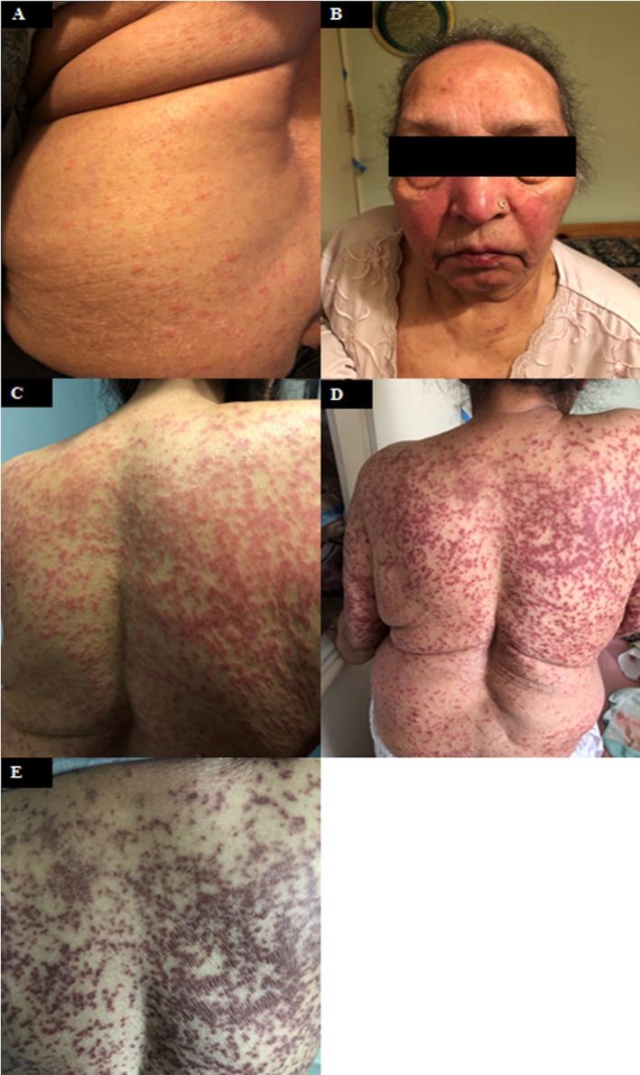
A week previously, the patient's daughter had noticed a widespread non‐pruritic, erythematous blanching maculopapular eruption which appeared on the trunk. There was evidence of vesicles and urticaria consistent with a viral exanthem. The daughter also reported facial angioedema with drooling and throat discomfort but without stridor. In addition, there was a malar distribution rash of similar morphology. These lasted for 2 days (Fig. 1a–c). On Day 4, the rash progressed to a purpuric appearance (Fig. 1d). Skin examination on Day 6 (admission Day 1) revealed hyperpigmented patches residual of the previous erythematous rash with surrounding scaling and flaking (Fig. 1e).
Figure 1.

(a) Day 1 – diffuse maculopapular rash with evidence of vesicles and urticarial lesions on truncal area. (b) Day 3 – malar rash with associated facial swelling. (c) Day 3 – dense progression of rash on posterior trunk. (d) Day 4 – progression of rash to a diffuse purpuric maculopapular appearance. (e) Day 6 – hyperpigmentation of residual rash with associated scaling and flaking of rash
Extended autoimmune and viral screens were found negative. A diagnosis of COVID‐19 rash was made, and an emollient was administered. The patient developed no other features of COVID‐19 infection and was discharged after 7 days.
Five cutaneous clinical patterns associated with COVID‐19 infection have been described: erythema with vesicles or pustules (pseudo‐chilblain), other vesicular eruptions, urticarial lesions, maculopapular eruptions, and livedo or necrosis. Polymorphism, especially in a single viral host, may be attributable to coinfection with other viruses, multiple viral strains, or different immunomodulatory responses to SARS‐CoV‐2 between hosts. 1
Our patient demonstrated three or more of the recognized patterns: vesicular eruptions, urticarial lesions, and papules which evolved to a purpuric appearance. These lasted for at least 6 days with gradual hyperpigmentation and scaling, consistent with previous descriptions of COVID‐19 rash. 1 , 2 Although the rash was extensive, it did not correlate with disease severity. Livedoid or necrotic lesions, which were not observed in our patient, may be associated with higher mortality and more aggressive disease in elderly patients. 1 This may be due to the vascular complications and prothrombotic tendency of COVID‐19 infection. 3 , 4
Our patient also developed facial angioedema and a malar rash, features similar to a case reported by Cohen and colleagues. 5 COVID‐19 appears to be optimized for binding to the human receptor angiotensin converting enzyme 2 (ACE2), and the significance of viral‐ACE2 binding and its degradation may provide the mechanism. ACE2 is involved in the cleavage of bradykinin and associated metabolites, and its downregulation results in enhancement of ACE activity leading to an increase in bradykinin which contributes to the development of angioedema. This pathomechanism may also explain the dry cough experienced by COVID‐19 patients. Cohen et al. proposed that modulation of the bradykinin pathway may be a potential therapeutic target in COVID‐19. 5
This is the first documentation of the natural history of cutaneous eruptions and angioedema within the context of COVID‐19 infection and their subsequent resolution. Although dermatological manifestations remain nonspecific to COVID‐19 infection, paucisymptomatic cases should be expectantly observed for the unexpected.
Acknowledgment
We thank the patient and her family members for allowing us to use the materials for publication.
Conflict of interest: None.
Funding source: None.
References
- 1. Galván Casas C, Catalá A, Carretero Hernández G, et al. Classification of the cutaneous manifestations of COVID‐19: a rapid prospective nationwide consensus study in Spain with 375 cases. Br J Dermatol 2020; 183: 71–77. [DOI] [PMC free article] [PubMed] [Google Scholar]
- 2. Recalcati S. Cutaneous manifestations in COVID‐19: a first perspective. J Eur Acad Dermatol Venereol 2020; 34: e212–e213. [DOI] [PubMed] [Google Scholar]
- 3. Varga Z, Flammer AJ, Steiger P, et al. Endothelial cell infection and endotheliitis in COVID‐19. Lancet 2020; 395: 1417–1418. [DOI] [PMC free article] [PubMed] [Google Scholar]
- 4. Zhang Y, Xiao M, Zhang S, et al. Coagulopathy and antiphospholipid antibodies in patients with Covid‐19. N Engl J Med 2020; 382: e38. [DOI] [PMC free article] [PubMed] [Google Scholar]
- 5. Cohen AJ, DiFrancesco MF, Solomon SD, et al. Angioedema in COVID‐19. Eur Heart J 2020. [DOI] [PMC free article] [PubMed] [Google Scholar]


