PLOS ONE logoLink to PLOS ONE
. 2020 Aug 5;15(8):e0236569. doi: 10.1371/journal.pone.0236569

Elevated serum YKL-40, IL-6, CRP, CEA, and CA19-9 combined as a prognostic biomarker panel after resection of colorectal liver metastases

Reetta Peltonen 1,*,#, Mathias H Gramkow 2,#, Christian Dehlendorff 3, Pia J Osterlund 4,5,, Julia S Johansen 2,6,7,, Helena Isoniemi 1,
Editor: Eugene J Koay8
PMCID: PMC7406016  PMID: 32756596

Abstract

Background

The inflammatory biomarkers, YKL-40 and interleukin-6 (IL-6), are elevated in patients with metastatic colorectal cancer. We examined their associations with relapse-free survival and overall survival in combination with serum C-reactive protein (CRP), carcinoembryonic antigen (CEA), and carbohydrate antigen 19–9 (CA19-9) in patients with colorectal liver metastases.

Methods

Altogether 441 consecutive patients undergoing liver resection at Helsinki University Hospital between 1998 and 2013 were included in the study. Pre- and postoperative YKL-40 and IL-6 were determined from serum samples with commercially available enzyme-linked immunosorbent assay (ELISA) kits, and CRP, CEA, and CA19-9 by routine methods. Associations between these biomarkers and relapse-free and overall survival were examined using Cox regression analysis.

Results

Patients with 2–5 elevated biomarkers were at an increased risk of relapse compared to those with 0–1 elevated biomarkers, preoperatively (HR 1.37, 95% CI 1.1–1.72) or postoperatively (HR 1.54, 95% CI 1.23–1.92). Patients with 2–5 elevated biomarkers were also at an increased risk of death compared to those with 0–1 elevated biomarkers, preoperatively (HR 1.76, 95% CI 1.39–2.24) or postoperatively (HR 1.83, 95% CI 1.44–2.33).

Conclusion

The results suggest that a protein panel of the inflammatory biomarkers YKL-40, IL-6, and CRP, and the cancer biomarkers CEA and CA19-9 might identify patients that benefit from more aggressive treatment and surveillance, although the additional value of IL-6 and CRP in this aspect is limited.

Introduction

In 2018, more than 860 000 patients died from colorectal cancer (CRC) worldwide [1]. A large number, up to 75%, of patients with CRC develop colorectal liver metastases (CRLM) [2]. If the metastases are confined to the liver and other resectable extrahepatic sites, resection with curative attempt is recommended, if technically and physiologically feasible [3]. Five- and ten-year survival rates after liver resection have been in the range of 38% and 26%, respectively, but a significant number of the resected experience an early recurrence [3]. As relapses are frequent and median overall survival (OS) short (median 3.6 years in a review), prognostic scoring systems have been developed for identifying patients who might benefit from intensified adjuvant therapy and follow-up, combined with a possible re-resection upon recurrence [35]. A well-defined, universally accepted adjuvant therapy and surveillance regimen after liver resection is yet to be established for this patient group.

Serum carcinoembryonic antigen (CEA) is widely used as a biomarker for detection of recurrent CRC and for monitoring the response to systemic therapy [6], but its prognostic value leaves room for improvement and added arsenal, as around 20% of CRC tumors are CEA negative [7]. Carbohydrate antigen 19–9 (CA19-9), also called sialyl-LewisA, is a tetrasaccharide carbohydrate synthesized by the gastrointestinal epithelium. Several studies have shown that elevated CA19-9 levels were strong negative prognostic factors in different stages of CRC [8, 9]. Finding new biomarkers in addition to CEA and CA19-9 would help estimate the risk of recurrence and determine a suitable adjuvant therapy and follow-up protocol for patients with metastatic CRC (mCRC) after liver resection.

Tumor-promoting inflammation is an enabling characteristic of cancer and enhanced systemic inflammatory response has been linked to impaired survival [10, 11]. Inflammatory biomarkers are of special interest in the search for new valuable prognostic biomarkers on top of established pathologic and demographic factors in patients resected for CRLM. Serum C-reactive protein (CRP), YKL-40 (also called chitinase-3-like-1 protein, CHI3L1) and interleukin-6 (IL-6) are established biomarkers of inflammation [9, 12, 13]. In a recent study, serum C-reactive protein (CRP), the production of which is stimulated by IL-6 [14], was found to be significantly associated with shorter OS in patients undergoing liver resection [15]. IL-6 and YKL-40 are secreted by cancer cells and macrophages [12, 16], and the latter also by neutrophils [12]. YKL-40 could play an important role in promoting tumor metastasis and impairing survival via multiple mechanisms, such as upregulating the secretion of matrix metalloproteinases, activating the TGFẞ pathway [17], inducing angiogenesis via its receptor IL-13α2 and membrane protein TMEM219, and binding to RAGE (receptor for advanced glycation end products) [12]. IL-6 stimulates the production of YKL-40 and angiogenesis via its receptors IL-6R and sIL-6R [12, 1820]. Elevated serum YKL-40 and IL-6 levels are seen in patients with diagnosed CRC or those later developing gastrointestinal cancers [13, 21], and higher levels are linked to impaired survival [12, 15].

The early detection of cancer with emerging technologies, such as liquid biopsies that enable the detection of circulating tumor cells, cell-free DNA (cfDNA) or circulating tumor DNA (ctDNA) with specific mutations in blood or other body fluids, is under active investigation [22]. However, protein biomarkers are at present an integral part of clinical work and may be more easily accessed.

In this retrospective biomarker study, we examined the prognostic value of preoperative and postoperative serum YKL-40, IL-6, CRP, CEA, and CA19-9 in a cohort of 441 consecutive patients who had undergone liver resection for CRLM. Our hypothesis was that the patients with combined high serum concentrations of YKL-40, IL-6, CRP, CEA, and CA19-9 would have the worst relapse-free survival (RFS) and OS in patients with mCRC treated with curative-intent liver resection.

Materials and methods

Patients

Altogether 455 consecutive patients undergoing liver resection for mCRC were enrolled in this study between March 1998 and February 2013 at the Helsinki University Hospital, Helsinki, Finland (S1 Fig). All patients included were diagnosed with resectable CRLM, and those with possible other malignancies were excluded. Liver metastases were defined as synchronous, if they were diagnosed at the same time or within 6 months after the operation of the primary tumor, and as metachronous, if diagnosed later. The liver resections were considered major if more than two Couinaud segments were resected. Extrahepatic disease was excluded with whole-body contrast-enhanced computed tomography (CT) and in some cases with positron emission tomography (PET). Neoadjuvant and/or adjuvant treatment were given according to guidelines at the time of surgery. Fourteen patients were excluded from the statistical analyses due to the following reasons: 7 patients had extrahepatic metastases that eventually could not be surgically removed, in 6 cases a radical liver resection could not be performed, and 1 patient died within 20 days after surgery, and thus, the prognostic value of the serum markers could not be evaluated.

Serum samples were taken before liver resection and approximately 3 months after operation. Median time from preoperative blood sampling to surgery was 16 days (interquartile range (IQR) 9–29 days). Median time from surgery to postoperative sampling was 94 days (IQR 89–98 days).

The study was conducted in accordance with the Declaration of Helsinki, and it was approved by the Ethics Committee at Helsinki University Hospital (IRB99/07/01, HUS531/E6/01, HUS460/E6/05, HUS323/13/3/2008, HUS242/13/03/02/2011). Collection and analysis of blood samples were approved by the National Supervisory Authority for Welfare and Health (Valvira) of Finland (STM Dno 4858/04/047/08), and information about the dates of death was obtained from the Central Statistical Office of Finland (TK-53-1004-9). A verbal informed consent was obtained from all the patients included in the study, and the collection of the research samples signified consent. The collection of blood samples was initiated over twenty years ago, and written consent was not required at that time according to the Finnish law. The REMARK guidelines have been used in reporting [23].

Serum samples

The pre- and postoperative venous blood samples were collected in gel tubes. The samples were centrifuged within 30–120 minutes, and serum was stored at -80°C. YKL-40 and IL-6 were determined in duplicate in both pre- and postoperative serum samples using commercially available enzyme-linked immunosorbent assays (ELISAs); YKL-40: MicroVue YKL-40 ELISA (Catalog #8020), Quidel Corporation, San Diego, CA, USA; and IL-6: Quantikine HS600B, R&D Systems, Abingdon, OX, UK; according to the manufacturer’s instructions. For YKL-40, the detection limit was 20 ng/ml, and intra- and inter-assay coefficients of variation (CVs) <5% and <6% [24]. For IL-6, the detection limit was 0.01 pg/ml, and intra- and inter-assay CVs ≤8% and ≤11% [25]. The pre- and postoperative serum levels of CRP, CEA and CA19-9 were determined with automatic analyzers as follows: CRP: immunoturbidimetric method (1998–2013) at Huslab laboratories, Helsinki University Hospital; CEA and CA19-9: immunoenzymatic assay, Bayer Immuno 1 (CEA: 1998–10/2005; and CA19-9: 1998–1/2006), or immunochemiluminometric assay, Abbott Architect (CEA: 10/2005–2013; and CA19-9: 1/2006–2013). All measurements were performed by technicians blinded to the study endpoints.

An age-corrected 95th percentile for serum YKL-40 in healthy individuals served as the cut-off level for elevated values, and it was calculated using the formula proposed by Bojesen et al. [24]:

Pi%=100(1+YKL403(1.062Age)5000

The cut-off values for the other biomarkers were the following: 4.95 pg/ml (P95% for serum IL-6 in healthy individuals [25]); 5 mg/l for CRP; 5 μg/l for CEA; and 37 kU/l for CA19-9.

Statistical analyses

Wilcoxon signed-rank test was used for estimating differences between pre- and postoperative serum values and Spearman’s rank test for testing correlations. For OS, the time to event was defined as time from the date of liver surgery to the date of death from any cause or censored at the end of follow-up, and for RFS as time from surgery to recurrence or censored at the end of follow-up. The cut-off date for follow-up was October 31st, 2017.

Serum YKL-40, IL-6, CRP, CEA, and CA19-9 were included in the Cox regression analyses as log2-transformed continuous variables due to non-normality and as elevated or normal values according to the above-mentioned cut-off levels in the Kaplan-Meier plots. Biomarker values below the limit of detection (LOD) were imputed as LOD/2 as a pragmatic approach, and serum values of zero seemed unlikely. Crude and adjusted hazard ratios (HR) with 95% confidence interval (CI) were estimated for RFS and OS using Cox regression. Survival curves were estimated using the Kaplan-Meier method and equality of strata was tested for by a likelihood ratio test. The variables included in the multivariate analyses (Tables 3 and 4) were the following: serum values of YKL-40, IL-6, CRP, CEA, CA19-9, age, gender, location of the primary tumor, presentation of liver metastases (synchronous/metachronous), type of liver resection (minor/major), the number and size of the liver metastases, and the resection margins (R0/1/2).

Table 3. Results of the Cox regression analyses for relapse-free survival.

Univariate p-value Multivariate1 p-value
HR (95% CI) HR (95% CI)
Preoperatively measured biomarkers
YKL-40 1.19 (1.07–1.32) <0.001 1.08 (0.96–1.22) 0.212
IL-6 1.15 (1.03–1.28) 0.010 1.04 (0.91–1.18) 0.566
CRP 1.08 (0.96–1.21) 0.219 1.03 (0.88–1.20) 0.756
CEA 1.01 (0.96–1.07) 0.583 1.00 (0.93–1.07) 0.952
CA19-9 1.08 (1.03–1.14) <0.001 1.12 (1.06–1.19) <0.001
0 elevated (N = 111) Reference Reference
1 elevated (N = 113) 0.81 (0.58–1.12) 0.202 0.81 (0.58–1.12) 0.202
2 elevated (N = 101) 1.13 (0.83–1.56) 0.438 1.21 (0.87–1.68) 0.252
3 elevated (N = 44) 1.60 (1.08–2.36) 0.018 1.63 (1.08–2.47) 0.021
4 elevated (N = 29) 1.57 (0.99–2.49) 0.055 1.88 (1.12–3.16) 0.016
5 elevated (N = 1) 2.86 (0.40–20.7) 0.298 3.60 (0.48–26.8) 0.211
0–1 elevated (N = 224) Reference Reference
2–5 elevated (N = 175) 1.37 (1.10–1.72) 0.005 1.40 (1.11–1.78) 0.005
Postoperatively measured biomarkers
YKL-40 1.21 (1.09–1.34) <0.001 1.06 (0.94–1.19) 0.323
IL-6 1.11 (1.01–1.23) 0.033 0.98 (0.87–1.11) 0.755
CRP 1.07 (0.99–1.15) 0.075 1.02 (0.92–1.12) 0.741
CEA 1.24 (1.14–1.34) <0.001 1.17 (1.07–1.29) <0.001
CA19-9 1.12 (1.04–1.20) 0.002 1.09 (1.01–1.17) 0.029
0 elevated (N = 137) Reference Reference
1 elevated (N = 99) 1.18 (0.87–1.62) 0.290 1.00 (0.73–1.38) 0.993
2 elevated (N = 106) 1.41 (1.05–1.91) 0.023 1.30 (0.95–1.78) 0.102
3 elevated (N = 48) 1.98 (1.37–2.86) <0.001 1.84 (1.25–2.71) 0.002
4 elevated (N = 10) 2.94 (1.48–5.86) 0.002 2.50 (1.22–5.12) 0.012
5 elevated (N = 3) 3.67 (1.16–11.7) 0.028 3.14 (0.97–10.1) 0.055
0–1 elevated (N = 236) Reference Reference
2–5 elevated (N = 167) 1.54 (1.23–1.92) <0.001 1.57 (1.24–1.98) <0.001

1 Variables included in the multivariate analyses: serum values of IL-6, YKL-40, CRP, CEA, and CA19-9; age; gender; location of the primary tumor; type of liver metastases (synchronous/metachronous; Table 1); type of liver resection (minor/major; Table 1); the number and size of the liver metastases; and the resection margins (R0/1/2). The biomarkers are presented as log2-transformed continuous variables.

Table 4. Results of the Cox regression analyses in relation to overall survival.

Univariate p-value Multivariate1 p-value
HR (95% CI) HR (95% CI)
Preoperatively measured biomarkers
YKL-40 1.29 (1.16–1.44) <0.001 1.19 (1.04–1.35) 0.010
IL-6 1.16 (1.03–1.30) 0.011 1.03 (0.90–1.18) 0.685
CRP 1.07 (0.95–1.21) 0.240 0.91 (0.77–1.06) 0.215
CEA 1.07 (1.01–1.12) 0.019 1.02 (0.95–1.09) 0.602
CA19-9 1.12 (1.06–1.18) <0.001 1.13 (1.06–1.20) <0.001
0 elevated (N = 111) Reference Reference
1 elevated (N = 113) 0.86 (0.60–1.24) 0.433 0.82 (0.57–1.18) 0.284
2 elevated (N = 101) 1.63 (1.16–2.30) 0.005 1.58 (1.11–2.24) 0.011
3 elevated (N = 44) 1.82 (1.19–2.79) 0.006 1.60 (1.02–2.50) 0.042
4 elevated (N = 29) 1.78 (1.08–2.93) 0.023 1.75 (1.01–3.04) 0.046
5 elevated (N = 1) 3.24 (0.45–23.5) 0.245 2.90 (0.39–21.7) 0.299
0–1 elevated (N = 224) Reference Reference
2–5 elevated (N = 175) 1.76 (1.39–2.24) <0.001 1.71 (1.33–2.21) <0.001
Postoperatively measured biomarkers
YKL-40 1.26 (1.13–1.41) <0.001 1.10 (0.97–1.24) 0.131
IL-6 1.13 (1.02–1.26) 0.022 0.95 (0.83–1.09) 0.498
CRP 1.11 (1.02–1.20) 0.011 1.09 (0.99–1.20) 0.096
CEA 1.33 (1.22–1.46) <0.001 1.24 (1.12–1.36) <0.001
CA19-9 1.22 (1.13–1.31) <0.001 1.17 (1.08–1.28) <0.001
0 elevated (N = 137) Reference Reference
1 elevated (N = 99) 1.03 (0.72–1.47) 0.882 0.88 (0.61–1.27) 0.488
2 elevated (N = 106) 1.57 (1.13–2.17) 0.007 1.40 (0.99–1.97) 0.055
3 elevated (N = 48) 2.42 (1.64–3.56) <0.001 2.33 (1.56–3.48) <0.001
4 elevated (N = 10) 4.45 (2.21–8.96) <0.001 4.42 (2.16–9.05) <0.001
5 elevated (N = 3) 10.2 (3.16–32.9) <0.001 8.91 (2.71–29.2) <0.001
0–1 elevated (N = 236) Reference Reference
2–5 elevated (N = 167) 1.83 (1.44–2.33) <0.001 1.84 (1.43–2.37) <0.001

1 Variables included in the multivariate analyses: serum values of IL-6, YKL-40, CRP, CEA, and CA19-9; age; gender; location of the primary tumor; type of liver metastases (synchronous/metachronous; Table 1); type of liver resection (minor/major; Table 1); the number and size of the liver metastases; and the resection margins (R0/1/2). The biomarkers are presented as log2-transformed continuous variables.

Proportional hazards assumption was evaluated by testing for trends in the scaled Schoenfeld residuals. A cut point analysis was carried out for all investigated markers with regards to their discriminatory power for 3-year mortality and 3-year recurrence using receiver operating characteristic (ROC) curves and calculating the area under the curve (AUC). The cut point with a sensitivity closest to 80% was chosen. All statistical analyses were carried out using the statistical software R version 3.3.3 (R Core Team, Vienna, Austria). A p-value of less than 0.05 was considered significant, and only two-sided tests were used.

Results

Patient characteristics and the pre- and postoperative serum concentrations of YKL-40, IL-6, CRP, CEA, and CA19-9 are shown in Tables 1 and 2, respectively. The median age of the patients was 64.9 years, and 59% of them were men. During the follow-up period, 265 patients (60%) experienced a recurrence, of which 242 (91%) were diagnosed within 3 years after liver resection. The median RFS was 1.7 years (IQR: 0.6–6.6 years). Median OS was 5.8 years (IQR: 5.4–8.4 years), and 268 patients (61%) died during follow-up. Median follow-up time was 5.7 years.

Table 1. Baseline patient characteristics and outcome data.

Variable Overall (N = 441)
Sex  
    Male 260 (59.0%)
    Female 181 (41.0%)
Age in years, median (min.–max.) 64.9 (33–84)
Location of the primary tumor  
    Right colon 84 (19.0%)
    Left colon 174 (39.5%)
    Rectum 183 (41.5%)
Presentation of liver metastases1  
    Synchronous 254 (57.6%)
    Metachronous 187 (42.4%)
Number of liver metastases, median (max.) 1 (16)
Size of the largest metastasis, median (max.) 2.5 cm (15.0)
Type of liver resection2:
    Minor 201 (45.6%)
    Major 239 (54.2%)
    Missing 1
Resection margins
    R0 412 (93.6%)
    R1 19 (4.3%)
    R2 9 (2.0%)
OS after liver resection, median (min.–max.) 5.8 years (0.0–19.7)
RFS after liver resection, median (min.–max.) 1.7 years (0.04–19.3)
Alive at the end of follow-up 172 (39.0%)
Alive without recurrence 123 (27.9%)
Recurrence or death 318 (72.1%)

1 Diagnosed within 6 months after the operation of the primary tumor (synchronous) or later (metachronous).

2 1–2 (minor) or ≥3 (major) Couinaud segments resected.

Table 2. Pre- and postoperative biomarker concentrations.

Biomarker Preoperative Postoperative
YKL-40 (number) 413 413
    Median (min.–max.) (ng/ml) 74 (20–1504) 87 (20–744)
    Elevated (>age-corrected 95th percentile), n (%) 57 (13.8) 78 (18.9)
IL-6 (number) 413 413
    Median (min.–max.) (pg/ml) 3.4 (0.3–33.5) 5.1 (0.6–80.7)
    Elevated (>4.95 pg/ml), n (%) 142 (34.4) 214 (51.8)
CRP (number) 429 434
    Median (min.–max.) (mg/l) <5 (<5–149) <5 (<5–153)
    Elevated (>5 mg/l), n (%) 86 (20.0) 129 (29.7)
CEA (number) 440 440
    Median (min.–max.) (μg/l) 5.2 (0.5–853) 2.5 (0.5–294)
    Elevated (>5 μg/l), n (%) 221 (50.2) 74 (16.8)
CA19-9 (number) 437 436
    Median (min.–max.) (kU/l) 13 (<2–11 310) 9 (<2–33 800)
    Elevated (>37 kU/l), n (%) 99 (22.7) 43 (9.9)

The results of the cut point analysis showed that only elevated postoperative CEA had an AUC larger than 0.7 (AUCCEA = 0.73, 95% CI 0.66–0.79) for predicting mortality within 3 years (S1 Table and S2 Fig). Based on the cut point analyses of the markers, we chose to use the pre-specified cut-offs as the optimal cut points did not differ markedly from them (see S1 TableS4 Table and S1 Fig for results of the cut point analyses).

The characteristics of the pre- and postoperative biomarker values are shown in Table 2. YKL-40 and IL-6 increased after liver resection: the median increase in YKL-40 levels was 13 ng/ml (range -1261–571, p = 0.015) and in IL-6 levels 1.7 pg/ml (range -19.4–73.9, p<0.001).

Preoperative YKL-40 correlated with preoperative IL-6 (rho = 0.39, p<0.001) and preoperative CEA (rho = 0.11, p = 0.024), and postoperative YKL-40 with postoperative IL-6 (rho = 0.28, p<0.001) and postoperative CEA (rho = 0.15, p = 0.002). Preoperative CRP correlated with preoperative IL-6 (rho = 0.31, p<0.001), YKL-40 (rho = 0.21, p<0.001) and CEA (rho = 0.25, p<0.001). Preoperative CEA correlated with preoperative CA19-9 (rho = 0.41, p<0.001) and CRP (rho = 0.25, p<0.001), and postoperative CEA with postoperative CA19-9 (rho = 0.22, p<0.001). Postoperative CA19-9 correlated with postoperative IL-6 (rho = 0.13, p = 0.008). Age correlated with pre- and postoperative YKL-40 (rho = 0.25, p<0.001; and rho = 0.33, p<0.001, respectively), with preoperative IL-6 (rho = 0.16, p = 0.001), and with postoperative CA19-9 (rho = 0.11, p = 0.027). There was no significant correlation between age and CEA.

Association of relapse-free survival with YKL-40, IL-6, CRP, CEA, and CA19-9

The results of the univariate and multivariate analyses for RFS are shown in Table 3. All the coefficients in the Cox regression analyses for pre- and postoperative variables in regard to RFS and OS can be found in the S5 Table.

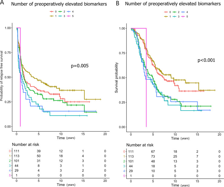
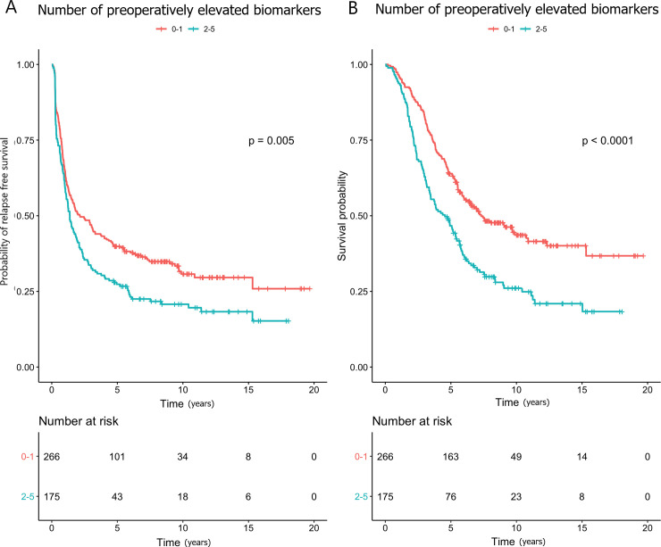
Higher preoperative log2-transformed YKL-40 (HR 1.19), IL-6 (HR 1.15) and CA19-9 (HR 1.08) were associated with shorter RFS in the univariate analysis, and CA19-9 in the multivariate analysis (HR 1.12). Patients with 3 elevated biomarkers concurrently were at an increased risk of relapse compared to those with no elevated biomarkers in the univariate (HR 1.60; Fig 1A) and in the multivariate analyses (HR 1.63). Patients with 2–5 elevated markers compared to those with 0–1 had shorter RFS in univariate (HR 1.37; Fig 2A) and in multivariate (HR 1.40) analyses.

Fig 1.

Fig 1

Kaplan-Meier curves showing the associations between the numbers of elevated preoperative biomarkers and (A) relapse-free survival and (B) overall survival.

Fig 2.

Fig 2

Kaplan-Meier curves showing the associations between 0–1 and 2–5 elevated preoperative biomarkers and (A) relapse-free survival and (B) overall survival.

To investigate whether YKL-40, IL-6 and/or CRP drove this association, an explorative multivariate analysis was carried out. It showed that neither preoperatively elevated CEA and/or CA19-9 nor elevated YKL-40, IL-6 and/or CRP associated with shorter RFS (S6 Table).

Higher postoperative log2-transformed YKL-40 (HR 1.21), IL-6 (HR 1.11), CEA (HR 1.24), and CA19-9 (HR 1.12) all were associated with shorter RFS in the univariate analyses, but CRP was not. In the multivariate analysis, the association between shorter RFS and higher CEA and CA19-9 remained statistically significant (Table 3).

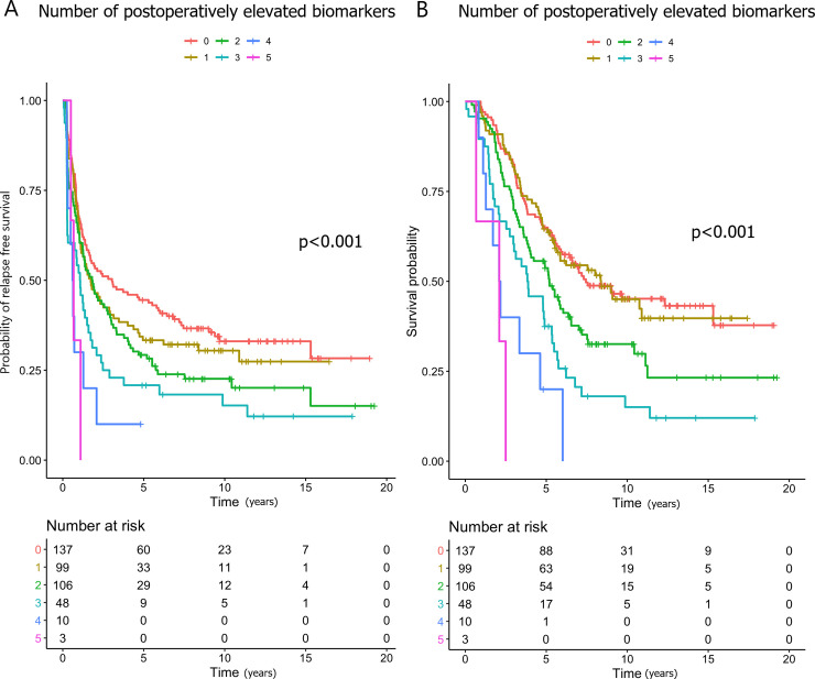
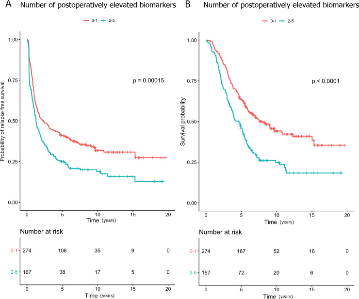
The patients with 2–5 elevated biomarkers postoperatively were at an increased risk of relapse compared to those with 0–1 elevated (HR 1.54; Figs 3A and 4A), and this association remained statistically significant in the multivariate analysis (HR 1.57). Compared with zero elevated biomarkers, having CEA and/or CA19-9 elevated was significantly associated with shorter RFS, while elevated YKL-40, IL-6 and/or CA19-9 were not (S6 Table).

Fig 3.

Fig 3

Kaplan-Meier curves showing the associations between the numbers of elevated postoperative biomarkers and (A) relapse-free survival and (B) overall survival.

Fig 4.

Fig 4

Kaplan-Meier curves showing the associations between 0–2 and 2–5 elevated postoperative biomarkers and (A) relapse-free survival and (B) overall survival.

Association of overall survival with YKL-40, IL-6, CRP, CEA, and CA19-9

The results of the univariate and multivariate analyses are shown in Table 4.

Higher preoperative log2-transformed YKL-40 (HR 1.29), IL-6 (HR 1.16), CEA (HR 1.07), and CA19-9 (HR 1.12) were associated with shorter OS in the univariate analyses, but CRP was not. Higher YKL-40 and CA19-9 remained significant in the multivariate analysis (HR 1.19, and HR 1.13, respectively).

The patients with 2–5 elevated biomarkers preoperatively were at an increased risk of death compared to those with 0–1 elevated biomarkers (HR 1.76; Figs 1B and 2B), and this association remained statistically significant in the multivariate analysis (HR 1.71). Neither preoperatively elevated CEA and/or CA19-9 nor elevated YKL-40, IL-6 and/or CRP associated significantly with shorter OS, when taken in combination (S6 Table).

Higher postoperative log2-transformed values of all biomarkers associated with shorter OS in the univariate analyses (YKL-40: HR 1.26; IL-6: HR 1.13; CRP: HR 1.11; CEA: HR 1.33; and CA19-9: HR 1.22), and CEA (HR 1.24) and CA19-9 (HR 1.17) remained significant in the multivariate analysis (Table 4).

The patients with 2–5 elevated biomarkers postoperatively were at an increased risk of death compared to those with 0–1 elevated (HR: 1.83; Figs 3B and 4B). In the multivariate analysis, this association remained significant (HR 1.84). Compared with zero elevated biomarkers, having CEA and/or CA19-9 elevated was significantly associated with shorter OS, while elevated YKL-40, IL-6 and/or CA19-9 were not (S6 Table).

Discussion

In this biomarker study of patients with colorectal liver metastases, we found that higher serum values of YKL-40 and CA19-9 preoperatively were associated with shorter OS. Similar results were found for postoperatively higher serum values of CEA and CA19-9 postoperatively. Interestingly, we found that patients with two or more elevated biomarkers pre- or postoperatively had a shorter OS, suggesting that a combined panel of inflammatory biomarkers could be used in combination with the tumor biomarkers CEA and CA19-9. Higher serum values of YKL-40 and CA19-9 preoperatively and higher serum values of CEA and CA19-9 postoperatively associated with shorter RFS, and a combination of all the biomarkers showed that patients with two or more elevated biomarkers postoperatively had a shorter RFS. No previous studies have evaluated the combined prognostic value of these 5 biomarkers.

Our results are in line with recent studies examining the prognostic value of serum YKL-40 [26], CEA [8, 9, 27, 28], and CA19-9 [8, 9, 27] as biomarkers in patients with mCRC. CRP has recently been found to have prognostic value in patients with mCRC, although our results do not directly support this finding [29]. The association between serum IL-6 and OS in patients with CRC is unclear, since some previous studies have found higher IL-6 levels to be significantly associated with shorter OS, while others have failed to show any association [16, 29, 30]. We did not find a direct association between IL-6 and RFS or OS, when adjusted for other biomarkers.

YKL-40 has recently been found to be involved in the formation of cancer metastases through regulation of epithelial to mesenchymal transformation and subsequent migration/invasion [31]. In addition, YKL-40 has been shown to regulate VEGF in tumor cells and to promote angiogenesis, enabling tumors to spread via a hematological pathway [12]. This may explain why we see high serum YKL-40 levels in patients with mCRC and, furthermore, why high serum YKL-40 seems to reflect relapse after liver resection. In a recent study it was shown that paracrine signaling of IL-6 in synergy with IL-8 directly promoted cell migration and is thus important in the formation of metastasis [32]. Recently, IL-6 has also been found to be involved in the formation of a metastatic niche in the liver [33], which indicates that it could be associated with a negative outcome in CRC. The symptoms of inflammation, i.e. sarcopenia and cachexia, that accompany many cancers may be related to higher IL-6 levels in serum, and they are presently being investigated relative to treatment efficacy also in mCRC [34]. One option that has been suggested is treatment with the monoclonal antibody tocilizumab, which targets the IL-6 receptor, given in combination with standard chemotherapy in other indications [35, 36].

The reason for investigating IL-6, CRP and YKL-40 in combination, in spite of the fact that IL-6 primarily stimulates YKL-40 and the production of CRP in the liver [14], is that YKL-40 is also stimulated by other biological processes and proinflammatory cytokines [12]. In addition, we can assume that the production of CRP is affected by liver metastases. This makes studying all biomarkers in our patient cohort interesting, as the normal pathways may be interrupted by the pathophysiology of the metastases. Our results show that while there are clear associations between the studied inflammatory biomarkers, YKL-40 in particular seems to be singularly associated with RFS, which cannot be accounted for by IL-6. Several studies have shown an association between high serum YKL-40 and IL-6 and shorter OS in different types of cancers and in patients with severe inflammatory diseases [12, 37, 38], which limits their specificity. In addition, serum YKL-40 has been shown to be increased until the 21st postoperative day merely due to the surgical trauma related to primary tumor resection [39]. We sampled blood a median of 94 (IQR 89–98) days after surgery. Thus, the postoperative increase seen in serum YKL-40 and IL-6 in some of the patients could be caused by micro-metastatic disease rather than the large surgical trauma in the liver, although it is possible that some effect of the surgical trauma persisted. We assume that the postoperative changes in inflammatory biomarkers would have normalized before the postoperative sampling.

Elevated postoperative CEA was associated with shorter RFS and OS, as has also been found in previous studies [9, 28]. A systematic review by Spelt et al. [40] showed a large variation in cut-off values and in the prognostic value of CEA measured before liver resection for CRLM, which naturally leads to the conclusion that different cut-off values may influence the results. We used a cut-off value of 5 μg/l since higher cut-offs have no impact on sensitivity in our cut-point analysis.

High preoperative and postoperative CA19-9 associated with poor prognosis, which is in line with previous studies [8, 9, 27, 28]. Despite these findings, the role of adding CA19-9 to CEA in the follow-up of CRC patients has not yet been established. Our results strengthen the importance of CA19-9 as a prognostic and predictive marker in mCRC.

In our explorative analysis, further investigating the observed enhanced risk of relapse or death when having an increased number of elevated markers, it seemed that CA19-9 and CEA were the main drivers of the association. These biomarkers were also more consistently associated with a negative outcome, when analyzed on a continuous scale. We acknowledge that the use of 2–5 elevated biomarkers is non-specific, but it suggests which combinations of markers most reliably predict the outcome of patients. The additive value of the inflammatory markers to a standard biomarker may thus be limited, but they could identify selected patients, since the risk of relapse or death increases with each additional elevated marker. Further research will be needed to elucidate this aspect, and we are aware that the added value of IL-6 and CRP is limited in the present study.

It is probable that new technologies, liquid biopsies in particular, will be applied to clinical use quite shortly. They have potential in the detection and management of CRC, as they offer a non-invasive method for early detection of cancer, prognostic and predictive information, monitoring of treatment response, and identification of minimal residual disease [41]. In conjunction with resection of CRLM, elevated pre- [42] and postoperative [43] ctDNA levels have shown high concordance with relapse in ctDNA-positive patients. However, it seems unlikely that liquid biopsies could provide information on inflammatory responses, which affect the prognosis of CRC. Thus, the inflammatory biomarkers have an important role also in this era of advances in these new technologies.

A limitation of our study is the lack of a validation cohort. We did not consider the diurnal variation of IL-6 [44], which may confound our results. It would also be interesting to study the value of YKL-40, IL-6, CRP, CEA and CA19-9 in consecutive blood samples after surgery to see whether repeated measurements give more information on the patients’ prognoses and lead-time to relapse. We did not adjust for multiple testing by for example Bonferroni-Holm correction of p-values. This represents a drawback of the study and limits the confidence in our findings. The strengths of our study are the fairly large number of consecutive patients included, the long follow-up time, and the reliable follow-up data with no patients lost to follow-up. Information about the KRAS, NRAS and BRAF mutational status as well as the microsatellite instability (MSI) status could have been valuable, since the mutational status has been shown to predict recurrence patterns after liver resection [45, 46], but the mutational status was not routinely analyzed at Helsinki University Hospital prior to 2013 and the MSI not prior to 2018.

Conclusions

Pre- and postoperative serum levels of a panel of three inflammatory biomarkers YKL-40, IL-6, and CRP with the two cancer biomarkers CEA and CA19-9 showed that the patients with 2 or more elevated biomarkers had shorter RFS and OS after resection of liver metastases. In the future, this panel might be used to select the patients that could benefit from more aggressive perioperative chemotherapy and follow-up, although the role of IL-6 and CRP needs to be explored further.

Supporting information

S1 Fig. Study flow diagram.

(DOC)

S2 Fig. ROC curves describing true positive (TP) and false positive (FP) rates for predicting mortality within 3 years from baseline for a random subpopulation of 25% of the entire cohort.

(DOCX)

S1 Table. AUC and sensitivity analysis for predicting relapse-free survival 3 years after liver resection for pre-specified cut-off values as defined in the legend.

(DOCX)

S2 Table. Cut point analysis for all biomarkers investigated.

The cut-off was optimized as the one closest to providing a sensitivity of 80% for predicting progression-free survival 3 years after liver resection.

(DOCX)

S3 Table. AUC and sensitivity analysis for predicting mortality 3 years after liver resection for pre-specified cut-off values as defined in the legend.

(DOCX)

S4 Table. Cut point analysis for all biomarkers investigated.

The cut-off was optimized as the one closest to providing a sensitivity of 80% for predicting mortality 3 years after liver resection.

(DOCX)

S5 Table

(A-D) Cox regression analysis estimates for all variables for both pre- and postoperative biomarker values and associations with (A and B) overall survival and (C and D) relapse-free survival.

(DOCX)

S6 Table. Explorative Cox regression analysis of combinations of elevated markers.

(DOCX)

S1 File

(XLSX)

Acknowledgments

We thank Noora Ask (Department of Transplantation and Liver Surgery, Abdominal Center, University of Helsinki and Helsinki University Hospital, Helsinki, Finland) for her valuable help with collecting the previously stored serum samples, and Ulla Kjærulff-Hansen and Marianne Sørensen (Department of Medicine, Herlev and Gentofte Hospital, Copenhagen University Hospital, Denmark) and Mie Barthold Krüger (Department of Oncology, Herlev and Gentofte Hospital, Copenhagen University Hospital, Denmark) for excellent technical assistance with the YKL-40 and IL-6 ELISA measurements.

Data Availability

All relevant data are within the manuscript and its Supporting Information files.

Funding Statement

This study was financially supported by the Competitive State Research Financing of the Expert Responsibility Area of Helsinki University Hospital (RP, PO, HI) and Tampere University Hospital (PO), the Cancer Foundation Finland (RP, PO, HI), Suomen Onkologiayhdistys (RP), the Danish Cancer Society (MG), and Finska Läkaresällskapet (PO). The YKL-40 and IL-6 kits and analysis were funded by the Department of Oncology, Herlev and Gentofte Hospital, Copenhagen University Hospital, Herlev, Denmark. The funders had no role in study design, data collection and analysis, decision to publish, or preparation of the manuscript.

References

  • 1.Bray F, Ferlay J, Soerjomataram I, Siegel RL, Torre LA, Jemal A. Global cancer statistics 2018: GLOBOCAN estimates of incidence and mortality worldwide for 36 cancers in 185 countries. CA Cancer J Clin. 2018;68(6):394–424. Epub 2018/09/13. 10.3322/caac.21492 . [DOI] [PubMed] [Google Scholar]
  • 2.Sorbye H, Kohne CH, Sargent DJ, Glimelius B. Patient characteristics and stratification in medical treatment studies for metastatic colorectal cancer: a proposal for standardization of patient characteristic reporting and stratification. Ann Oncol. 2007;18(10):1666–72. Epub 2007/07/31. 10.1093/annonc/mdm267 . [DOI] [PubMed] [Google Scholar]
  • 3.Kanas GP, Taylor A, Primrose JN, Langeberg WJ, Kelsh MA, Mowat FS, et al. Survival after liver resection in metastatic colorectal cancer: review and meta-analysis of prognostic factors. Clin Epidemiol. 2012;4:283–301. Epub 2012/11/16. 10.2147/CLEP.S34285 [DOI] [PMC free article] [PubMed] [Google Scholar]
  • 4.Fong Y, Fortner J, Sun RL, Brennan MF, Blumgart LH. Clinical score for predicting recurrence after hepatic resection for metastatic colorectal cancer: analysis of 1001 consecutive cases. Ann Surg. 1999;230(3):309–18; discussion 18–21. Epub 1999/09/24. 10.1097/00000658-199909000-00004 [DOI] [PMC free article] [PubMed] [Google Scholar]
  • 5.Vigano L, Capussotti L, Lapointe R, Barroso E, Hubert C, Giuliante F, et al. Early recurrence after liver resection for colorectal metastases: risk factors, prognosis, and treatment. A LiverMetSurvey-based study of 6,025 patients. Ann Surg Oncol. 2014;21(4):1276–86. Epub 2013/12/19. 10.1245/s10434-013-3421-8 . [DOI] [PubMed] [Google Scholar]
  • 6.Locker GY, Hamilton S, Harris J, Jessup JM, Kemeny N, Macdonald JS, et al. ASCO 2006 update of recommendations for the use of tumor markers in gastrointestinal cancer. J Clin Oncol. 2006;24(33):5313–27. Epub 2006/10/25. 10.1200/JCO.2006.08.2644 . [DOI] [PubMed] [Google Scholar]
  • 7.Nicholson BD, Shinkins B, Pathiraja I, Roberts NW, James TJ, Mallett S, et al. Blood CEA levels for detecting recurrent colorectal cancer. Cochrane Database Syst Rev. 2015;(12):CD011134 Epub 2015/12/15. 10.1002/14651858.CD011134.pub2 . [DOI] [PMC free article] [PubMed] [Google Scholar]
  • 8.Sakamoto Y, Miyamoto Y, Beppu T, Nitta H, Imai K, Hayashi H, et al. Post-chemotherapeutic CEA and CA19-9 are prognostic factors in patients with colorectal liver metastases treated with hepatic resection after oxaliplatin-based chemotherapy. Anticancer Res. 2015;35(4):2359–68. Epub 2015/04/12. . [PubMed] [Google Scholar]
  • 9.Peltonen R, Osterlund P, Lempinen M, Nordin A, Stenman UH, Isoniemi H. Postoperative CEA is a better prognostic marker than CA19-9, hCGbeta or TATI after resection of colorectal liver metastases. Tumour Biol. 2018;40(1):1010428317752944 Epub 2018/01/31. 10.1177/1010428317752944 . [DOI] [PubMed] [Google Scholar]
  • 10.Nasr R, Salim Hammoud M, Nassar F, Mukherji D, Shamseddine A, Temraz S. Inflammatory Markers and MicroRNAs: The Backstage Actors Influencing Prognosis in Colorectal Cancer Patients. Int J Mol Sci. 2018;19(7). Epub 2018/06/29. 10.3390/ijms19071867 [DOI] [PMC free article] [PubMed] [Google Scholar]
  • 11.Hanahan D, Weinberg RA. Hallmarks of cancer: the next generation. Cell. 2011;144(5):646–74. Epub 2011/03/08. 10.1016/j.cell.2011.02.013 . [DOI] [PubMed] [Google Scholar]
  • 12.Yeo IJ, Lee CK, Han SB, Yun J, Hong JT. Roles of chitinase 3-like 1 in the development of cancer, neurodegenerative diseases, and inflammatory diseases. Pharmacol Ther. 2019;203:107394 Epub 2019/07/30. 10.1016/j.pharmthera.2019.107394 . [DOI] [PubMed] [Google Scholar]
  • 13.Vainer N, Dehlendorff C, Johansen JS. Systematic literature review of IL-6 as a biomarker or treatment target in patients with gastric, bile duct, pancreatic and colorectal cancer. Oncotarget. 2018;9(51):29820–41. Epub 2018/07/25. 10.18632/oncotarget.25661 [DOI] [PMC free article] [PubMed] [Google Scholar]
  • 14.Del Giudice M, Gangestad SW. Rethinking IL-6 and CRP: Why they are more than inflammatory biomarkers, and why it matters. Brain Behav Immun. 2018;70:61–75. Epub 2018/03/03. 10.1016/j.bbi.2018.02.013 . [DOI] [PubMed] [Google Scholar]
  • 15.Kostner AH, Kersten C, Lowenmark T, Ydsten KA, Peltonen R, Isoniemi H, et al. The prognostic role of systemic inflammation in patients undergoing resection of colorectal liver metastases: C-reactive protein (CRP) is a strong negative prognostic biomarker. J Surg Oncol. 2016;114(7):895–9. Epub 2016/10/30. 10.1002/jso.24415 . [DOI] [PubMed] [Google Scholar]
  • 16.Knupfer H, Preiss R. Serum interleukin-6 levels in colorectal cancer patients—a summary of published results. Int J Colorectal Dis. 2010;25(2):135–40. Epub 2009/11/10. 10.1007/s00384-009-0818-8 . [DOI] [PubMed] [Google Scholar]
  • 17.Qiu QC, Wang L, Jin SS, Liu GF, Liu J, Ma L, et al. CHI3L1 promotes tumor progression by activating TGF-beta signaling pathway in hepatocellular carcinoma. Sci Rep. 2018;8(1):15029 Epub 2018/10/12. 10.1038/s41598-018-33239-8 ; PubMed Central PMCID: PMC6177412. [DOI] [PMC free article] [PubMed] [Google Scholar]
  • 18.Wolf J, Rose-John S, Garbers C. Interleukin-6 and its receptors: a highly regulated and dynamic system. Cytokine. 2014;70(1):11–20. Epub 2014/07/06. 10.1016/j.cyto.2014.05.024 . [DOI] [PubMed] [Google Scholar]
  • 19.Nielsen AR, Plomgaard P, Krabbe KS, Johansen JS, Pedersen BK. IL-6, but not TNF-alpha, increases plasma YKL-40 in human subjects. Cytokine. 2011;55(1):152–5. Epub 2011/04/12. 10.1016/j.cyto.2011.03.014 . [DOI] [PubMed] [Google Scholar]
  • 20.Nagasaki T, Hara M, Nakanishi H, Takahashi H, Sato M, Takeyama H. Interleukin-6 released by colon cancer-associated fibroblasts is critical for tumour angiogenesis: anti-interleukin-6 receptor antibody suppressed angiogenesis and inhibited tumour-stroma interaction. Br J Cancer. 2014;110(2):469–78. Epub 2013/12/19. 10.1038/bjc.2013.748 [DOI] [PMC free article] [PubMed] [Google Scholar]
  • 21.Johansen JS, Bojesen SE, Mylin AK, Frikke-Schmidt R, Price PA, Nordestgaard BG. Elevated plasma YKL-40 predicts increased risk of gastrointestinal cancer and decreased survival after any cancer diagnosis in the general population. J Clin Oncol. 2009;27(4):572–8. Epub 2008/12/17. 10.1200/JCO.2008.18.8367 . [DOI] [PubMed] [Google Scholar]
  • 22.Normanno N, Cervantes A, Ciardiello F, De Luca A, Pinto C. The liquid biopsy in the management of colorectal cancer patients: Current applications and future scenarios. Cancer Treat Rev. 2018;70:1–8. Epub 2018/07/28. 10.1016/j.ctrv.2018.07.007 . [DOI] [PubMed] [Google Scholar]
  • 23.McShane LM, Altman DG, Sauerbrei W, Taube SE, Gion M, Clark GM, et al. REporting recommendations for tumour MARKer prognostic studies (REMARK). Br J Cancer. 2005;93(4):387–91. Epub 2005/08/18. 10.1038/sj.bjc.6602678 [DOI] [PMC free article] [PubMed] [Google Scholar]
  • 24.Bojesen SE, Johansen JS, Nordestgaard BG. Plasma YKL-40 levels in healthy subjects from the general population. Clin Chim Acta. 2011;412(9–10):709–12. Epub 2011/01/29. 10.1016/j.cca.2011.01.022 . [DOI] [PubMed] [Google Scholar]
  • 25.Knudsen LS, Christensen IJ, Lottenburger T, Svendsen MN, Nielsen HJ, Nielsen L, et al. Pre-analytical and biological variability in circulating interleukin 6 in healthy subjects and patients with rheumatoid arthritis. Biomarkers. 2008;13(1):59–78. Epub 2007/09/14. 10.1080/13547500701615017 . [DOI] [PubMed] [Google Scholar]
  • 26.Tarpgaard LS, Guren TK, Glimelius B, Christensen IJ, Pfeiffer P, Kure EH, et al. Plasma YKL-40 in patients with metastatic colorectal cancer treated with first line oxaliplatin-based regimen with or without cetuximab: RESULTS from the NORDIC VII Study. PLoS One. 2014;9(2):e87746 Epub 2014/02/06. 10.1371/journal.pone.0087746 [DOI] [PMC free article] [PubMed] [Google Scholar]
  • 27.Thomsen M, Skovlund E, Sorbye H, Bolstad N, Nustad KJ, Glimelius B, et al. Prognostic role of carcinoembryonic antigen and carbohydrate antigen 19–9 in metastatic colorectal cancer: a BRAF-mutant subset with high CA 19–9 level and poor outcome. Br J Cancer. 2018;118(12):1609–16. Epub 2018/06/07. 10.1038/s41416-018-0115-9 [DOI] [PMC free article] [PubMed] [Google Scholar]
  • 28.Bredt LC, Rachid AF. Predictors of recurrence after a first hepatectomy for colorectal cancer liver metastases: a retrospective analysis. World J Surg Oncol. 2014;12:391 Epub 2014/12/22. 10.1186/1477-7819-12-391 [DOI] [PMC free article] [PubMed] [Google Scholar]
  • 29.Thomsen M, Kersten C, Sorbye H, Skovlund E, Glimelius B, Pfeiffer P, et al. Interleukin-6 and C-reactive protein as prognostic biomarkers in metastatic colorectal cancer. Oncotarget. 2016;7(46):75013–22. Epub 2016/10/16. 10.18632/oncotarget.12601 [DOI] [PMC free article] [PubMed] [Google Scholar]
  • 30.Yeh KY, Li YY, Hsieh LL, Lu CH, Chou WC, Liaw CC, et al. Analysis of the effect of serum interleukin-6 (IL-6) and soluble IL-6 receptor levels on survival of patients with colorectal cancer. Jpn J Clin Oncol. 2010;40(6):580–7. Epub 2010/03/03. 10.1093/jjco/hyq010 . [DOI] [PubMed] [Google Scholar]
  • 31.Jefri M, Huang YN, Huang WC, Tai CS, Chen WL. YKL-40 regulated epithelial-mesenchymal transition and migration/invasion enhancement in non-small cell lung cancer. BMC Cancer. 2015;15:590 Epub 2015/08/16. 10.1186/s12885-015-1592-3 [DOI] [PMC free article] [PubMed] [Google Scholar]
  • 32.Jayatilaka H, Tyle P, Chen JJ, Kwak M, Ju J, Kim HJ, et al. Synergistic IL-6 and IL-8 paracrine signalling pathway infers a strategy to inhibit tumour cell migration. Nat Commun. 2017;8:15584 Epub 2017/05/27. 10.1038/ncomms15584 [DOI] [PMC free article] [PubMed] [Google Scholar]
  • 33.Lee JW, Stone ML, Porrett PM, Thomas SK, Komar CA, Li JH, et al. Hepatocytes direct the formation of a pro-metastatic niche in the liver. Nature. 2019;567(7747):249–52. Epub 2019/03/08. 10.1038/s41586-019-1004-y [DOI] [PMC free article] [PubMed] [Google Scholar]
  • 34.Porporato PE. Understanding cachexia as a cancer metabolism syndrome. Oncogenesis. 2016;5(2):e200 Epub 2016/02/24. 10.1038/oncsis.2016.3 [DOI] [PMC free article] [PubMed] [Google Scholar]
  • 35.Rossi J-F, Lu Z-Y, Jourdan M, Klein B. Interleukin-6 as a Therapeutic Target. Clin Cancer Res. 2015;21(6):1248 10.1158/1078-0432.CCR-14-2291 [DOI] [PubMed] [Google Scholar]
  • 36.Dijkgraaf EM, Santegoets SJ, Reyners AK, Goedemans R, Wouters MC, Kenter GG, et al. A phase I trial combining carboplatin/doxorubicin with tocilizumab, an anti-IL-6R monoclonal antibody, and interferon-alpha2b in patients with recurrent epithelial ovarian cancer. Ann Oncol. 2015;26(10):2141–9. Epub 2015/07/29. 10.1093/annonc/mdv309 . [DOI] [PubMed] [Google Scholar]
  • 37.Lippitz BE, Harris RA. Cytokine patterns in cancer patients: A review of the correlation between interleukin 6 and prognosis. Oncoimmunology. 2016;5(5):e1093722 Epub 2016/07/29. 10.1080/2162402X.2015.1093722 [DOI] [PMC free article] [PubMed] [Google Scholar]
  • 38.Gabay C. Interleukin-6 and chronic inflammation. Arthritis Res Ther. 2006;8 Suppl 2(Suppl 2):S3 Epub 2006/08/11. 10.1186/ar1917 [DOI] [PMC free article] [PubMed] [Google Scholar]
  • 39.Shantha Kumara HM, Gaita D, Miyagaki H, Yan X, Hearth SA, Njoh L, et al. Plasma chitinase 3-like 1 is persistently elevated during first month after minimally invasive colorectal cancer resection. World J Gastrointest Oncol. 2016;8(8):607–14. Epub 2016/08/31. 10.4251/wjgo.v8.i8.607 [DOI] [PMC free article] [PubMed] [Google Scholar]
  • 40.Spelt L, Andersson B, Nilsson J, Andersson R. Prognostic models for outcome following liver resection for colorectal cancer metastases: A systematic review. Eur J Surg Oncol. 2012;38(1):16–24. Epub 2011/11/15. 10.1016/j.ejso.2011.10.013 . [DOI] [PubMed] [Google Scholar]
  • 41.Yamada T, Matsuda A, Koizumi M, Shinji S, Takahashi G, Iwai T, et al. Liquid Biopsy for the Management of Patients with Colorectal Cancer. Digestion. 2019;99(1):39–45. 10.1159/000494411 [DOI] [PubMed] [Google Scholar]
  • 42.He Y, Ma X, Chen K, Liu F, Cai S, Han-Zhang H, et al. Perioperative Circulating Tumor DNA in Colorectal Liver Metastases: Concordance with Metastatic Tissue and Predictive Value for Tumor Burden and Prognosis. Cancer Manag Res. 2020;12:1621–30. 10.2147/CMAR.S240869 . [DOI] [PMC free article] [PubMed] [Google Scholar]
  • 43.Benešová L, Hálková T, Ptáčková R, Semyakina A, Menclová K, Pudil J, et al. Significance of postoperative follow-up of patients with metastatic colorectal cancer using circulating tumor DNA. World J Gastroenterol. 2019;25(48):6939–48. 10.3748/wjg.v25.i48.6939 . [DOI] [PMC free article] [PubMed] [Google Scholar]
  • 44.Nilsonne G, Lekander M, Akerstedt T, Axelsson J, Ingre M. Diurnal Variation of Circulating Interleukin-6 in Humans: A Meta-Analysis. PLoS One. 2016;11(11):e0165799 Epub 2016/11/11. 10.1371/journal.pone.0165799 [DOI] [PMC free article] [PubMed] [Google Scholar]
  • 45.Vauthey JN, Zimmitti G, Kopetz SE, Shindoh J, Chen SS, Andreou A, et al. RAS mutation status predicts survival and patterns of recurrence in patients undergoing hepatectomy for colorectal liver metastases. Ann Surg. 2013;258(4):619–26; discussion 26–7. Epub 2013/09/11. 10.1097/SLA.0b013e3182a5025a [DOI] [PMC free article] [PubMed] [Google Scholar]
  • 46.Margonis GA, Buettner S, Andreatos N, Kim Y, Wagner D, Sasaki K, et al. Association of BRAF Mutations With Survival and Recurrence in Surgically Treated Patients With Metastatic Colorectal Liver Cancer. JAMA surgery. 2018;153(7):e180996 Epub 2018/05/26. 10.1001/jamasurg.2018.0996 [DOI] [PMC free article] [PubMed] [Google Scholar]

Decision Letter 0

Eugene J Koay

15 Jan 2020

PONE-D-19-31995

Elevated serum YKL-40, IL-6, CRP, CEA, and CA19-9 combined as a prognostic biomarker panel after resection of colorectal liver metastases

PLOS ONE

Dear Dr. Peltonen,

Thank you for submitting your manuscript to PLOS ONE. After careful consideration, we feel that it has merit but does not fully meet PLOS ONE’s publication criteria as it currently stands. Therefore, we invite you to submit a revised version of the manuscript that addresses the points raised during the review process.

We would appreciate receiving your revised manuscript by Feb 29 2020 11:59PM. When you are ready to submit your revision, log on to https://www.editorialmanager.com/pone/ and select the 'Submissions Needing Revision' folder to locate your manuscript file.

If you would like to make changes to your financial disclosure, please include your updated statement in your cover letter.

To enhance the reproducibility of your results, we recommend that if applicable you deposit your laboratory protocols in protocols.io, where a protocol can be assigned its own identifier (DOI) such that it can be cited independently in the future. For instructions see: http://journals.plos.org/plosone/s/submission-guidelines#loc-laboratory-protocols

Please include the following items when submitting your revised manuscript:

  • A rebuttal letter that responds to each point raised by the academic editor and reviewer(s). This letter should be uploaded as separate file and labeled 'Response to Reviewers'.

  • A marked-up copy of your manuscript that highlights changes made to the original version. This file should be uploaded as separate file and labeled 'Revised Manuscript with Track Changes'.

  • An unmarked version of your revised paper without tracked changes. This file should be uploaded as separate file and labeled 'Manuscript'.

Please note while forming your response, if your article is accepted, you may have the opportunity to make the peer review history publicly available. The record will include editor decision letters (with reviews) and your responses to reviewer comments. If eligible, we will contact you to opt in or out.

We look forward to receiving your revised manuscript.

Kind regards,

Eugene J. Koay, M.D., Ph.D.

Academic Editor

PLOS ONE

Journal Requirements:

When submitting your revision, we need you to address these additional requirements.

1.  Please ensure that your manuscript meets PLOS ONE's style requirements, including those for file naming. The PLOS ONE style templates can be found at http://www.journals.plos.org/plosone/s/file?id=wjVg/PLOSOne_formatting_sample_main_body.pdf and http://www.journals.plos.org/plosone/s/file?id=ba62/PLOSOne_formatting_sample_title_authors_affiliations.pdf

2. Please provide additional details regarding participant consent. In the ethics statement in the Methods and online submission information, please ensure that you have specified whether consent was written or verbal/oral. If consent was verbal/oral, please specify: 1) whether the ethics committee approved the verbal/oral consent procedure, 2) why written consent could not be obtained, and 3) how verbal/oral consent was recorded. If your study included minors, please state whether you obtained consent from parents or guardians in these cases. If the need for consent was waived by the ethics committee, please include this information.

3. We note that you have indicated that data from this study are available upon request. PLOS only allows data to be available upon request if there are legal or ethical restrictions on sharing data publicly. For information on unacceptable data access restrictions, please see http://journals.plos.org/plosone/s/data-availability#loc-unacceptable-data-access-restrictions.

In your revised cover letter, please address the following prompts:

a) If there are ethical or legal restrictions on sharing a de-identified data set, please explain them in detail (e.g., data contain potentially identifying or sensitive patient information) and who has imposed them (e.g., an ethics committee). Please also provide contact information for a data access committee, ethics committee, or other institutional body to which data requests may be sent.

b) If there are no restrictions, please upload the minimal anonymized data set necessary to replicate your study findings as either Supporting Information files or to a stable, public repository and provide us with the relevant URLs, DOIs, or accession numbers. Please see http://www.bmj.com/content/340/bmj.c181.long for guidelines on how to de-identify and prepare clinical data for publication. For a list of acceptable repositories, please see http://journals.plos.org/plosone/s/data-availability#loc-recommended-repositories.

We will update your Data Availability statement on your behalf to reflect the information you provide.

Additional Editor Comments (if provided):

[Note: HTML markup is below. Please do not edit.]

Reviewers' comments:

Reviewer's Responses to Questions

Comments to the Author

1. Is the manuscript technically sound, and do the data support the conclusions?

The manuscript must describe a technically sound piece of scientific research with data that supports the conclusions. Experiments must have been conducted rigorously, with appropriate controls, replication, and sample sizes. The conclusions must be drawn appropriately based on the data presented.

Reviewer #1: No

Reviewer #2: Yes

**********

2. Has the statistical analysis been performed appropriately and rigorously?

Reviewer #1: Yes

Reviewer #2: Yes

**********

3. Have the authors made all data underlying the findings in their manuscript fully available?

The PLOS Data policy requires authors to make all data underlying the findings described in their manuscript fully available without restriction, with rare exception (please refer to the Data Availability Statement in the manuscript PDF file). The data should be provided as part of the manuscript or its supporting information, or deposited to a public repository. For example, in addition to summary statistics, the data points behind means, medians and variance measures should be available. If there are restrictions on publicly sharing data—e.g. participant privacy or use of data from a third party—those must be specified.

Reviewer #1: Yes

Reviewer #2: No

**********

4. Is the manuscript presented in an intelligible fashion and written in standard English?

PLOS ONE does not copyedit accepted manuscripts, so the language in submitted articles must be clear, correct, and unambiguous. Any typographical or grammatical errors should be corrected at revision, so please note any specific errors here.

Reviewer #1: Yes

Reviewer #2: Yes

**********

5. Review Comments to the Author

Please use the space provided to explain your answers to the questions above. You may also include additional comments for the author, including concerns about dual publication, research ethics, or publication ethics. (Please upload your review as an attachment if it exceeds 20,000 characters)

Reviewer #1: The authors here present data from an impressively large number of patients who underwent resection of liver metastases for their colorectal cancer. They examine the role of 5 biomarkers and conclude elevated expression of a combination of these biomarkers associate with worse survival outcomes. These findings are consistent with prior efforts and fit into the general understanding of clinical management of metastatic colorectal cancer. I think that the message could be strengthened with additional points to be addressed:

1. None of the markers for systemic inflammation are associated with clinical outcomes as a single marker in the multivariate analysis, and the significance is seen only when analyzed "in combination." It is unclear which markers, when "2 or more" were elevated, were actually elevated. If one is to translate these to the clinic, should he or she order all five, or were there specific ones which seemed to be more associated with outcome? As presented, the applicability here is very difficult for the reader.

2. The authors mention that these findings support the use of these markers as a predictive biomarker and "might identify patients who benefit from more adjuvant chemotherapy." However, while there analyses may support the combination of markers PROGNOSTICALLY, the authors provide zero detail of receipt of any adjuvant chemotherapy for these patients. Therefore these claims are unsupported and should be removed as presented from the manuscript.

3. One could argue that if this combination of markers is associated with a poor prognosis, the biology of the given CRC tumor is especially poor, and these might identify the patients who undergo resection that will have a higher risk of relapse and therefore NOT benefit from more surgery if at especially high risk for relapse and shortened OS. I disagree that re-resection would be warranted based on the data presented.

4. If IL-6 is responsible for producing CRP and YKL-40, then if IL-6 is elevated, should the other downstream markers be as well? If so, what is the reason for needed to test all three? IL-6 alone did not identify high-risk patients in the multivariate survival analysis. The authors could consider addressing this in their discussion.

5. Clarification of "major" and "minor" hepatic resections (how are these defined?) is needed in the methods section.

6. YKL-40, IL-6 both increased from the preoperative to postoperative setting. One would expect that if these were associated with cancer only, there would be some decrease in median levels following resection, unless most patients recurred in the time before the first postoperative blood specimen was drawn. How do the authors interpret the fact that %patients with elevated inflammatory markers rose after surgery? Could these nonspecific markers be reflective of other factors?

7. Without knowing which markers were elevated (2 of 5, 3 of 5 -which ones???), the clinical applicability of these findings is low, especially since, as the authors mention, these markers can be attributed to other cancers, autoimmune diseases, etc. The cost effectiveness of this approach is unclear, and the omission of alternate emerging approaches as a prognostic/predictive biomarker such as ctDNA in this setting by the authors is glaring. I am not sure the conclusions from the authors are supported by their data.

Reviewer #2: I would like the commend the authors on a extremely well written manuscript which seeks to evaluate whether elevated serum biomarker levels of YKL-40, IL-6, and CRP may improve upon prognostic significance beyond CEA and CA 19-9. The authors perform a retrospective evaluation of biomarker study of 441 consecutive patients undergoing resection for colorectal liver metastases, where the collected samples prospectively preoperatively and postoperatively. With a median follow-up of 5+ years, the authors found that patients with 2-5 biomarkers elevated were at increased risk of relapse and worse OS compared to those with 0-1 biomarkers elevated. This is a large cohort of patients with a well defined population of patients, with potential for important prognostic and therapeutic implications of treatment. I would ask the authors to clarify the following points to further strengthen their manuscript:

1) Given the awareness and prognostic significance of mutational status, as the authors acknowledged, this manuscript would be further improved if mutational status were available to correlate with inflammatory biomarkers. Was microsatellite status available on any of these patients?

2) From a statistical standpoint, was Bonferroni correction applied to adjust for multiple testing, given the authors evaluation of outcomes categorized as: 0 elevated/1 elevated/ 2 elevated/3 elevated/ 4 elevated/5 elevated and then subsequently as: 0-1 elevated/2-5 elevated. Also, were other categorizations evaluated as well (i.e 0-2 elevated/3-5 elevated) and if so, it further strengthens the role for application of Bonferroni correction.

3) How was 3 months postop chosen as the optimal time point for this analysis? Was there subsequent serial draws at other time points as part of the study as well?

4) Given increasing data evaluating early detection of disease recurrence with liquid biopsies, the authors should mention it as emerging technologies that are challenging the prognostic significance of traditional biomarkers like CEA/CA 19-9.

5) Interesting that CA 19-9 was the only significant biomarker on MVA in the preoperative setting for RFS and only CEA and CA 19-9 were significant in the postoperative setting on MVA. This suggests that perhaps CEA and CA 19-9 (known prognostic factors) are what is driving the significance of the 2-5 biomarkers, and not YKL-40, IL-6 and CRP. Where any of the other clinical variables on MVA significant?

6) Table 1 as current presented is confusing and hard to follow. I would suggest that the authors remove outcome data from the Table 1 (alive/dead), and then present the biomarker data in the more concise and readable manner. Also please add R0/R1/R2 rates to Table. 1

7) On page 10, line 201: Please edit to include "WERE".....Ca 19-9 (HR 1.08) WERE associated

8) Similalry, on page 12: Please edit to include "WERE"....CA 19-9 (HR 1.12) WERE associated...

**********

6. PLOS authors have the option to publish the peer review history of their article (what does this mean?). If published, this will include your full peer review and any attached files.

If you choose “no”, your identity will remain anonymous but your review may still be made public.

Do you want your identity to be public for this peer review? For information about this choice, including consent withdrawal, please see our Privacy Policy.

Reviewer #1: No

Reviewer #2: No

[NOTE: If reviewer comments were submitted as an attachment file, they will be attached to this email and accessible via the submission site. Please log into your account, locate the manuscript record, and check for the action link "View Attachments". If this link does not appear, there are no attachment files to be viewed.]

While revising your submission, please upload your figure files to the Preflight Analysis and Conversion Engine (PACE) digital diagnostic tool, https://pacev2.apexcovantage.com/. PACE helps ensure that figures meet PLOS requirements. To use PACE, you must first register as a user. Registration is free. Then, login and navigate to the UPLOAD tab, where you will find detailed instructions on how to use the tool. If you encounter any issues or have any questions when using PACE, please email us at figures@plos.org. Please note that Supporting Information files do not need this step.

PLoS One. 2020 Aug 5;15(8):e0236569. doi: 10.1371/journal.pone.0236569.r002

Author response to Decision Letter 0


28 Feb 2020

Responses to the comments of the Academic Editor and the Reviewers

We thank the Academic Editor and the Reviewers for thorough and accurate comments and constructive criticism. We have revised the manuscript according to the suggestions and responded to the comments point by point (please see below). We strongly feel that these changes have improved the manuscript.

Journal Requirements:

When submitting your revision, we need you to address these additional requirements.

1. Please ensure that your manuscript meets PLOS ONE's style requirements, including those for file naming. The PLOS ONE style templates can be found at http://www.journals.plos.org/plosone/s/file?id=wjVg/PLOSOne_formatting_sample_main_body.pdf and http://www.journals.plos.org/plosone/s/file?id=ba62/PLOSOne_formatting_sample_title_authors_affiliations.pdf

- We have edited our manuscript, where necessary, to ensure that it meets the style requirements.

2. Please provide additional details regarding participant consent. In the ethics statement in the Methods and online submission information, please ensure that you have specified whether consent was written or verbal/oral. If consent was verbal/oral, please specify: 1) whether the ethics committee approved the verbal/oral consent procedure, 2) why written consent could not be obtained, and 3) how verbal/oral consent was recorded. If your study included minors, please state whether you obtained consent from parents or guardians in these cases. If the need for consent was waived by the ethics committee, please include this information.

- We have added these details to our manuscript and online submission information as follows:

1) The Ethics Committee at Helsinki University Hospital has approved the verbal consent procedure.

2) The collection of blood samples was initiated over twenty years ago, and at that time, verbal consent was generally accepted. Many of the patients included in our study have already died, and the permission to use the samples in recent studies has been given by the National Supervisory Authority for Welfare and Health (Valvira) of Finland.

3) The verbal consent was taken by the treating physician(s) and passed on to the nurse in charge of ordering all the necessary laboratory specimens. The collection of the research samples signified participant consent.

4) No minors were included in this study.

3. We note that you have indicated that data from this study are available upon request. PLOS only allows data to be available upon request if there are legal or ethical restrictions on sharing data publicly. For information on unacceptable data access restrictions, please see http://journals.plos.org/plosone/s/data-availability#loc-unacceptable-data-access-restrictions.

In your revised cover letter, please address the following prompts:

a) If there are ethical or legal restrictions on sharing a de-identified data set, please explain them in detail (e.g., data contain potentially identifying or sensitive patient information) and who has imposed them (e.g., an ethics committee). Please also provide contact information for a data access committee, ethics committee, or other institutional body to which data requests may be sent.

b) If there are no restrictions, please upload the minimal anonymized data set necessary to replicate your study findings as either Supporting Information files or to a stable, public repository and provide us with the relevant URLs, DOIs, or accession numbers. Please see http://www.bmj.com/content/340/bmj.c181.long for guidelines on how to de-identify and prepare clinical data for publication. For a list of acceptable repositories, please see http://journals.plos.org/plosone/s/data-availability#loc-recommended-repositories.

- We have now uploaded a fully anonymized data set as a Supporting Information file, as requested.

Reviewers' comments:

Reviewer's Responses to Questions

Comments to the Author

1. Is the manuscript technically sound, and do the data support the conclusions?

The manuscript must describe a technically sound piece of scientific research with data that supports the conclusions. Experiments must have been conducted rigorously, with appropriate controls, replication, and sample sizes. The conclusions must be drawn appropriately based on the data presented.

Reviewer #1: No

Reviewer #2: Yes

2. Has the statistical analysis been performed appropriately and rigorously?

Reviewer #1: Yes

Reviewer #2: Yes

3. Have the authors made all data underlying the findings in their manuscript fully available?

The PLOS Data policy requires authors to make all data underlying the findings described in their manuscript fully available without restriction, with rare exception (please refer to the Data Availability Statement in the manuscript PDF file). The data should be provided as part of the manuscript or its supporting information, or deposited to a public repository. For example, in addition to summary statistics, the data points behind means, medians and variance measures should be available. If there are restrictions on publicly sharing data—e.g. participant privacy or use of data from a third party—those must be specified.

Reviewer #1: Yes

Reviewer #2: No

4. Is the manuscript presented in an intelligible fashion and written in standard English?

PLOS ONE does not copyedit accepted manuscripts, so the language in submitted articles must be clear, correct, and unambiguous. Any typographical or grammatical errors should be corrected at revision, so please note any specific errors here.

Reviewer #1: Yes

Reviewer #2: Yes

5. Review Comments to the Author

Please use the space provided to explain your answers to the questions above. You may also include additional comments for the author, including concerns about dual publication, research ethics, or publication ethics. (Please upload your review as an attachment if it exceeds 20,000 characters)

Reviewer #1: The authors here present data from an impressively large number of patients who underwent resection of liver metastases for their colorectal cancer. They examine the role of 5 biomarkers and conclude elevated expression of a combination of these biomarkers associate with worse survival outcomes. These findings are consistent with prior efforts and fit into the general understanding of clinical management of metastatic colorectal cancer. I think that the message could be strengthened with additional points to be addressed:

1. None of the markers for systemic inflammation are associated with clinical outcomes as a single marker in the multivariate analysis, and the significance is seen only when analyzed "in combination." It is unclear which markers, when "2 or more" were elevated, were actually elevated. If one is to translate these to the clinic, should he or she order all five, or were there specific ones which seemed to be more associated with outcome? As presented, the applicability here is very difficult for the reader.

- We thank the Reviewer for this comment. We have now explored this further by studying three groups of patients in an additional explorative analysis, namely patients with zero elevated markers, patients with CEA and/or CA19-9 elevated, and patients with YKL-40, IL-6 and/or CRP elevated. The results of this analysis are included as S6 Table in the Supporting Information files. They showed that patients with elevated CEA and/or CA19-9 elevated postoperatively had a significantly higher risk of relapse or death, while elevated YKL-40, IL-6, and/or CRP was not significantly associated with increased risk of relapse or death in any instance. We have already analyzed the biomarkers together in a multivariable analysis, although on a continuous scale, which showed that mainly CEA and CA19-9 were the drivers behind the negative prognostic value of having several elevated biomarkers, although YKL-40 was also associated with a negative prognostic outcome as a preoperative marker in relation to OS.

We still conclude that the risk of relapse and death associated with an increase in the number of elevated markers, although unspecific, could be due to an additive effect of the markers, each highlighting a different aspect of disease relapse or imminent death. The sections “Results” and “Discussion” have been updated with comments on these explorative findings.

2. The authors mention that these findings support the use of these markers as a predictive biomarker and "might identify patients who benefit from more adjuvant chemotherapy." However, while there analyses may support the combination of markers PROGNOSTICALLY, the authors provide zero detail of receipt of any adjuvant chemotherapy for these patients. Therefore these claims are unsupported and should be removed as presented from the manuscript.

- We thank the Reviewer for this accurate observation. The manuscript text has been modified as follows (section “Abstract”):

“The results suggest that a protein panel of the inflammatory biomarkers YKL-40, IL-6, and CRP, and the cancer biomarkers CEA and CA19-9 might identify patients that benefit from more aggressive treatment and surveillance.”

3. One could argue that if this combination of markers is associated with a poor prognosis, the biology of the given CRC tumor is especially poor, and these might identify the patients who undergo resection that will have a higher risk of relapse and therefore NOT benefit from more surgery if at especially high risk for relapse and shortened OS. I disagree that re-resection would be warranted based on the data presented.

- We fully agree with the Reviewer. We have not included the information on possible re-resections in our data, and thus, their role cannot be evaluated based on our findings. Therefore, we have removed the claim in question from the manuscript and edited the text as follows:

Section “Abstract”: “The results suggest that a protein panel of the inflammatory biomarkers YKL-40, IL-6, and CRP, and the cancer biomarkers CEA and CA19-9 might identify patients that benefit from more aggressive treatment and surveillance.”

Section “Conclusions”: “In the future, this panel might be used to select the patients that could benefit from more aggressive perioperative chemotherapy and follow-up.”

4. If IL-6 is responsible for producing CRP and YKL-40, then if IL-6 is elevated, should the other downstream markers be as well? If so, what is the reason for needed to test all three? IL-6 alone did not identify high-risk patients in the multivariate survival analysis. The authors could consider addressing this in their discussion.

- We thank the reviewer for this thoughtful question. It is true that IL-6 primarily stimulates the production of CRP in the liver, but YKL-40 is also stimulated by other biological processes. Since the studied cohort has metastases to the liver, we must assume some dysfunction of the hepatic production of CRP. This makes studying all parameters in this particular cohort interesting. We have updated the section “Discussion” addressing this aspect in a more elaborate manner.

5. Clarification of "major" and "minor" hepatic resections (how are these defined?) is needed in the methods section.

- We have defined the meaning of “minor” and “major” 1) in Materials and Methods, in the section “Patients” (“The liver resections were considered major if more than two Couinaud segments were resected.”), and 2) in Table 1 (“2 1–2 (minor) or ≥3 (major) Couinaud segments resected”).

6. YKL-40, IL-6 both increased from the preoperative to postoperative setting. One would expect that if these were associated with cancer only, there would be some decrease in median levels following resection, unless most patients recurred in the time before the first postoperative blood specimen was drawn. How do the authors interpret the fact that %patients with elevated inflammatory markers rose after surgery? Could these nonspecific markers be reflective of other factors?

- We thank the Reviewer for this interesting reflection. It is possible that some of the increase is explained by the surgical trauma, although the publication that we have as a reference in the Discussion suggests that this increase wanes after approximately 3 weeks. That is why we attribute – at least in part – the increase to micro-metastatic disease, although we acknowledge that this is partially only speculative.

We have now addressed the Reviewer’s questions in the section “Discussion” as follows:

“In addition, serum YKL-40 has been shown to be increased until the 21st postoperative day merely due to the surgical trauma related to primary tumor resection [34]. We sampled blood a median of 94 (IQR 89–98) days after surgery. Thus, the postoperative increase seen in serum YKL-40 and IL-6 in some of the patients could be caused by micro-metastatic disease rather than the large surgical trauma in the liver, although it is possible that some effect of the surgical trauma persisted. We assume that the postoperative changes in inflammatory biomarkers would have normalized before the postoperative sampling.”

7. Without knowing which markers were elevated (2 of 5, 3 of 5 -which ones???), the clinical applicability of these findings is low, especially since, as the authors mention, these markers can be attributed to other cancers, autoimmune diseases, etc. The cost effectiveness of this approach is unclear, and the omission of alternate emerging approaches as a prognostic/predictive biomarker such as ctDNA in this setting by the authors is glaring. I am not sure the conclusions from the authors are supported by their data.

- We thank the Reviewer for this comment. We have now defined, which biomarkers were elevated and thus, which combinations were prognostic.

In our data, all the patients had only colorectal cancer, and other malignancies were an exclusion criterion. We have now mentioned this in the section “Patients” (“All patients included were diagnosed with resectable CRLM, and those with possible other malignancies were excluded.”). Our results suggest that the biomarkers are prognostic in colorectal cancer, but we cannot generalize this to apply to other cancers.

We have not included the information concerning possible autoimmune diseases in the database, because there were only a few – if any – patients with those diseases. We assume that if the increase in the biomarker levels would be related to autoimmune diseases instead of colorectal cancer, the number of patients with those diseases should be considerable to confound the biomarker-mortality association.

Concerning ctDNA, its role has by far been equivocal, and further research is needed. As the Reviewer suggests, it would have been interesting to investigate ctDNA in our material, but unfortunately, our samples do not fulfill the technical criteria for these analyses. We have now updated the section “Discussion” including ctDNA as an alternate approach in this field.

Reviewer #2: I would like the commend the authors on a extremely well written manuscript which seeks to evaluate whether elevated serum biomarker levels of YKL-40, IL-6, and CRP may improve upon prognostic significance beyond CEA and CA 19-9. The authors perform a retrospective evaluation of biomarker study of 441 consecutive patients undergoing resection for colorectal liver metastases, where the collected samples prospectively preoperatively and postoperatively. With a median follow-up of 5+ years, the authors found that patients with 2-5 biomarkers elevated were at increased risk of relapse and worse OS compared to those with 0-1 biomarkers elevated. This is a large cohort of patients with a well defined population of patients, with potential for important prognostic and therapeutic implications of treatment. I would ask the authors to clarify the following points to further strengthen their manuscript:

1) Given the awareness and prognostic significance of mutational status, as the authors acknowledged, this manuscript would be further improved if mutational status were available to correlate with inflammatory biomarkers. Was microsatellite status available on any of these patients?

- We fully agree with the Reviewer. Unfortunately, the microsatellite status was not routinely analyzed at our hospital prior to 2018. We have now specified this in the manuscript text as follows (section “Discussion”):

“Information about the KRAS, NRAS and BRAF mutational status as well as the microsatellite instability (MSI) status could have been valuable, since the mutational status has been shown to predict recurrence patterns after liver resection [42, 43], but the mutational status was not routinely analyzed at Helsinki University Hospital prior to 2013 and the MSI not prior to 2018.”

2) From a statistical standpoint, was Bonferroni correction applied to adjust for multiple testing, given the authors evaluation of outcomes categorized as: 0 elevated/1 elevated/ 2 elevated/3 elevated/ 4 elevated/5 elevated and then subsequently as: 0-1 elevated/2-5 elevated. Also, were other categorizations evaluated as well (i.e 0-2 elevated/3-5 elevated) and if so, it further strengthens the role for application of Bonferroni correction.

- Bonferroni correction was not taken into account, and thus the results may be due to chance. The large number of patients included in the material increases the statistical validity of the results, but as stated in our Discussion, we acknowledge the statistical uncertainty surrounding our findings and state that our results will need external validation, whereby an independent cohort can show the same results in order to attach certainty to the results we obtain. We have tried to investigate our cohort using different approaches. The only comparison of number of elevated markers was 0–1 vs. 2–5, which can be seen as an exploration of testing each number of elevated markers against zero elevated markers. No other combinations were tested, which is why the use of Bonferroni correction is not justified by this particular aspect. We have updated the sections “Results” and “Discussion” mentioning the additional testing of 0–1 vs. 2–5 elevated markers, thus attaching less certainty to this finding, and we hope that this will alleviate the Reviewer’s concern.

3) How was 3 months postop chosen as the optimal time point for this analysis? Was there subsequent serial draws at other time points as part of the study as well?

- We thank the Reviewer for this question. We chose that time point, because we hypothesized that 3 months after liver resection, the possible postoperative complications would have resolved and the liver tissue would have regenerated, and thus, the surgical trauma would not affect the tumor marker levels anymore. We also estimated that earlier increase in the biomarker levels would most probably be due to either residual disease or surgical trauma and not recurrence.

Serum samples were also drawn one week after resection, but those samples were not included in this study. Their prognostic value would have been controversial, since the levels of the inflammatory biomarkers rise postoperatively merely due to surgical trauma (Shantha Kumara HM et al. World J Gastrointest Oncol. 2016;8(8):607-14.).

4) Given increasing data evaluating early detection of disease recurrence with liquid biopsies, the authors should mention it as emerging technologies that are challenging the prognostic significance of traditional biomarkers like CEA/CA 19-9.

- We thank the Reviewer for this observation. We have now mentioned this in the section “Introduction” as follows:

“The early detection of cancer with emerging technologies, such as liquid biopsies that consist of circulating tumor cells, DNA, and exosomes in the peripheral blood, is under active investigation [22]. However, protein biomarkers are at present an integral part of clinical work and may be more easily accessed.”

5) Interesting that CA 19-9 was the only significant biomarker on MVA in the preoperative setting for RFS and only CEA and CA 19-9 were significant in the postoperative setting on MVA. This suggests that perhaps CEA and CA 19-9 (known prognostic factors) are what is driving the significance of the 2-5 biomarkers, and not YKL-40, IL-6 and CRP. Where any of the other clinical variables on MVA significant?

- To respond to the Reviewer’s concerns in this aspect, we have explored this further by studying three groups of patients in an additional explorative analysis, namely patients with zero elevated markers, patients with elevated CEA and/or CA19-9, and patients with elevated YKL-40, IL-6 and/or CRP. The results of this analysis are included as the S6 Table in the Supporting Information files. They showed that patients with elevated CEA and/or CA19-9 postoperatively had a significantly higher risk of relapse or death, while elevated YKL-40, IL-6, and/or CRP was not significantly associated with increased risk of relapse or death in any instance. We have already analyzed the biomarkers together in a multivariate analysis, although on a continuous scale, which showed that mainly CEA and CA19-9 were the drivers behind the negative prognostic value of having several elevated biomarkers, although YKL-40 was also associated with a negative prognostic outcome as a preoperative marker in relation to OS. The sections “Results” and “Discussion” have been updated with comments on these explorative findings.

6) Table 1 as current presented is confusing and hard to follow. I would suggest that the authors remove outcome data from the Table 1 (alive/dead), and then present the biomarker data in the more concise and readable manner. Also please add R0/R1/R2 rates to Table. 1

- We have now edited the Table 1 according to the Reviewer’s suggestions. To present the biomarker data in a more readable manner, we separated them from the patient characteristics and created a new Table 2.

However, we decided to keep the outcome data included in Table 1, since we find that the information helps the readers to get a better overall understanding of the patient material.

7) On page 10, line 201: Please edit to include "WERE".....Ca 19-9 (HR 1.08) WERE associated

- We have corrected the sentence according to the Reviewer’s suggestion.

8) Similalry, on page 12: Please edit to include "WERE"....CA 19-9 (HR 1.12) WERE associated...

- We have corrected the sentence according to the Reviewer’s suggestion.

Attachment

Submitted filename: Response to Reviewers - PLOS ONE 2020.docx

Decision Letter 1

Eugene J Koay

29 Apr 2020

PONE-D-19-31995R1

Elevated serum YKL-40, IL-6, CRP, CEA, and CA19-9 combined as a prognostic biomarker panel after resection of colorectal liver metastases

PLOS ONE

Dear Dr. Peltonen,

Thank you for submitting your manuscript to PLOS ONE. After careful consideration, we feel that it has merit but does not fully meet PLOS ONE’s publication criteria as it currently stands. Therefore, we invite you to submit a revised version of the manuscript that addresses the points raised during the review process.

We would appreciate receiving your revised manuscript by Jun 13 2020 11:59PM. When you are ready to submit your revision, log on to https://www.editorialmanager.com/pone/ and select the 'Submissions Needing Revision' folder to locate your manuscript file.

If you would like to make changes to your financial disclosure, please include your updated statement in your cover letter.

To enhance the reproducibility of your results, we recommend that if applicable you deposit your laboratory protocols in protocols.io, where a protocol can be assigned its own identifier (DOI) such that it can be cited independently in the future. For instructions see: http://journals.plos.org/plosone/s/submission-guidelines#loc-laboratory-protocols

Please include the following items when submitting your revised manuscript:

  • A rebuttal letter that responds to each point raised by the academic editor and reviewer(s). This letter should be uploaded as separate file and labeled 'Response to Reviewers'.

  • A marked-up copy of your manuscript that highlights changes made to the original version. This file should be uploaded as separate file and labeled 'Revised Manuscript with Track Changes'.

  • An unmarked version of your revised paper without tracked changes. This file should be uploaded as separate file and labeled 'Manuscript'.

Please note while forming your response, if your article is accepted, you may have the opportunity to make the peer review history publicly available. The record will include editor decision letters (with reviews) and your responses to reviewer comments. If eligible, we will contact you to opt in or out.

We look forward to receiving your revised manuscript.

Kind regards,

Eugene J. Koay, M.D., Ph.D.

Academic Editor

PLOS ONE

Additional Editor Comments (if provided):

The reviewers have identified some concerns about the statistical approach. Please address these along with their other concerns if you wish to revise the manuscript again.

[Note: HTML markup is below. Please do not edit.]

Reviewers' comments:

Reviewer's Responses to Questions

Comments to the Author

1. If the authors have adequately addressed your comments raised in a previous round of review and you feel that this manuscript is now acceptable for publication, you may indicate that here to bypass the “Comments to the Author” section, enter your conflict of interest statement in the “Confidential to Editor” section, and submit your "Accept" recommendation.

Reviewer #1: All comments have been addressed

Reviewer #2: (No Response)

Reviewer #3: (No Response)

**********

2. Is the manuscript technically sound, and do the data support the conclusions?

The manuscript must describe a technically sound piece of scientific research with data that supports the conclusions. Experiments must have been conducted rigorously, with appropriate controls, replication, and sample sizes. The conclusions must be drawn appropriately based on the data presented.

Reviewer #1: Yes

Reviewer #2: Partly

Reviewer #3: Partly

**********

3. Has the statistical analysis been performed appropriately and rigorously?

Reviewer #1: Yes

Reviewer #2: I Don't Know

Reviewer #3: No

**********

4. Have the authors made all data underlying the findings in their manuscript fully available?

The PLOS Data policy requires authors to make all data underlying the findings described in their manuscript fully available without restriction, with rare exception (please refer to the Data Availability Statement in the manuscript PDF file). The data should be provided as part of the manuscript or its supporting information, or deposited to a public repository. For example, in addition to summary statistics, the data points behind means, medians and variance measures should be available. If there are restrictions on publicly sharing data—e.g. participant privacy or use of data from a third party—those must be specified.

Reviewer #1: Yes

Reviewer #2: Yes

Reviewer #3: Yes

**********

5. Is the manuscript presented in an intelligible fashion and written in standard English?

PLOS ONE does not copyedit accepted manuscripts, so the language in submitted articles must be clear, correct, and unambiguous. Any typographical or grammatical errors should be corrected at revision, so please note any specific errors here.

Reviewer #1: Yes

Reviewer #2: Yes

Reviewer #3: Yes

**********

6. Review Comments to the Author

Please use the space provided to explain your answers to the questions above. You may also include additional comments for the author, including concerns about dual publication, research ethics, or publication ethics. (Please upload your review as an attachment if it exceeds 20,000 characters)

Reviewer #1: The authors have addressed all comments with the submitted revision, and limitations to the generalizability to the findings have been adequately detailed in the Discussion section.

Reviewer #2: Overall, I appreciate the reviewer's responses to commentary.

The one persistent issue I would raise is why the authors place emphasis and suggest benefit of IL-6 and CRP when I am not sure the data shows this. When you tease out the results, it appears that CEA and CA 19-9 are driving the prognostic significance. CRP is not significant on any UVA or MVA either in preop or postop. IL-6 and YLK-40 are significant on UVA, but not MVA. And the authors even perform a requested analysis at reviewers' request that demonstrates: "We have now explored this further by studying three groups of patients in an additional explorative analysis, namely patients with zero elevated markers, patients with CEA and/or CA19-9 elevated, and patients with YKL-40, IL-6 and/or CRP elevated. The results of this analysis are included as S6

Table in the Supporting Information files. They showed that patients with elevated CEA

and/or CA19-9 elevated postoperatively had a significantly higher risk of relapse or

death, while elevated YKL-40, IL-6, and/or CRP was not significantly associated with

increased risk of relapse or death in any instance."

All in all, I somehow have a hard time wrapping my head around the conclusion that the "combined prognostic value of all 5 markers has benefit" as the results of the study do not support these findings.

Reviewer #3: The manuscript entitled ‘Elevated serum YKL-40, IL-6, CRP, CEA, and CA19-9 combined as a prognostic biomarker panel after resection of colorectal liver metastases’ with the aim to examine their associations with relapse-free survival and overall survival in combination with serum C-reactive protein (CRP), carcinoembryonic antigen (CEA), and carbohydrate antigen 19-9 (CA19-9) in patients with colorectal liver metastases.

This is an interesting study, however, the manuscript requires further improvement.

Comments

Abstract, all presentation of CI95% to be written as 95% CI. This apply throughout the manuscript i.e Page 10 Line 195, Page 24 Table 3, Page 27 Table 4, Supplementary Table 5A, 5B, 5C and 5D, 6

The abstract to be labelled with heading Background, Methods, Results, Conclusion.

Materials and methods

It would be good to include a study flowchart.

There was no sample size calculation or power of study from the sample size used was discussed in the manuscript.

Page 7 Line 151. P95% (subscript 95%) to be written as P95%.

Statistical analyses

Page 7 Line 155-158, the sentence on the function of statistical tests (Wilcoxon signed-rank test and Spearman’s rank correlation coefficient) incomplete and requires improvement.

Page 7 Line 163, the reason to use LOD/2 as one of the imputation methods for values below the LOD to be stated.

Page 7 Line 173, full name for AUC to be stated.

It would be good to include a brief description on the variables coding which were used in the analysis.

Results

Page 21 Table 1, the title is too short. Decimal points for percentages to be standardized.

Page 21 Table 1 and Page 22 Table 2 and text, the word range to be replaced or to be denoted as min-max.

Page 22 Table 2, symbol N to be replaced with n. N to be used for overall/Total sample size. Total sample size for each marker at pre and post operative to be stated.

Page 10 Line 202, 205, 205, 207, 210 the word pre operative or post operatively to be stated for YKL-40, CEA, CEA, CA19-9, CEA.

Page 24 Table 3, Page 27 Table 4, the 95%CI for the 5 elevated are wide. Perhaps data for 4 elevated and 5 elevated could be merged as one.

Page 25 & 26 Fig 1 & 2 and Fig 3 & 4 title, the number elevated to be stated in order to differentiate the titles.

Supplementary Table 5A-5D & Table 6, (0) to be omitted from Synchronous (0).

Model fit information for the multivariate analysis to be stated.

The statement/reason on 'Bonferroni correction' to be clearly stated in the discussion.

References did not conform to the journal format.

**********

7. PLOS authors have the option to publish the peer review history of their article (what does this mean?). If published, this will include your full peer review and any attached files.

If you choose “no”, your identity will remain anonymous but your review may still be made public.

Do you want your identity to be public for this peer review? For information about this choice, including consent withdrawal, please see our Privacy Policy.

Reviewer #1: No

Reviewer #2: No

Reviewer #3: No

[NOTE: If reviewer comments were submitted as an attachment file, they will be attached to this email and accessible via the submission site. Please log into your account, locate the manuscript record, and check for the action link "View Attachments". If this link does not appear, there are no attachment files to be viewed.]

While revising your submission, please upload your figure files to the Preflight Analysis and Conversion Engine (PACE) digital diagnostic tool, https://pacev2.apexcovantage.com/. PACE helps ensure that figures meet PLOS requirements. To use PACE, you must first register as a user. Registration is free. Then, login and navigate to the UPLOAD tab, where you will find detailed instructions on how to use the tool. If you encounter any issues or have any questions when using PACE, please email us at figures@plos.org. Please note that Supporting Information files do not need this step.

PLoS One. 2020 Aug 5;15(8):e0236569. doi: 10.1371/journal.pone.0236569.r004

Author response to Decision Letter 1


1 Jun 2020

Responses to the comments of the Academic Editor and the Reviewers

We thank the Reviewers for accurate comments and constructive criticism. We have revised the manuscript according to the suggestions, and responses to the comments and questions point by point are shown below. We strongly feel that these changes have improved the manuscript.

Additional Editor Comments (if provided):

The reviewers have identified some concerns about the statistical approach. Please address these along with their other concerns if you wish to revise the manuscript again.

Reviewers' comments:

Reviewer's Responses to Questions

Comments to the Author

1. If the authors have adequately addressed your comments raised in a previous round of review and you feel that this manuscript is now acceptable for publication, you may indicate that here to bypass the “Comments to the Author” section, enter your conflict of interest statement in the “Confidential to Editor” section, and submit your "Accept" recommendation.

Reviewer #1: All comments have been addressed

Reviewer #2: (No Response)

Reviewer #3: (No Response)

2. Is the manuscript technically sound, and do the data support the conclusions?

The manuscript must describe a technically sound piece of scientific research with data that supports the conclusions. Experiments must have been conducted rigorously, with appropriate controls, replication, and sample sizes. The conclusions must be drawn appropriately based on the data presented.

Reviewer #1: Yes

Reviewer #2: Partly

Reviewer #3: Partly

3. Has the statistical analysis been performed appropriately and rigorously?

Reviewer #1: Yes

Reviewer #2: I Don't Know

Reviewer #3: No

4. Have the authors made all data underlying the findings in their manuscript fully available?

The PLOS Data policy requires authors to make all data underlying the findings described in their manuscript fully available without restriction, with rare exception (please refer to the Data Availability Statement in the manuscript PDF file). The data should be provided as part of the manuscript or its supporting information, or deposited to a public repository. For example, in addition to summary statistics, the data points behind means, medians and variance measures should be available. If there are restrictions on publicly sharing data—e.g. participant privacy or use of data from a third party—those must be specified.

Reviewer #1: Yes

Reviewer #2: Yes

Reviewer #3: Yes

5. Is the manuscript presented in an intelligible fashion and written in standard English?

PLOS ONE does not copyedit accepted manuscripts, so the language in submitted articles must be clear, correct, and unambiguous. Any typographical or grammatical errors should be corrected at revision, so please note any specific errors here.

Reviewer #1: Yes

Reviewer #2: Yes

Reviewer #3: Yes

6. Review Comments to the Author

Please use the space provided to explain your answers to the questions above. You may also include additional comments for the author, including concerns about dual publication, research ethics, or publication ethics. (Please upload your review as an attachment if it exceeds 20,000 characters)

Reviewer #1: The authors have addressed all comments with the submitted revision, and limitations to the generalizability to the findings have been adequately detailed in the Discussion section.

Reviewer #2: Overall, I appreciate the reviewer's responses to commentary.

The one persistent issue I would raise is why the authors place emphasis and suggest benefit of IL-6 and CRP when I am not sure the data shows this. When you tease out the results, it appears that CEA and CA 19-9 are driving the prognostic significance. CRP is not significant on any UVA or MVA either in preop or postop. IL-6 and YLK-40 are significant on UVA, but not MVA. And the authors even perform a requested analysis at reviewers' request that demonstrates: "We have now explored this further by studying three groups of patients in an additional explorative analysis, namely patients with zero elevated markers, patients with CEA and/or CA19-9 elevated, and patients with YKL-40, IL-6 and/or CRP elevated. The results of this analysis are included as S6 Table in the Supporting Information files. They showed that patients with elevated CEA and/or CA19-9 elevated postoperatively had a significantly higher risk of relapse or death, while elevated YKL-40, IL-6, and/or CRP was not significantly associated with increased risk of relapse or death in any instance."

All in all, I somehow have a hard time wrapping my head around the conclusion that the "combined prognostic value of all 5 markers has benefit" as the results of the study do not support these findings.

- We thank the Reviewer for again taking the time to review our work in this second revision. As for the above-mentioned concern, we agree to some extent with the Reviewer, and we are glad that the other changes made in the latest revision were of satisfactory quality. We have addressed the Reviewer’s concern by changing our wording to be less reflective of a perceived benefit of our biomarker panel as it, indeed, cannot be said that it unequivocally has benefit (please see lines 57–58 in the abstract, lines 357–359, and line 378 in the present revised manuscript).

Reviewer #3: The manuscript entitled ‘Elevated serum YKL-40, IL-6, CRP, CEA, and CA19-9 combined as a prognostic biomarker panel after resection of colorectal liver metastases’ with the aim to examine their associations with relapse-free survival and overall survival in combination with serum C-reactive protein (CRP), carcinoembryonic antigen (CEA), and carbohydrate antigen 19-9 (CA19-9) in patients with colorectal liver metastases.

This is an interesting study, however, the manuscript requires further improvement.

Comments

Abstract, all presentation of CI95% to be written as 95% CI. This apply throughout the manuscript i.e Page 10 Line 195, Page 24 Table 3, Page 27 Table 4, Supplementary Table 5A, 5B, 5C and 5D, 6.

- We have made the corrections according to the Reviewer’s suggestion.

The abstract to be labelled with heading Background, Methods, Results, Conclusion.

- We have corrected the Abstract according to the Reviewer’s suggestion.

Materials and methods

It would be good to include a study flowchart.

- We have now included in the manuscript a study flow diagram as a Supplementary Figure 1.

There was no sample size calculation or power of study from the sample size used was discussed in the manuscript.

- We did not perform a post-hoc power calculation nor do we find it necessary. Most post-hoc power estimations can be easily biased. As the present study was not planned prior to the collection of samples, an a priori sample size calculation was not needed for this specific study. Power estimations are essential when applying for ethics approval, but this is done at a much earlier stage in planning.

Page 7 Line 151. P95% (subscript 95%) to be written as P95%.

- The correction is made according to the Reviewer’s suggestion.

Statistical analyses

Page 7 Line 155-158, the sentence on the function of statistical tests (Wilcoxon signed-rank test and Spearman’s rank correlation coefficient) incomplete and requires improvement.

- We thank the Reviewer for bringing our attention to this issue, although we do not appreciate in the same way what is incomplete about the sentence. We explicitly state that these tests were used to test for pre- and postoperative differences in serum values, and we use a Spearman’s rank test for testing for correlations. Later in the manuscript we mention the results of correlations and p-values for differences in the serum values pre- and postoperatively. If the Reviewer continues to find the sentence erroneous, please direct us to what is specifically incomplete about the sentence and the specific improvements needed.

Page 7 Line 163, the reason to use LOD/2 as one of the imputation methods for values below the LOD to be stated.

- The reason is that we believed the most correct value was a non-zero value, and chose a pragmatic approach to dealing with “missing” values. Please see the lines 165–167.

Page 7 Line 173, full name for AUC to be stated.

- The correction is made according to the Reviewer’s suggestion.

It would be good to include a brief description on the variables coding which were used in the analysis.

- We only coded the variable number of elevated markers by simply counting the number of elevated markers a patient had according to the already mentioned cut points. We specifically state the investigated cut points in the Methods section, and we hope that this is sufficient for the Reviewer.

Results

Page 21 Table 1, the title is too short. Decimal points for percentages to be standardized.

- We have now standardized the percentages and lengthened the title.

Page 21 Table 1 and Page 22 Table 2 and text, the word range to be replaced or to be denoted as min-max.

- The word “range” is now replaced by “min.–max.”.

Page 22 Table 2, symbol N to be replaced with n. N to be used for overall/Total sample size. Total sample size for each marker at pre and post operative to be stated.

- The corrections are made according to the Reviewer’s suggestion.

Page 10 Line 202, 205, 205, 207, 210 the word pre operative or post operatively to be stated for YKL-40, CEA, CEA, CA19-9, CEA.

- The section is now edited according to the Reviewer’s suggestion.

Page 24 Table 3, Page 27 Table 4, the 95%CI for the 5 elevated are wide. Perhaps data for 4 elevated and 5 elevated could be merged as one.

- These confidence intervals are wide since very few patients have all five markers elevated. We do not believe that rearranging groups according to the confidence intervals they present in the analyses is a valid method of defining groups, and it could easily lead to statistical cosmetic improvement. We hope that the readers will interpret the confidence intervals warily and accordingly, and we ourselves do not put emphasis in these findings.

Page 25 & 26 Fig 1 & 2 and Fig 3 & 4 title, the number elevated to be stated in order to differentiate the titles.

- The corrections are made according to the Reviewer’s suggestion.

Supplementary Table 5A-5D & Table 6, (0) to be omitted from Synchronous (0).

- The corrections are made according to the Reviewer’s suggestion.

Model fit information for the multivariate analysis to be stated.

- We state in the section “Methods” that the “Proportional hazards assumption was evaluated by testing for trends in the scaled Schoenfeld residuals” (lines 174–175). This should suffice for fitting of the models as no consensus exists on the correct model fit number to compare Cox regression models.

The statement/reason on 'Bonferroni correction' to be clearly stated in the discussion.

- We have now edited the section “Discussion” according to the suggestion (lines 364–365).

References did not conform to the journal format.

- We use the EndNote style format provided by the journal itself. If any issues should arise later in the editorial process, we trust it that the editorial staff will correct any mistakes in typesetting and direct us to the correct format of references.

7. PLOS authors have the option to publish the peer review history of their article. If published, this will include your full peer review and any attached files.

If you choose “no”, your identity will remain anonymous but your review may still be made public.

Do you want your identity to be public for this peer review? For information about this choice, including consent withdrawal, please see our Privacy Policy.

Reviewer #1: No

Reviewer #2: No

Reviewer #3: No

Attachment

Submitted filename: Response to Reviewers (re-revisions).docx

Decision Letter 2

Eugene J Koay

3 Jul 2020

PONE-D-19-31995R2

Elevated serum YKL-40, IL-6, CRP, CEA, and CA19-9 combined as a prognostic biomarker panel after resection of colorectal liver metastases

PLOS ONE

Dear Dr. Peltonen,

Thank you for submitting your manuscript to PLOS ONE. After careful consideration, we feel that it has merit but does not fully meet PLOS ONE’s publication criteria as it currently stands. Therefore, we invite you to submit a revised version of the manuscript that addresses the points raised during the review process.

==============================

The manuscript is acceptable with one minor revision in the Discussion as requested by Reviewer 1. Please address this point in the Discussion and resubmit.

==============================

Please submit your revised manuscript by Aug 17 2020 11:59PM. If you will need more time than this to complete your revisions, please reply to this message or contact the journal office at plosone@plos.org. When you're ready to submit your revision, log on to https://www.editorialmanager.com/pone/ and select the 'Submissions Needing Revision' folder to locate your manuscript file.

Please include the following items when submitting your revised manuscript:

  • A rebuttal letter that responds to each point raised by the academic editor and reviewer(s). You should upload this letter as a separate file labeled 'Response to Reviewers'.

  • A marked-up copy of your manuscript that highlights changes made to the original version. You should upload this as a separate file labeled 'Revised Manuscript with Track Changes'.

  • An unmarked version of your revised paper without tracked changes. You should upload this as a separate file labeled 'Manuscript'.

If you would like to make changes to your financial disclosure, please include your updated statement in your cover letter. Guidelines for resubmitting your figure files are available below the reviewer comments at the end of this letter.

If applicable, we recommend that you deposit your laboratory protocols in protocols.io to enhance the reproducibility of your results. Protocols.io assigns your protocol its own identifier (DOI) so that it can be cited independently in the future. For instructions see: http://journals.plos.org/plosone/s/submission-guidelines#loc-laboratory-protocols

We look forward to receiving your revised manuscript.

Kind regards,

Eugene J. Koay, M.D., Ph.D.

Academic Editor

PLOS ONE

[Note: HTML markup is below. Please do not edit.]

Reviewers' comments:

Reviewer's Responses to Questions

Comments to the Author

1. If the authors have adequately addressed your comments raised in a previous round of review and you feel that this manuscript is now acceptable for publication, you may indicate that here to bypass the “Comments to the Author” section, enter your conflict of interest statement in the “Confidential to Editor” section, and submit your "Accept" recommendation.

Reviewer #1: All comments have been addressed

Reviewer #2: All comments have been addressed

Reviewer #3: All comments have been addressed

**********

2. Is the manuscript technically sound, and do the data support the conclusions?

The manuscript must describe a technically sound piece of scientific research with data that supports the conclusions. Experiments must have been conducted rigorously, with appropriate controls, replication, and sample sizes. The conclusions must be drawn appropriately based on the data presented.

Reviewer #1: Partly

Reviewer #2: Yes

Reviewer #3: Partly

**********

3. Has the statistical analysis been performed appropriately and rigorously?

Reviewer #1: Yes

Reviewer #2: Yes

Reviewer #3: Yes

**********

4. Have the authors made all data underlying the findings in their manuscript fully available?

The PLOS Data policy requires authors to make all data underlying the findings described in their manuscript fully available without restriction, with rare exception (please refer to the Data Availability Statement in the manuscript PDF file). The data should be provided as part of the manuscript or its supporting information, or deposited to a public repository. For example, in addition to summary statistics, the data points behind means, medians and variance measures should be available. If there are restrictions on publicly sharing data—e.g. participant privacy or use of data from a third party—those must be specified.

Reviewer #1: Yes

Reviewer #2: Yes

Reviewer #3: Yes

**********

5. Is the manuscript presented in an intelligible fashion and written in standard English?

PLOS ONE does not copyedit accepted manuscripts, so the language in submitted articles must be clear, correct, and unambiguous. Any typographical or grammatical errors should be corrected at revision, so please note any specific errors here.

Reviewer #1: Yes

Reviewer #2: Yes

Reviewer #3: Yes

**********

6. Review Comments to the Author

Please use the space provided to explain your answers to the questions above. You may also include additional comments for the author, including concerns about dual publication, research ethics, or publication ethics. (Please upload your review as an attachment if it exceeds 20,000 characters)

Reviewer #1: I think that the authors have made their case about the five biomarkers here. It still remains very unclear the clinical practicality of these findings, as I could not envision these ultimately making it to the clinic for routine use, especially in the emerging era of ctDNA technologies, which are more sensitive and specific than the biomarker combinations detailed here. I think that acknowledgement of the ctDNA data for the metastatic CRC resections as a prognostic biomarker could help further put the authors' conclusions into a more accurate context. I think it is more likely that ctDNA would be the more feasible route for personalizing management of perioperative therapies. I would recommend the authors updating the discussion to include some of the ctDNA findings.

Reviewer #2: Thank you very much for the responses to my concerns. The authors have adequately addressed my concerns.

Reviewer #3: (No Response)

**********

7. PLOS authors have the option to publish the peer review history of their article (what does this mean?). If published, this will include your full peer review and any attached files.

If you choose “no”, your identity will remain anonymous but your review may still be made public.

Do you want your identity to be public for this peer review? For information about this choice, including consent withdrawal, please see our Privacy Policy.

Reviewer #1: No

Reviewer #2: No

Reviewer #3: No

[NOTE: If reviewer comments were submitted as an attachment file, they will be attached to this email and accessible via the submission site. Please log into your account, locate the manuscript record, and check for the action link "View Attachments". If this link does not appear, there are no attachment files.]

While revising your submission, please upload your figure files to the Preflight Analysis and Conversion Engine (PACE) digital diagnostic tool, https://pacev2.apexcovantage.com/. PACE helps ensure that figures meet PLOS requirements. To use PACE, you must first register as a user. Registration is free. Then, login and navigate to the UPLOAD tab, where you will find detailed instructions on how to use the tool. If you encounter any issues or have any questions when using PACE, please email PLOS at figures@plos.org. Please note that Supporting Information files do not need this step.

PLoS One. 2020 Aug 5;15(8):e0236569. doi: 10.1371/journal.pone.0236569.r006

Author response to Decision Letter 2


6 Jul 2020

Responses to the comments of the Academic Editor and the Reviewers

We thank the Reviewer #1 for the accurate comment and constructive criticism. We have revised the manuscript according to the suggestion, and our response is shown below.

Additional Editor Comments (if provided):

The manuscript is acceptable with one minor revision in the Discussion as requested by Reviewer 1. Please address this point in the Discussion and resubmit.

Reviewers' comments:

6. Review Comments to the Author

Reviewer #1: I think that the authors have made their case about the five biomarkers here. It still remains very unclear the clinical practicality of these findings, as I could not envision these ultimately making it to the clinic for routine use, especially in the emerging era of ctDNA technologies, which are more sensitive and specific than the biomarker combinations detailed here. I think that acknowledgement of the ctDNA data for the metastatic CRC resections as a prognostic biomarker could help further put the authors' conclusions into a more accurate context. I think it is more likely that ctDNA would be the more feasible route for personalizing management of perioperative therapies. I would recommend the authors updating the discussion to include some of the ctDNA findings.

- We thank the Reviewer for this observation. We have now edited the section "Discussion" and pondered the prognostic value of the biomarkers investigated in this study in relation to the new emerging technologies, such as liquid biopsies, as follows (lines 361–369):

"It is probable that new technologies, liquid biopsies in particular, will be applied to clinical use quite shortly. They have potential in the detection and management of CRC, as they offer a non-invasive method for early detection of cancer, prognostic and predictive information, monitoring of treatment response, and identification of minimal residual disease [41]. In conjunction with resection of CRLM, elevated pre- [42] and postoperative [43] ctDNA levels have shown high concordance with relapse in ctDNA-positive patients. However, it seems unlikely that liquid biopsies could provide information on inflammatory responses, which affect the prognosis of CRC. Thus, the inflammatory biomarkers have an important role also in this era of advances in these new technologies."

41. Yamada T, Matsuda A, Koizumi M, Shinji S, Takahashi G, Iwai T, et al. Liquid Biopsy for the Management of Patients with Colorectal Cancer. Digestion. 2019;99(1):39-45. doi: 10.1159/000494411.

42. He Y, Ma X, Chen K, Liu F, Cai S, Han-Zhang H, et al. Perioperative Circulating Tumor DNA in Colorectal Liver Metastases: Concordance with Metastatic Tissue and Predictive Value for Tumor Burden and Prognosis. Cancer Manag Res. 2020;12:1621-30. doi: 10.2147/CMAR.S240869. PubMed PMID: 32184665.

43. Benešová L, Hálková T, Ptáčková R, Semyakina A, Menclová K, Pudil J, et al. Significance of postoperative follow-up of patients with metastatic colorectal cancer using circulating tumor DNA. World J Gastroenterol. 2019;25(48):6939-48. doi: 10.3748/wjg.v25.i48.6939. PubMed PMID: 31908397.

Reviewer #2: Thank you very much for the responses to my concerns. The authors have adequately addressed my concerns.

Reviewer #3: (No Response)

Attachment

Submitted filename: Response to Reviewers.docx

Decision Letter 3

Eugene J Koay

10 Jul 2020

Elevated serum YKL-40, IL-6, CRP, CEA, and CA19-9 combined as a prognostic biomarker panel after resection of colorectal liver metastases

PONE-D-19-31995R3

Dear Dr. Peltonen,

We’re pleased to inform you that your manuscript has been judged scientifically suitable for publication and will be formally accepted for publication once it meets all outstanding technical requirements.

Within one week, you’ll receive an e-mail detailing the required amendments. When these have been addressed, you’ll receive a formal acceptance letter and your manuscript will be scheduled for publication.

An invoice for payment will follow shortly after the formal acceptance. To ensure an efficient process, please log into Editorial Manager at http://www.editorialmanager.com/pone/, click the 'Update My Information' link at the top of the page, and double check that your user information is up-to-date. If you have any billing related questions, please contact our Author Billing department directly at authorbilling@plos.org.

If your institution or institutions have a press office, please notify them about your upcoming paper to help maximize its impact. If they’ll be preparing press materials, please inform our press team as soon as possible -- no later than 48 hours after receiving the formal acceptance. Your manuscript will remain under strict press embargo until 2 pm Eastern Time on the date of publication. For more information, please contact onepress@plos.org.

Kind regards,

Eugene J. Koay, M.D., Ph.D.

Academic Editor

PLOS ONE

Additional Editor Comments (optional):

The authors have adequately addressed the minor revision. Thank you.

Reviewers' comments:

Acceptance letter

Eugene J Koay

15 Jul 2020

PONE-D-19-31995R3

Elevated serum YKL-40, IL-6, CRP, CEA, and CA19-9 combined as a prognostic biomarker panel after resection of colorectal liver metastases

Dear Dr. Peltonen:

I'm pleased to inform you that your manuscript has been deemed suitable for publication in PLOS ONE. Congratulations! Your manuscript is now with our production department.

If your institution or institutions have a press office, please let them know about your upcoming paper now to help maximize its impact. If they'll be preparing press materials, please inform our press team within the next 48 hours. Your manuscript will remain under strict press embargo until 2 pm Eastern Time on the date of publication. For more information please contact onepress@plos.org.

If we can help with anything else, please email us at plosone@plos.org.

Thank you for submitting your work to PLOS ONE and supporting open access.

Kind regards,

PLOS ONE Editorial Office Staff

on behalf of

Dr. Eugene J. Koay

Academic Editor

PLOS ONE

Associated Data

    This section collects any data citations, data availability statements, or supplementary materials included in this article.

    Supplementary Materials

    S1 Fig. Study flow diagram.

    (DOC)

    S2 Fig. ROC curves describing true positive (TP) and false positive (FP) rates for predicting mortality within 3 years from baseline for a random subpopulation of 25% of the entire cohort.

    (DOCX)

    S1 Table. AUC and sensitivity analysis for predicting relapse-free survival 3 years after liver resection for pre-specified cut-off values as defined in the legend.

    (DOCX)

    S2 Table. Cut point analysis for all biomarkers investigated.

    The cut-off was optimized as the one closest to providing a sensitivity of 80% for predicting progression-free survival 3 years after liver resection.

    (DOCX)

    S3 Table. AUC and sensitivity analysis for predicting mortality 3 years after liver resection for pre-specified cut-off values as defined in the legend.

    (DOCX)

    S4 Table. Cut point analysis for all biomarkers investigated.

    The cut-off was optimized as the one closest to providing a sensitivity of 80% for predicting mortality 3 years after liver resection.

    (DOCX)

    S5 Table

    (A-D) Cox regression analysis estimates for all variables for both pre- and postoperative biomarker values and associations with (A and B) overall survival and (C and D) relapse-free survival.

    (DOCX)

    S6 Table. Explorative Cox regression analysis of combinations of elevated markers.

    (DOCX)

    S1 File

    (XLSX)

    Attachment

    Submitted filename: Response to Reviewers - PLOS ONE 2020.docx

    Attachment

    Submitted filename: Response to Reviewers (re-revisions).docx

    Attachment

    Submitted filename: Response to Reviewers.docx

    Data Availability Statement

    All relevant data are within the manuscript and its Supporting Information files.


    Articles from PLoS ONE are provided here courtesy of PLOS

    RESOURCES