Abstract
Pancreatic ductal adenocarcinoma (PDAC) is an aggressive disease with high mortality rates. PDAC initiation and progression are promoted by genetic and epigenetic dysregulation. Here, we aimed to characterize the PDAC DNA methylome in search of novel altered pathways associated with tumor development. We examined the genome-wide DNA methylation profile of PDAC in an exploratory cohort including the comparative analyses of tumoral and non-tumoral pancreatic tissues (PT). Pathway enrichment analysis was used to choose differentially methylated (DM) CpGs with potential biological relevance. Additional samples were used in a validation cohort. DNA methylation impact on gene expression and its association with overall survival (OS) was investigated from PDAC TCGA (The Cancer Genome Atlas) data. Pathway analysis revealed DM genes in the calcium signaling pathway that is linked to the key pathways in pancreatic carcinogenesis. DNA methylation was frequently correlated with expression, and a subgroup of calcium signaling genes was associated with OS, reinforcing its probable phenotypic effect. Cluster analysis of PT samples revealed that some of the methylation alterations observed in the Calcium signaling pathway seemed to occur early in the carcinogenesis process, a finding that may open new insights about PDAC tumor biology.
Keywords: pancreatic ductal adenocarcinoma, calcium signaling pathway, DNA methylation, early methylation alterations, prognostic biomarkers
1. Introduction
Pancreatic cancer is a very aggressive disease, with 5-year survival rates below 8% and a strong ability to metastasize even before the primary tumor is clinically detected [1,2]. Tumor aggressiveness, lack of specific symptoms in early stages, and resistance to cytotoxic drugs, all contribute to the high mortality rates [3]. In fact, current estimates predict that pancreatic cancer will be the second most lethal tumor by 2030 [4]. Considering all pancreatic malignancies, pancreatic ductal adenocarcinoma (PDAC) is the most prevalent type, accounting for more than 90% of the cases [5]. PDAC initiation and progression are promoted by the interaction between driver mutations and the disruption of epigenetic regulatory circuits, such as DNA methylation [6].
DNA methylation is one of the best understood epigenetic mechanisms of transcriptional regulation. In cancer, the DNA methylation landscape often involves global hypomethylation mainly described at CpG sites located in intergenic regions, including repetitive elements [7]. Alternatively, studies using RefSeq gene analysis (such as the 450 K BeadChip Array platform) show that most CpGs are hypermethylated in cancer, affecting mainly tumor suppressor genes [8,9]. However, there are only limited data on the wide DNA methylation patterns of PDAC [10,11].
The discovery of new biomarkers including DNA methylation and other biologic processes for the development of novel target-driven therapies, and the definition of prognosis in PDAC are urgent needs. In this study, we aimed to characterize the PDAC DNA methylome in search of novel altered pathways associated with tumor development through a comparative analysis of tumor and non-tumor pancreatic tissue (PT) samples. An important new finding resulting from this analysis was the identification of several differentially methylated genes of the calcium (Ca2+) signaling pathway, linked to key pathways in pancreatic carcinogenesis. In addition, some of the methylation alterations observed in this pathway seem to occur early in the carcinogenesis process, a finding that may open new insights about PDAC tumor biology.
2. Results
2.1. Genome-Wide DNA Methylation Profile in Pancreatic Adenocarcinoma
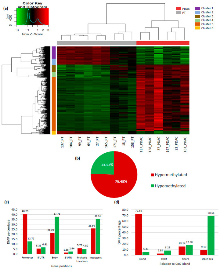
The genome-wide DNA methylation profile of six PDACs (with a minimum of 70% cellularity) compared to nine PT fresh frozen tissue samples was determined in an exploratory cohort using Infinium 450 K beadchips. The unsupervised hierarchical clustering analysis of the 10,361 differentially methylated probes (DMPs, adjusted p-value < 0.01 and ∆β ≥ 0.2) exhibited clear separation between the PDAC and PT. As shown in Figure 1a, two major clusters emerged: the first comprised PT samples and was predominantly hypomethylated, while the PDAC samples formed the second cluster that showed an overall hypermethylated profile (75.48% of the DMPs). PDAC hypermethylated probes were most frequently annotated to promoter regions (40.23%), while most hypomethylated probes were mapped to gene bodies (37.76%). Stratification by distance to CpG islands revealed that hypomethylated CpG sites most commonly encompassed open sea regions (68.66%), and hypermethylation was most common at CpG islands (72.68%) (Figure 1b–d).
Figure 1.
DNA methylation profile of pancreatic ductal adenocarcinoma. (a) Heatmap showing the unsupervised hierarchical clustering of pancreatic ductal adenocarcinoma (PDAC, with a minimum 70% cellularity) and non-tumoral adjacent pancreatic tissue (PT) according to the methylation profile of the 10,361 probes found to be differentially methylated between groups (adjusted p-value < 0.01 and ∆β ≥ 0.2). Hyper- and hypomethylation are represented in red and green, respectively. Colored bars on the left side of the heatmap represent probe clusters defined by their methylation similarities; (b) pie chart showing the percentage of hypomethylated and hypermethylated probes in PDAC relative to PT; (c) overall distribution of hypo- and hypermethylated probes according to their gene position; (d) overall distribution of hypo- and hypermethylated probes according to their relation to CpG islands.
2.2. Ca2+ Pathway Genes Are Epigenetically Altered in PDAC
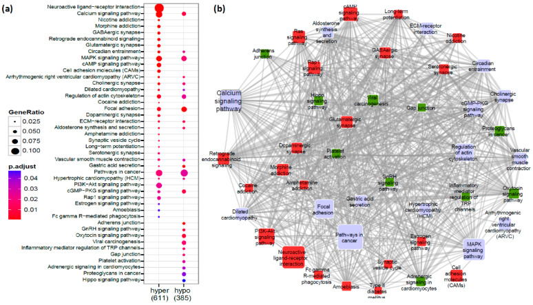
The next step was to analyze the potential biological relevance of the differentially methylated CpG sites identified in the supervised comparison between PDAC and PT samples. The DMPs mapped to 2715 genes, 1766 hypermethylated and 1100 hypomethylated, with an overlap of 151 genes among both sets. The set of 2715 genes had 611 hypermethylated and 386 hypomethylated genes annotated in the Kyoto Encyclopedia of Genes and Genomes (KEGG) database that were used as query gene sets to assess the functional enrichment of DMPs. Hypermethylated and hypomethylated genes were significantly associated with the enrichment of 36 and 25 cellular pathways, respectively (Figure S1a,b, Tables S1 and S2). Subsequently, we merged both analyses to identify the biological pathways that were frequently deregulated in PDAC by both hyper- and hypomethylation. As shown in Figure 2a, several pathways well known to be involved in cancer development were identified, such as the MAPK signaling pathway and the focal adhesion pathway [12,13]. Additionally, we observed that the Ca2+ signaling pathway had a large number of genes significantly hypo- and hypermethylated. This pathway is intrinsic to multiple aspects of cancer biology, such as tumor initiation, metastasis, and drug resistance [14]. The overlap between the differentially methylated genes of the Ca2+ signaling and other cellular pathways was investigated, and revealed that several of them were shared with other significantly enriched pathways, such as the Hippo and Ras signaling pathways (Figure 2b).
Figure 2.
Pathway enrichment analysis of differentially methylated genes (n = 996) in pancreatic ductal adenocarcinoma. (a) Functional enrichment of the hypermethylated (n = 611) and hypomethylated genes (n = 385) annotated from the differentially methylated probes (DMPs); (b) interactions between the enriched pathways evidencing the number of shared differentially methylated genes. The pathways in red and green are those enriched for hyper- and hypomethylated genes, respectively, and those in lilac are enriched for both methylation profiles.
2.3. Validation Cohort
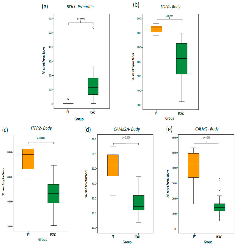
The methylation profile of five genes were assessed in a validation cohort made of 43 PDAC and 24 PT samples, with the clinical features of patients shown in Table S3. The five genes were selected with the following criteria: those with a previously described role in cancer and containing DMPs associated with a high |∆β|. One hypermethylated promoter (RYR3, Ryanodine Receptor 3) and four hypomethylated gene body probes (EGFR, Epidermal Growth Factor Receptor; ITPR2, Inositol 1,4,5-Trisphosphate Receptor Type 2; CAMK2A, Calcium/Calmodulin Dependent Protein Kinase II Alpha; and CALM2, Calmodulin 2) were chosen. Methylation levels at the CpG sites interrogated by the Infinium probe as well as surrounding CpG sites were evaluated, and the β-values obtained by pyrosequencing were strongly correlated with the Infinium assay (Figure 3 and Figure S2).
Figure 3.
Validation of the genome-wide DNA methylation results by pyrosequencing. Boxplots represent the overall methylation of pancreatic adenocarcinoma (PDAC, n = 43) and non-tumoral adjacent pancreatic tissue (PT, n = 24) samples for the following genes: (a) RYR3 (promoter); (b) EGFR (gene body); (c) ITPR2 (gene body), (d) CAMK2A (gene body); and (e) CALM2 (gene body).
2.4. Correlation between Methylation and Expression of Ca2+ Pathway Genes
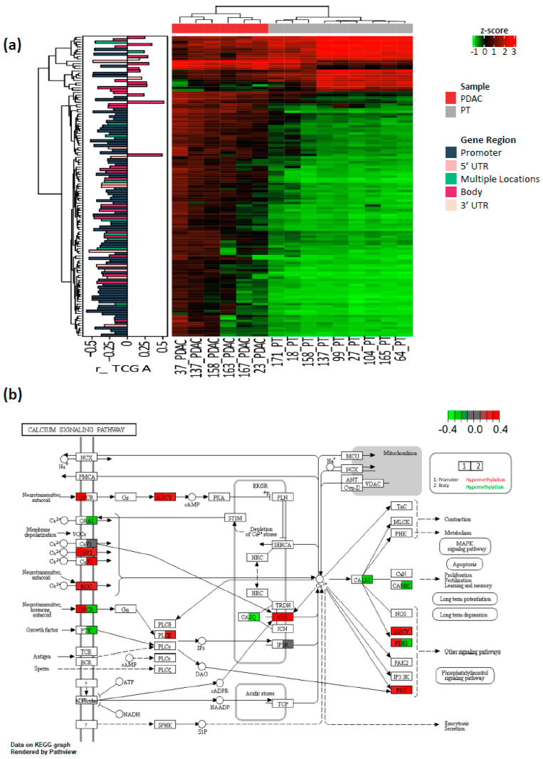
The methylation profile of five genes were assessed in a validation cohort made of 43 PDAC and 24 PT samples, with the clinical features of patients shown in Table S3. The information available in TCGA (The Cancer Genome Atlas) about PDAC was used to explore the impact of DNA methylation on the expression of the Ca2+ signaling pathway genes [15]. Among 173 DMPs in the Ca2+ pathway genes, 112 (64.73%) showed a significant correlation with gene expression (Table S4). The bars located on the left side of the heatmap (Figure 4a) indicate the correlation coefficient per gene region affected. The methylation levels of probes annotated to promoters were significantly and inversely correlated with the expression in 62.3% of the comparisons. Comparatively, the methylation profile of probes located in gene bodies showed a correlation with an expression of 59.6% in the comparisons, 64.3% inverse and 35.7% positive.
Figure 4.
Calcium (Ca2+) signaling pathway analysis. (a) Heatmap showing the methylation profile of the probes (n = 112 probes) annotated to genes involved in the Ca2+ signaling pathway for which a significant correlation (false discovery rate (FDR)-adjusted p-value < 0.05) with a gene expression was observed in the TCGA dataset (The Cancer Genome Atlas, n = 141). Heatmap was built based on the methylation data generated in the present study. The bars located on the left side of the heatmap indicate the correlation coefficient per gene region affected by differential methylation, and is based on the TCGA data; (b) schematic diagram of the Ca2+ signaling pathway. The network was built based on the Kyoto Encyclopedia of Genes and Genomes (KEGG) pathway map (KEGG: hsa04020). Each box represents a group of gene products (mostly protein) that have a common function, and its nomenclature can include one or many gene products. Gene products regarding each box are represented in Table S4. Ca2+ signaling pathway genes for which a significant correlation between methylation and expression was observed are highlighted showing the differential methylation in promoters and gene bodies identified in the present study. The red squares indicate hypermethylated genes while the green squares represent hypomethylated genes. UTR: Untranslated Region.
In Figure 4b, a schematic diagram of the Ca2+ signaling pathway based on the KEGG database shows groups of genes (grouped within boxes by function, as indicated in Table S4) with significant correlations between methylation and expression. Colored boxes indicate the differential methylation in the promoters and gene bodies identified in the present study. Genes that control intracellular Ca2+ storage, such as ryanodine receptors (RYR2, Ryanodine Receptor 2, and RYR3), were hypermethylated (promoters and gene bodies), while the inositol 1,4,5- trisphosphate receptor (ITPR1, Inositol 1,4,5-Trisphosphate Receptor Type 1) showed a heterogeneous DNA methylation profile in the body region.
2.5. Differential Expression of Ca2+ Pathway Genes Is Potentially Associated with Survival
Ca2+ pathway genes that displayed significant correlations between methylation and expression in TCGA pancreatic cancer samples (Table S4) were selected for survival analysis. We evaluated the expression impact of 40 genes on a PDAC patient’s overall survival (OS) (Table S5), including two genes that had already been investigated in the validation cohort (CALM2, and RYR3).
In the TCGA dataset (n = 146), pathologic N stage (N1 vs. N0, Hazard Ratio (HR) = 1.832 (1.039–3.23 95%CI), p = 0.0364) and residual tumor status after surgical resection (R1 vs. R0, HR = 1.899 (1.152–3.131 95%CI), p = 0.012) were associated with OS, and remained in the model.
In the univariate analysis, PDAC patients with high ADRA1A (Adrenoceptor Alpha 1A), CACNA1A (Calcium Voltage-Gated Channel Subunit Alpha1 A), CACNA1B (Calcium Voltage-Gated Channel Subunit Alpha1 B), CACNA1H (Calcium Voltage-Gated Channel Subunit Alpha1 H), CASQ2 (Calsequestrin 2) ORAI2 (ORAI Calcium Release-Activated Calcium Modulator 2), P2RX2 (Purinergic Receptor P2X 2), PDE1C (Phosphodiesterase 1C), and PRKCB (Protein Kinase C Beta) expression showed increased OS when compared to those with a low expression (Table S5). Conversely, decreased HRH1 (Histamine Receptor H1) expression was associated with increased OS (Table S5).
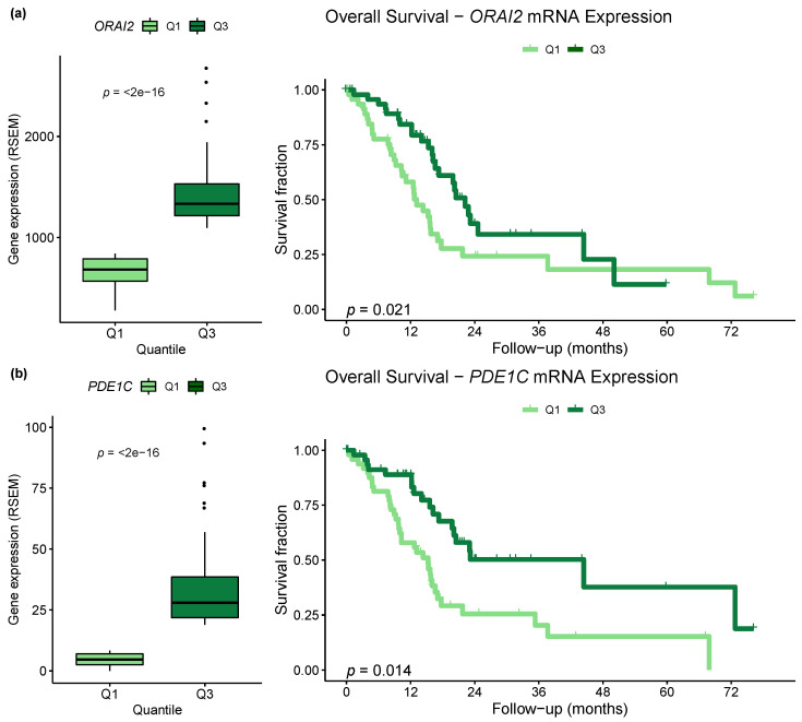
After COX regression, the ORAI2 (adjusted HR = 0.5004 (0.278–0.9009 95%CI), adjusted p = 0.021) and PDE1C (adjusted HR = 0.4625 (0.2493–0.8577 95%CI), adjusted p = 0.0144) higher expressions were shown to be independent variables associated with a better OS (Figure 5, Table S5).
Figure 5.
Impact of the expression of Ca2+ signaling genes on PDAC overall survival. mRNA expression of selected genes was divided by tertiles, and the lower (Q1) and higher (Q3) tertiles were used to classify the cases into low and high expression, respectively. The adjustments were performed for pathologic N stage and residual tumor status after surgical resection. (a) On the left side of the Figure, the boxplot representing the expression profile of ORAI2 in the low (Q1) and high (Q3) expression groups of PDAC patients. On the right side, the Kaplan–Meier survival curve shows the differences between the groups. Hazard Ratio (HR) = 0.5004 (0.278–0.9009 95%CI), p = 0.021. (b) On the left side of the Figure, the boxplot representing the expression profile of PDE1C in the low (Q1) and high (Q3) expression groups of PDAC patients. On the right side, the Kaplan–Meier survival curve shows the differences between the groups. HR = 0.4625 (0.2493–0.8577 95%CI), p = 0.0144. RSEM, RNA-Seq by Expectation-Maximization.
2.6. Aberrant Methylation in Ca2+ Genes Is an Early Change in Pancreatic Cancer
We investigated the overall methylation profile of three PT samples (18 PT, 158 PT, and 171 PT) with intermediate methylation levels at the DMPs between PDAC and PT (Figure 1a, these samples will be named PT-like from now on). To validate this discordant methylation profile, first a multidimensional scaling plot was used, showing that PT-like samples were more closely related to PDAC than PT samples, when considering principal component 1 (Figure S3a). This finding may be associated with molecular field cancerization, since these samples showed a high percentage of normal ducts (>80%), small focal fibrosis regions, and no evidence of neoplastic cell contamination (Figure S3b–d).
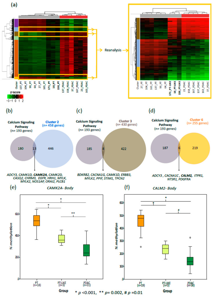
Field cancerization is defined by a set of genetic and epigenetic alterations that indicate that a specific tissue area is undergoing a transformation process, or has a predisposition to initiate such a process, and this may occur without overt morphological changes [16]. Considering that this process occurred in PT-like samples, we performed a deep analysis of DMP clusters, comparing the major similarities between PDAC and PT-like. Three clusters derived from the previous unsupervised hierarchical clustering analysis were selected for further investigation, namely, clusters 2, 3, and 6. After a new unsupervised hierarchical clustering analysis, using only the DMPs of these clusters, the PT-like samples were grouped with PDAC samples (Figure 6a). Then, we performed the pathway enrichment analysis with the corresponding annotated genes and observed that 24 differentially methylated genes were involved in the Ca2+ signaling pathway: thirteen genes from cluster 2, eight from cluster 3, and six from cluster 6; and three genes appeared in more than one cluster (Figure 6b–d, Table S6). The stepwise methylation profile in the PT, PT-like and the PDAC samples was observed in two previously validated genes (CALM2 and CAMK2A, Figure 6e,f) as well as in other 22 genes from the Ca2+ signaling pathway (Figure S4).
Figure 6.
Comparative DNA methylation analysis of the PT, PT-like and the PDAC samples. (a–d) Tissue samples with ≥70% cellularity. (a) Heatmaps highlighting the methylation profile of the probe clusters supposed to be altered in early stages of PDAC development. Heatmap on the left shows the 10,361 DMPs between the PDAC and PT. Clusters 2, 3 and 6 were selected for a detailed analysis (heatmap on the right) since their probes showed intermediate methylation levels (between PDAC and PT) in the PT-like samples; (b–d) the overlap between the genes involved in the calcium signaling pathway and the genes belonging to each selected probe cluster ((b) cluster 2; (c) cluster 3; and (d) cluster 6). (e,f) The boxplots representing the overall methylation level of CALM2 (e) and CAMK2A (f) in the PT, PT-like and the PDAC samples (≥20% cellularity). p-values were calculated using the generalized estimating equations.
3. Discussion
In this study, we used a genome-wide DNA methylation approach to identify new pathways associated with PDAC development. Pathway enrichment analysis revealed the differentially methylated genes of the Ca2+ signaling pathway. Moreover, we showed aberrant DNA methylation patterns in a few PT samples indicating the possible formation of field cancerization. Nones et al. and Mishra et al. previously used the 450 K BeadChip Array to build two PDAC methylome databases, and similar to our data (Figure 1b), they showed that the majority of DMPs in PDAC were hypermethylated (56.59% and 50.59%, respectively) and located in promoter regions [10,11]. CpG island methylation in promoters is frequently associated with gene silencing during tumorigenesis, providing an alternative mechanism to mutations by which tumor suppressor genes may be inactivated within a cancer cell [17,18]. In our exploratory cohort, as well as in other studies, the Tumor Suppressor Genes (TSGs) NPTX2 (Neuronal Pentraxin 2), CDO1 (Cysteine Dioxygenase Type 1), TFPI2 (Tissue Factor Pathway Inhibitor 2), SFRP1 (Secreted Frizzled Related Protein 1), SFRP2 (Secreted Frizzled Related Protein 2), PENK (Proenkephalin) and FOXE1 (Forkhead box E1) had a remarkable hypermethylation pattern in the PDAC group [19,20,21,22,23,24].
Key signaling pathways involved in cancer were identified as differentially methylated in our study, e.g., the MAPK signaling pathway, which is engaged in multiple proliferative cellular processes (cell differentiation, proliferation, and apoptosis) [12], and the focal adhesion pathway, which may play a role in the development and progression of cancer [13]. In addition, we observed that the Ca2+ signaling pathway had a high number of genes both significantly hypo- and hypermethylated (Figure 2a). Ca2+ is a ubiquitous intracellular messenger that controls diverse processes in cellular physiology, such as gene transcription, cell progression, cell motility, and apoptosis [25]. Although Ca2+ pathway methylation in PDAC is still poorly explored, many of its genes have already been described as differentially methylated in other solid tumors, including gastric [8], prostate [8,26], and breast cancer [8,26]. Resting cytosolic free Ca2+ is maintained at lower levels than those of extracellular space, and its equilibrium dynamics is carefully regulated by the plasmatic membrane, endoplasmic reticulum (ER), and mitochondria [27], using a “toolkit” of channels, pumps, and cytosolic buffers to control Ca2+ cell homeostasis [28]. The spatial and temporal dynamics of Ca2+ signaling results in specific cellular responses mediated by the activation of a subset of Ca2+-dependent effectors [29].
During carcinogenesis, several cellular metabolic functions become deregulated, including those related to Ca2+ signaling [30,31], and therefore, it is not surprising that changes in the expression or function of Ca2+ handling proteins impact tumorigenesis. In fact, different tumors present altered expression or mutations in the genes involved in Ca2+ signaling [32,33,34,35,36]. Different Ca2+ channels or pumps are potential therapeutic targets in different cancer subtypes, and are correlated with prognosis [8,32,37,38]. In our study, we showed that altered methylation levels of ADRA1A, CACNA1A, CACNA1B, CACNA1H, CASQ2, HRH1, ORAI2, P2RX2, PDE1C, and PRKCB, had an impact on their expression (Table S4). Furthermore, PDE1C promoter hypermethylation (Table S4, positive ∆beta, and negative r correlation) and ORAI2 gene body hypomethylation (Table S4, negative ∆beta, and negative r correlation) resulted in their downregulated expression in PDAC samples, identified as independent predictors of lower overall survival (Figure 5 and Table S5). Although we did not perform protein expression analyses (due to the limitation of the tissue samples available), the correlation between DNA methylation and mRNA expression, and the consequent impact on prognosis, which suggests a phenotypic importance of this finding. Interestingly, ORAI2 controls Ca2+ influx through the plasmatic membrane [32,39,40].
Scarce information is available regarding the methylation-related expression deregulation of genes involved in the Ca2+ signaling pathway in PDAC. In one of the few studies, the methylation profile of S100A4 (S100 Calcium Binding Protein A4), a Ca2+-binding protein previously implicated in metastasis [41], was evaluated in PDAC samples and cell lines. Hypomethylation was detected in tumors, whereas all normal pancreatic tissue samples analyzed were hypermethylated in the same region. Moreover, gene and protein expression patterns were correlated with the methylation profile, and were associated with poor tumor differentiation [42]. In addition, methylation patterns of PCDH10 (Protocadherin 10), a member of the non-clustered protocadherin family that plays an important role in Ca2+-dependent cell–cell signal transduction and adhesion, was investigated in PDAC cell lines (Capan-1, Panc-1, AsPC-1 and BxPC-3). PCDH10 promoter methylation was observed in 50% of the cell lines studied and resulted in a marked reduction of its expression [43]. Later, this tumor suppressor gene was investigated in other PDAC cell lines and, as expected, the PCDH10 promoter methylation again correlated with reduced protein expression. This study also showed high methylation levels in clinical samples (n = 23), and the presence of this methylation pattern was associated with reduced progression-free survival [44]. Using the TCGA data, Mishra et al. analyzed a genome-wide DNA methylation profile, and although the genes of the Ca2+ signaling pathway occupied the fourth position in the functional enrichment of differentially methylated probes, this finding was not further explored [11]. In addition, also using the TCGA data, another study combined the methylation and expression data of twelve solid tumors (no PDAC included) in order to identify common patterns of methylation. Alterations in the Ca2+ signaling pathway were observed in nine cancer types and AGTR1 GRIN2A (Glutamate Ionotropic Receptor NMDA Type Subunit 2A), ITPKB (Inositol-Trisphosphate 3-Kinase B), and SLC8A3 (Solute Carrier Family 8 Member A3) were repressed by hypermethylation in six of them [8].
Cytoplasmic Ca2+ concentrations rise in response to a variety of stimuli that activate Ca2+ channels in the plasma membrane (as ORAI, CaV1,2 and 3), or by release from intracellular stores through stromal interaction molecule (STIM), inositol 1,4,5-trisphosphate receptors (IP3R), and ryanodine receptors (RYRs). ORAI2, CACNA1C (Calcium Voltage-Gated Channel Subunit Alpha1 C), CACNA1G (Calcium Voltage-Gated Channel Subunit Alpha1 D), STIM1 (Stromal interaction molecule 1) and ITPR1 (Inositol 1,4,5-Trisphosphate Receptor Type 1) had an altered methylation profile in the PT-like samples (Figure 6b–d and Figure S4). ORAI1 and STIM1 are known as calcium release-activated calcium (CRAC) channels [45]. In PDAC cell lines, they are overexpressed and play a pro-survival anti-apoptotic role in this tumor [46]. In a recent study, Khan et al. have demonstrated that PDAC proliferation suppression is possible by targeting the CRAC channel with RP4010, a CRAC channel inhibitor [47]. Growth factors binding to tyrosine kinase receptors (e.g., epidermal growth factor receptor, EGFR) and G protein-coupled receptors (GPCRs; e.g., histamine receptor H1, HRH1) control the intracellular Ca2+ by plasma membrane. After binding, phospholipase C is activated and promotes the generation of inositol-1,4,5-trisphosphate (IP3) and diacylglycerol, which lead to the release of Ca2+ from the ER into the cytosol by intracellular channels (such as ITPR1 and ITPR2) [48]. In addition to ITPR2, EGFR is frequently mutated or overexpressed in various cancer types [49,50], and in PDAC particularly, EGFR is essential for KRAS-driven pancreatic carcinogenesis [51,52,53]. It is known that KRAS-activating mutations are early events in PDAC carcinogenesis and occur in ~90% of cases [54]. RAS proteins can also activate phospholipase C and generate IP3, leading to Ca2+ influx, so the most frequently mutated gene in PDAC is directly linked to Ca2+ signaling.
RYRs represent another way to control Ca2+ ER store, and in this study, RYR2 and RYR3 were found hypermethylated in PDAC, and their methylation levels were inversely correlated with mRNA expression (Table S4). These receptors are regulated by Ca2+ voltage channels and by various ions, molecules and proteins, e.g., Ca2+, Mg2+, calmodulin (CALM), Ca2+/calmodulin- dependent protein kinase II (CAMK2), and nicotine [55,56]. Once Ca2+ levels rise in the cytoplasm, they are strictly controlled by CALM. Ca2+ binding dramatically changes the conformation of CALM and increases its affinity for a large number of CALM-binding proteins, including the multifunctional CALM kinases such as CAMK2 [57]. CAMK2 phosphorylates nearly 40 different proteins, including enzymes, ion channels, kinases, and transcription factors [58], and it is overexpressed in digestive cancers, such as colorectal cancer [59,60]. Epithelial–mesenchymal transition (EMT), one of the cancer hallmarks, is also controlled by Ca2+ levels through CAMK2A. Focal adhesion kinase (FAK), which increases the turnover of cell–cell contacts, is phosphorylated by CAMK2 and is consequently upregulated [61]. It is important to mention that the FAK pathway was differentially methylated in our analysis (Figure 2a).
On top of its association with different aspects of cancer cell biology and its crosstalk with other key signaling pathways, a role for the activation of Ca2+ signaling in therapy resistance has also been proposed. The mechanisms involved include the induction of multi-drug resistance (MDR) proteins via the Ca2+-dependent transcription factor NFAT and the acquisition of stemness phenotypes by the induction of pluripotency genes and EMT [62,63,64]. In pancreatic cancer, ORAI3 and STIM1 (key components of store operated calcium entry—SOCE) have been shown to be required for TGF-β-dependent SNAIL transcription [65] and TGF-β signaling has already been associated with stroma-mediated drug resistance [66]. The EMT induction of pancreatic cells can also be mediated by acid-sensing ion channels (ASICs). ASICs sense the extracellular acidification (common in pancreatic cancer) and in response, increase intracellular Ca2+ levels and activate RhoA signaling [64]. Furthermore, Na(+)/H(+) exchangers, such as NHE1 (sodium/proton exchanger 1), interact with calmodulin in a Ca2+-dependent manner and become activated, promoting intracellular alkalization and extracellular acidification [67]. Therefore, a complex positive feedback loop involving Ca2+ signaling, TGF-β, and microenvironment acidification might be involved in EMT induction and, consequently, therapy resistance in pancreatic cancer.
It is important to mention that extracellular acidification is also involved in immune escape, which has been associated with lymph node metastasis in pancreatic cancer and is being explored as one of the most promising targets for therapy in cancer [64,68,69]. Besides the previously mentioned mechanisms of microenvironment acidification, KRAS mutations and the consequent activation of MAPK and PI3K-mTOR signaling can lead to GLUT1 upregulation. This glucose transporter can also be induced by nutrient deprivation and hypoxia. Consequently, high rates of aerobic glycolysis result in lactate production and microenvironment acidification. This not only blocks lactate export by T-cells, which also use aerobic glycolysis as an energetic source, but also downregulates interferon γ (IFN-γ) production by T-cells and natural killer cells, and polarizes macrophages to an immunosuppressive phenotype [70]. At the same time, tumor cells can cope with high extracellular acidification by modulating the expression of pH-regulating proteins and they can also use lactate as an alternative energetic source [70]. Hypoxia and the glycolytic metabolism were also associated with a wide range of epigenetic alterations both in tumor and immune cells within the tumor microenvironment, as shown in different cancer models, and HIF-2α (hypoxia-inducible factor 2 alpha) interaction with beta-catenin was recently shown in pancreatic cancer [71,72]. Indeed, immune tolerance and lymph node invasion have also been linked to the activation of the Wnt signaling pathway in PDAC [68,73]. This pathway is one of the central players in the acquisition of stemness phenotypes and one of its non-canonical forms involves Ca2+ signaling [74]. Altogether, these data suggest another connection between the alterations identified in the present study, immune escape and local dissemination in PDAC.
Based on the widespread dysregulation of Ca2+ signaling in tumors and on its impact on different steps of tumor development and progression, Ca2+ signaling receptors have been suggested as therapeutic targets for different types of cancer. The blockage of TRPVs (transient receptor potential vanilloid channels), T-type Ca2+ channels, ORAIs and TRPCs (transient receptor potential channels) have been shown to suppress the proliferation/invasion and induce cell death in different cancer models [75]. In this context, the drug repurposing of already available blockers of SOCE and T-type Ca2+ channels have been evaluated [76,77]. Conversely, the activation of overexpressed targets may also be an alternative for inducing cell death, as shown in breast and prostate cancer [78,79]. Therapy targeting the Ca2+ signaling pathway in combination with conventional chemo or radiotherapy has also been proposed. Chemotherapy drugs approved for pancreatic cancer treatment, such as Fluorouracil (5-FU) and platins, have been tested in combination with ORAI1 (ORAI Calcium Release-Activated Calcium Modulator 1) silencing and T-type Ca2+ channels blockers in hepatocellular carcinoma and ovarian cancer, respectively, with promising effects [80,81]. Nevertheless, our findings suggest that in vitro studies should be carried out to understand the role of Ca2+ pathway altered genes in pancreatic carcinogenesis before pre-clinical assays with some of these drugs are developed.
Another relevant aspect to be considered before proposing a targeted therapy against the Ca2+ signaling pathway is its participation in the maintenance of different physiological systems. Therefore, although Ca2+ signaling represents a complex pathway involved in the establishment of different cancer phenotypes, and its therapeutic targeting seems promising, it might result in a plethora of side effects. In this context, and based on the widespread alterations of DNA methylation affecting different central signaling pathways in pancreatic cancer as shown here, epigenetic therapy may represent an alternative option. This is especially relevant because a crosstalk between Ca2+ signaling and DNA methylation has already been described. Ca2+ signaling is essential to initiating epigenetic reprogramming in early embryogenesis [82], and candidate epigenetic drugs have been shown to induce the reactivation of TSGs and cell death via a Ca2+/CAMK-dependent pathway [38]. In addition to TSGs reactivation, DNA methylation inhibitors have also been shown to induce the expression of cancer-testis genes and transposable elements in cancer cells, resulting in the expression of neoantigens and in a viral mimicry state [83,84,85]. These effects, together with the reprogramming of exhausted T-cells, put epigenetic therapy as a very promising approach in combination with immune checkpoint blockade for PDAC [85].
Finally, our data suggest that the dysregulation of Ca+2 signaling by epigenetic mechanisms is an early event in pancreatic carcinogenesis, reinforcing its relevance in this process. The stepwise methylation profile of HRH1, CASQ2, and ORAI2, together with twenty-one other genes that control the Ca2+ influx, were found in PT-like samples (Figure 4b–d and Figure S4, Table S6). However, these alterations need to be validated in an independent longitudinal study, since they may not only shed light on the early molecular mechanisms driving PDAC development, but also represent potential biomarkers. DNA methylation alterations have been proposed as useful biomarkers due to their high sensitivity and specificity, and because they can be assessed by minimally invasive approaches, such as in liquid biopsies [86]. Therefore, our results bring the epigenetic dysregulation of the Ca+2 signaling pathway as a promising tool for PDAC management, either as diagnostic and prognostic biomarkers, or as therapeutic targets.
4. Materials and Methods
4.1. Patients and Sample Collection
Patients enrolled in this study were treated at Hospital de Clínicas de Porto Alegre (HCPA/UFRGS), in Southern Brazil between 2012 and 2018. Inclusion criteria were: pathology-proven diagnosis of PDAC, and no history of previous or current chemo- or radiation therapy. PDAC and PT samples were obtained during surgery or diagnostic biopsy procedures, and stored according to the biobank protocols from the hospital. Hematoxylin–eosin slides were prepared for all cases to confirm the diagnosis and assess the sample quality, which was performed by two pathologists (R.R. and S.M.). Samples with pancreatitis, necrosis, fibrosis and less than 20% cellularity were excluded [10]. The study was conducted in accordance with the Declaration of Helsinki, and the protocol was approved by the Ethics Committee of Hospital de Clínicas de Porto Alegre (Project Number: 2014–0526). All subjects gave their informed consent for inclusion before they participated in the study. Information about age at diagnosis, gender, TNM Classification of Malignant Tumors (TNM), classification, tumor location, and differentiation grade were obtained from the patient’s electronic medical record. Tumor location was categorized as pancreatic head vs. non-head.
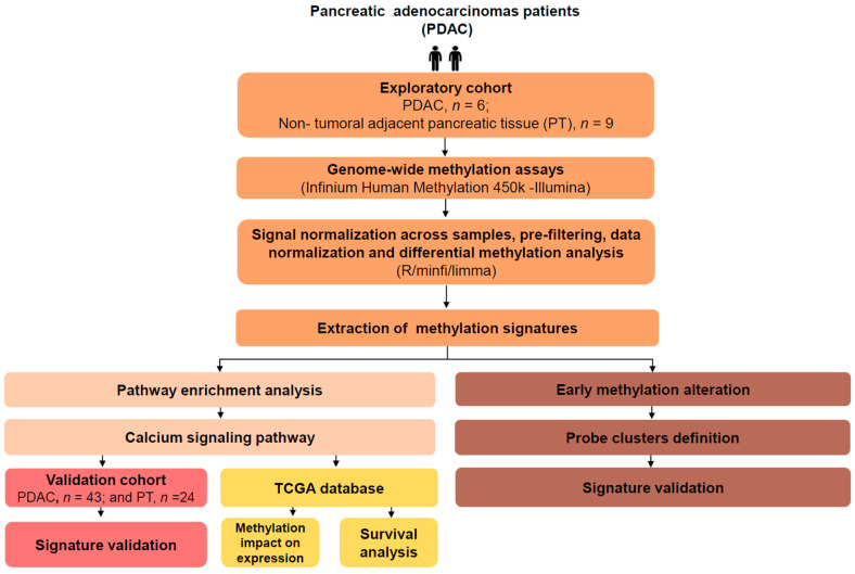
We performed a genome-wide DNA methylation profiling in an exploratory cohort using the 450 K BeadChip Array platform (Illumina Infinium Human Methylation, San Diego, CA, USA). Six PDAC and nine PT samples with content (cellularity) ≥70% were included. Differentially methylated genes were selected, and validation was performed by pyrosequencing in a biological and technical validation cohort comprising a different set including 43 PDAC and 24 PT samples. The overall experimental design is summarized in Scheme 1.
Scheme 1.
Flow chart illustrating the overall design of the study.
4.2. DNA Isolation and Bisulfite Conversion
The PureLink Genomic DNA Kit (Thermo Fisher Scientific) was used to isolate the DNA from fresh frozen tissues according to the manufacturer’s protocol and eluted DNA was quantified using Qubit V2.0 (Invitrogen, Carlsbad, USA). DNA from each sample was bisulfite converted using the EZ-DNA methylation kit (Zymo Research Corporation, California) according to the manufacturers’ protocol.
4.3. Human Methylation 450 K Array and Data Preprocessing
PDAC and PT tissues were used to establish an exploratory cohort. Illumina Infinium Human Methylation450 k (HM450 K) Bead-Chips (Illumina, San Diego, CA, USA) were used for investigating the genome-wide DNA methylation profile. Raw data were subjected to quality control, prefiltering, signal normalization across samples, and normalization using the funnorm function (R packages minfi) [87,88]. The Infinium data generated in this study were deposited in Gene Expression Omnibus (GEO) database and are available under accession number (GSE149250).
4.4. Differential Methylation Analysis
Differential methylation analysis was performed with limma R/Bioconductor package, using the M-values matrix as input [89]. Correction for multiple testing was conducted with the Benjamini–Hochberg false discovery rate (FDR) procedure. Probes were annotated for approved gene symbols using annotation files provided by the manufacturer, and all probes with ambiguous annotations were removed from further analyses. The identification of DMPs in PDAC and PT samples was performed as previously described [90]. The DMPs were identified by using a cut-off of FDR-corrected p-values < 0.01 and an absolute difference between the means of the β-values (∆β) ≥ 0.2. To verify the methylation patterns in cases and controls, a hierarchical clustering analysis was applied to the M-values from DMPs using the complete linkage method and Euclidean distance, as implemented by the heatmap.2 function from gplots R package.
4.5. Functional Enrichment Analysis
DMPs were subject to functional enrichment analysis using pathway annotations from the KEGG [91,92] and the clusterProfiler package [93] in the R/Bioconductor environment. For this purpose, the gene symbol annotation of hyper- and hypomethylated probes were analyzed separately. Only the KEGG pathways with a minimum size of 30 annotated genes were considered for further analyses. Statistical significance for the enrichment of the KEGG pathways was estimated with a hypergeometric test and adjusted to account for the multiple hypotheses testing using the FDR procedure. Pathways with a FDR-corrected p-value < 0.05 were highlighted as potentially enriched for hypo- or hypermethylated genes. Results were visualized using Cytoscape v3.4.0.
4.6. Technical and Biologic Validation
In order to biologically and technically validate the array data, we performed the pyrosequencing (PyroMark Q96 ID-Qiagen) of selected Ca2+ pathway genes in tissue samples with ≥ 20% cellularity. Genes were chosen according to the following selection criteria: only genes with DMP which were exclusively hypo- or hypermethylated; genes associated with a high |∆β|; and/or genes with previously described roles in carcinogenesis. After applying this filter, five genes were selected: RYR3 (hypermethylated) and CALM2, CAMK2A, ITPR2 and EGFR (all of them hypomethylated).
4.7. TCGA Analysis
RNA-seq and methylation microarray (HM450 K) data were downloaded using the GDC Data Transfer Tool. Only patients who had both methylation and expression data for all the target genes were included. The unsupervised hierarchical clustering (heatmap with dendrograms) and KEGG pathway figures were built with the Complex Heatmap v.2.0 and KEGG graph v.1.44.0 packages, respectively, and the correlations (FDR-adjusted p-value < 0.05) were performed with the psych v.1.8.12 package. The promoter category includes probes located in the genomic region TSS1500 and TSS200. All the PDAC samples with available mRNA data (n = 146) were used to analyze the mRNA expression prognostic values of selected Ca2+ pathway genes in PDCA samples. Only stage IV cases (n = 4) were excluded for being unresectable. The mRNA expression of selected genes was divided by tertiles and the lower and higher tertiles were used to classify cases into low and high expression, respectively. Genes for which more than one third of the cases had no detectable expression were analyzed by comparing the groups of patients with no detectable expression vs. patients with detectable expression. For the estimation of univariate survival, we used the Kaplan–Meier survival curve and statistical significance between the two groups was calculated by the log-rank test. Only genes with log-rank p-values < 0.05 remained in the model. Lymph node invasion and residual tumor status after surgical resection were selected for multivariate analysis due to their statistically significant association with OS in the univariate analysis. Cox regression using the stepwise forward method was applied. All survival analyses were performed using the package “Survival” for R.
5. Conclusions
In summary, our results show that DNA methylation alterations are involved in the deregulation of the Ca2+ signaling pathway genes. These alterations impact gene expression, the overall survival of patients, and are already seen in tumor-adjacent tissue. Future studies should be performed to validate our findings, but the data presented here indicate a significant role of epigenetic alterations in the Ca2+ signaling pathway in PDAC.
Acknowledgments
The authors thank Eduardo Chiela, Yasminne Rocha, Ivaine Sauthier, Tiago Finger Andreis, Igor Araujo Vieira, Marina Nicolau and Paula Vieira for technical support and stimulating discussions, and CNPq/Rede Genoprot (#559814/2009-7).
Abbreviations
| ADCY8 | Adenylate Cyclase 8 |
| ADRA1A | Adrenoceptor Alpha 1A |
| AGTR1 | Angiotensin II Receptor Type 1 |
| ASICs | Acid-Sensing Ion Channels |
| Ca2+ | Calcium |
| CACNA1A | Calcium Voltage-Gated Channel Subunit Alpha1 A |
| CACNA1B | Calcium Voltage-Gated Channel Subunit Alpha1 B |
| CACNA1C | Calcium Voltage-Gated Channel Subunit Alpha1 C |
| CACNA1D | Calcium Voltage-Gated Channel Subunit Alpha1 D |
| CACNA1H | Calcium Voltage-Gated Channel Subunit Alpha1 H |
| CALM2 | Calmodulin 2 |
| CAMK2A | Calcium/Calmodulin Dependent Protein Kinase II Alpha |
| CAMKK1 | Calcium/Calmodulin Dependent Protein Kinase Kinase 1 |
| CASQ2 | Calsequestrin 2 |
| CDO1 | Cysteine Dioxygenase Type 1 |
| CRAC | Calcium Release-Activated Calcium Channels |
| DMPs | Differentially Methylated Probes |
| EGFR | Epidermal Growth Factor Receptor |
| ER | Endoplasmic Reticulum |
| FAK | Focal Adhesion Kinase |
| FDR | False Discovery Rate |
| FOXE1 | Forkhead Box E1 |
| GEO | Gene Expression Omnibus |
| GRIN2A | Glutamate Ionotropic Receptor NMDA Type Subunit 2A |
| HM450K | Human Methylation 450k |
| HR | Hazard Ratio |
| HRH1 | Histamine Receptor H1 |
| IP3 | Inositol-1,4,5-Trisphosphate |
| IP3Rs | Inositol 1,4,5-Trisphosphate Receptors |
| ITPKB | Inositol-Trisphosphate 3-Kinase B |
| ITPR1 | Inositol 1,4,5-Trisphosphate Receptor Type 1 |
| ITPR2 | Inositol 1,4,5-Trisphosphate Receptor Type 2 |
| KEGG | Kyoto Encyclopedia of Genes and Genomes |
| KRAS | KRAS Proto-Oncogene, GTPase |
| Mg2+ | Magnesium |
| NHE1 | Sodium/Proton Exchanger-1 |
| NPTX2 | Neuronal Pentraxin 2 |
| ORAI1 | ORAI Calcium Release-Activated Calcium Modulator 1 |
| ORAI2 | ORAI Calcium Release-Activated Calcium Modulator 2 |
| OS | Overall Survival |
| P2RX2 | Purinergic Receptor P2X 2 |
| PCDH10 | Protocadherin 10 |
| PDAC | Pancreatic Ductal Adenocarcinoma |
| PDE1C | Phosphodiesterase 1C |
| PENK | Proenkephalin |
| PLCB1 | Phospholipase C Beta 1 |
| PRKCB | Protein Kinase C Beta |
| PT | Pancreatic Non-Tumoral Tissue |
| PT-LIKE | Pancreatic Non-Tumoral-Like Tissue |
| RSEM | RNA-Seq by Expectation-Maximization |
| RYR2 | Ryanodine Receptor 2 |
| RYR3 | Ryanodine Receptor 3 |
| RyRs | Ryanodine Receptors |
| S1004A | S100 Calcium Binding Protein A4 |
| SERCA | Sarcoendoplasmic Reticulum Ca2+ ATPases |
| SFRP1 | Secreted Frizzled Related Protein 1 |
| SFRP2 | Secreted Frizzled Related Protein 2 |
| SLC8A3 | Solute Carrier Family 8 Member A3 |
| SOCE | Store Operated Calcium Entry |
| STIM | Stromal Interaction Molecule |
| STIM | Stromal Interaction Molecule 1 |
| TCGA | Cancer Genome Atlas |
| TFPI2 | Tissue Factor Pathway Inhibitor 2 |
| TNM | TNM Classification of Malignant Tumors |
| TRPCs | Transient Receptor Potential Channels |
| TRPVs | Transient Receptor Potential Vanilloid Channels |
| TSGs | Tumor Suppressor Genes |
| UTR | Untranslated Region |
Supplementary Materials
The following are available online at https://www.mdpi.com/2072-6694/12/7/1735/s1, Figure S1: Functional enrichment of differentially methylated genes in pancreatic adenocarcinoma, Figure S2: Validation of the genome-wide DNA methylation results with pyrosequencing, Figure S3: Non-tumoral pancreatic tissue -like (PT-like) analysis, Figure S4: Methylation levels of 24 Ca2+ signaling pathway genes in PT, PT-like and PDAC samples, Table S1: Enriched KEGG terms for hypermethylated genes, Table S2: Enriched KEGG terms for hypomethylated genes, Table S3: PDAC clinicopathological features of the validation cohort (N = 43), Table S4: Correlation between probe methylation and gene expression, Table S5: Evaluation of the impact of the expression of Ca2+ signaling genes on pancreatic ductal adenocarcinoma overall survival, Table S6: Analysis of early DNA methylation alterations, Table S7: Clinicopathological features of pancreatic ductal adenocarcinoma patients included in the exploratory and validation cohort.
Author Contributions
Conceptualization C.G., S.C.S.-L., and B.A.; methodology C.G., S.C.S.-L., B.A., R.R., S.M.; and A.O.; software S.C.S.-L., M.R.-M., D.C. and P.T.d.S.-S.; validation C.G., S.C.S.-L., and B.A.; formal analysis C.G., S.C.S.-L., and B.A.; data curation C.G., S.C.S.-L., and B.A.; writing—original draft preparation C.G., S.C.S.-L., B.A., M.R.-M., and P.A.-P.; writing—review and editing C.G., S.C.S.-L., B.A., M.R.-M., D.C., P.T.d.S.-S., R.R., S.M., A.O., P.A.-P., and L.F.R.P.; supervision P.A.-P.; and L.F.R.P.; All authors read and contributed, with a critical revision of the paper, to the final version of the manuscript. All authors have read and agreed to the published version of the manuscript.
Funding
The study was supported by Fundo de Incentivo à Pesquisa—FIPE, Hospital de Clínicas de Porto Alegre (GPPG Grant #14-0526), Instituto Nacional do Câncer, Conselho Nacional de Pesquisa (CNPq) universal (Grant #424646/2016-1), scholarships from CNPq, Coordenação de aperfeiçoamento de pessoal de nível superior (CAPES), Fundação de Amparo a Pesquisa do Estado do Rio de Janeiro (FAPERJ, Grant #E-26/010.001856/2015), and Swiss Bridge Foundation (Grant # 302404).
Conflicts of Interest
All authors declare no potential financial or ethical conflicts of interest regarding the contents of this submission.
References
- 1.Collisson E.A., Maitra A. Pancreatic Cancer Genomics 2.0: Profiling Metastases. Cancer Cell. 2017;31:309–310. doi: 10.1016/j.ccell.2017.02.014. [DOI] [PubMed] [Google Scholar]
- 2.Siegel R.L., Miller K.D., Jemal A. Cancer statistics, 2018. CA Cancer J. Clin. 2018;68:7–30. doi: 10.3322/caac.21442. [DOI] [PubMed] [Google Scholar]
- 3.Chakraborty S., Baine M.J., Sasson A.R., Batra S.K. Current status of molecular markers for early detection of sporadic pancreatic cancer. Biochim. Biophys. Acta. 2011;1815:44–64. doi: 10.1016/j.bbcan.2010.09.002. [DOI] [PMC free article] [PubMed] [Google Scholar]
- 4.Rahib L., Smith B.D., Aizenberg R., Rosenzweig A.B., Fleshman J.M., Matrisian L.M. Projecting cancer incidence and deaths to 2030: The unexpected burden of thyroid, liver, and pancreas cancers in the United States. Cancer Res. 2014;74:2913–2921. doi: 10.1158/0008-5472.CAN-14-0155. [DOI] [PubMed] [Google Scholar]
- 5.Kleeff J., Korc M., Apte M., La Vecchia C., Johnson C.D., Biankin A.V., Neale R.E., Tempero M., Tuveson D.A., Hruban R.H., et al. Pancreatic cancer. Nat. Rev. Dis Prim. 2016;2:16022. doi: 10.1038/nrdp.2016.22. [DOI] [PubMed] [Google Scholar]
- 6.Orth M., Metzger P., Gerum S., Mayerle J., Schneider G., Belka C., Schnurr M., Lauber K. Pancreatic ductal adenocarcinoma: Biological hallmarks, current status, and future perspectives of combined modality treatment approaches. Radiat. Oncol. 2019;14:141. doi: 10.1186/s13014-019-1345-6. [DOI] [PMC free article] [PubMed] [Google Scholar]
- 7.Ehrlich M. DNA hypomethylation in cancer cells. Epigenomics. 2009;1:239–259. doi: 10.2217/epi.09.33. [DOI] [PMC free article] [PubMed] [Google Scholar]
- 8.Wang X.-X., Xiao F.-H., Li Q.-G., Liu J., He Y.-H., Kong Q.-P. Large-scale DNA methylation expression analysis across 12 solid cancers reveals hypermethylation in the calcium-signaling pathway. Oncotarget. 2017;8:11868–11876. doi: 10.18632/oncotarget.14417. [DOI] [PMC free article] [PubMed] [Google Scholar]
- 9.Ahuja N., Sharma A.R., Baylin S.B. Epigenetic Therapeutics: A New Weapon in the War Against Cancer. Annu. Rev. Med. 2016;67:73–89. doi: 10.1146/annurev-med-111314-035900. [DOI] [PMC free article] [PubMed] [Google Scholar]
- 10.Nones K., Waddell N., Song S., Patch A.-M., Miller D., Johns A., Wu J., Kassahn K.S., Wood D., Bailey P., et al. Genome-wide DNA methylation patterns in pancreatic ductal adenocarcinoma reveal epigenetic deregulation of SLIT-ROBO, ITGA2 and MET signaling. Int. J. cancer. 2014;135:1110–1118. doi: 10.1002/ijc.28765. [DOI] [PubMed] [Google Scholar]
- 11.Mishra N.K., Guda C. Genome-wide DNA methylation analysis reveals molecular subtypes of pancreatic cancer. Oncotarget. 2017;8:28990–29012. doi: 10.18632/oncotarget.15993. [DOI] [PMC free article] [PubMed] [Google Scholar]
- 12.Boutros T., Chevet E., Metrakos P. Mitogen-activated protein (MAP) kinase/MAP kinase phosphatase regulation: Roles in cell growth, death, and cancer. Pharmacol. Rev. 2008;60:261–310. doi: 10.1124/pr.107.00106. [DOI] [PubMed] [Google Scholar]
- 13.Maziveyi M., Alahari S.K. Cell matrix adhesions in cancer: The proteins that form the glue. Oncotarget. 2017;8:48471–48487. doi: 10.18632/oncotarget.17265. [DOI] [PMC free article] [PubMed] [Google Scholar]
- 14.Xu M., Seas A., Kiyani M., Ji K.S.Y., Bell H.N. A temporal examination of calcium signaling in cancer- from tumorigenesis, to immune evasion, and metastasis. Cell Biosci. 2018;8:25. doi: 10.1186/s13578-018-0223-5. [DOI] [PMC free article] [PubMed] [Google Scholar]
- 15.Network C.G.A.R. Integrated Genomic Characterization of Pancreatic Ductal Adenocarcinoma. Cancer Cell. 2017;32:185.e13–203.e13. doi: 10.1016/j.ccell.2017.07.007. [DOI] [PMC free article] [PubMed] [Google Scholar]
- 16.Curtius K., Wright N.A., Graham T.A. An evolutionary perspective on field cancerization. Nat. Rev. Cancer. 2018;18:19–32. doi: 10.1038/nrc.2017.102. [DOI] [PubMed] [Google Scholar]
- 17.Baylin S.B., Jones P.A. A decade of exploring the cancer epigenome-biological and translational implications. Nat. Rev. Cancer. 2011;11:726–734. doi: 10.1038/nrc3130. [DOI] [PMC free article] [PubMed] [Google Scholar]
- 18.Yamashita K., Hosoda K., Nishizawa N., Katoh H., Watanabe M. Epigenetic biomarkers of promoter DNA methylation in the new era of cancer treatment. Cancer Sci. 2018;109:3695–3706. doi: 10.1111/cas.13812. [DOI] [PMC free article] [PubMed] [Google Scholar]
- 19.Nishizawa N., Harada H., Kumamoto Y., Kaizu T., Katoh H., Tajima H., Ushiku H., Yokoi K., Igarashi K., Fujiyama Y., et al. Diagnostic potential of hypermethylation of the cysteine dioxygenase 1 gene (CDO1) promoter DNA in pancreatic cancer. Cancer Sci. 2019 doi: 10.1111/cas.14134. [DOI] [PMC free article] [PubMed] [Google Scholar]
- 20.Kinugawa Y., Uehara T., Sano K., Matsuda K., Maruyama Y., Kobayashi Y., Nakajima T., Hamano H., Kawa S., Higuchi K., et al. Methylation of Tumor Suppressor Genes in Autoimmune Pancreatitis. Pancreas. 2017;46:614–618. doi: 10.1097/MPA.0000000000000804. [DOI] [PubMed] [Google Scholar]
- 21.Matsubayashi H., Canto M., Sato N., Klein A., Abe T., Yamashita K., Yeo C.J., Kalloo A., Hruban R., Goggins M. DNA methylation alterations in the pancreatic juice of patients with suspected pancreatic disease. Cancer Res. 2006;66:1208–1217. doi: 10.1158/0008-5472.CAN-05-2664. [DOI] [PubMed] [Google Scholar]
- 22.Yang L., Yang H., Li J., Hao J., Qian J. ppENK Gene Methylation Status in the Development of Pancreatic Carcinoma. Gastroenterol. Res. Pr. 2013;2013:130927. doi: 10.1155/2013/130927. [DOI] [PMC free article] [PubMed] [Google Scholar]
- 23.Brune K., Hong S.M., Li A., Yachida S., Abe T., Griffith M., Yang D., Omura N., Eshleman J., Canto M., et al. Genetic and epigenetic alterations of familial pancreatic cancers. Cancer Epidemiol. Biomarkers Prev. 2008;17:3536–3542. doi: 10.1158/1055-9965.EPI-08-0630. [DOI] [PMC free article] [PubMed] [Google Scholar]
- 24.Sato N., Fukushima N., Maitra A., Matsubayashi H., Yeo C.J., Cameron J.L., Hruban R.H., Goggins M. Discovery of novel targets for aberrant methylation in pancreatic carcinoma using high-throughput microarrays. Cancer Res. 2003;63:3735–3742. [PubMed] [Google Scholar]
- 25.Berridge M.J., Lipp P., Bootman M.D. The versatility and universality of calcium signalling. Nat. Rev. Mol. Cell Biol. 2000;1:11–21. doi: 10.1038/35036035. [DOI] [PubMed] [Google Scholar]
- 26.Li H., Liu J.-W., Liu S., Yuan Y., Sun L.-P. Bioinformatics-Based Identification of Methylated-Differentially Expressed Genes and Related Pathways in Gastric Cancer. Dig. Dis. Sci. 2017;62:3029–3039. doi: 10.1007/s10620-017-4740-6. [DOI] [PubMed] [Google Scholar]
- 27.Pedriali G., Rimessi A., Sbano L., Giorgi C., Wieckowski M.R., Previati M., Pinton P. Regulation of Endoplasmic Reticulum-Mitochondria Ca. Front. Oncol. 2017;7:180. doi: 10.3389/fonc.2017.00180. [DOI] [PMC free article] [PubMed] [Google Scholar]
- 28.Bootman M.D. Calcium signaling. Cold Spring Harb. Perspect Biol. 2012;4:a011171. doi: 10.1101/cshperspect.a011171. [DOI] [PMC free article] [PubMed] [Google Scholar]
- 29.Eichberg J., Zhu M.X. In: Calcium Entry Channels in Non-Excitable Cells. 1st ed. Kozak J.A., Putney J.W.J., editors. CRC Press; Boca Raton, FL, USA: 2018. [Google Scholar]
- 30.Feber A., Clark J., Goodwin G., Dodson A.R., Smith P.H., Fletcher A., Edwards S., Flohr P., Falconer A., Roe T., et al. Amplification and overexpression of E2F3 in human bladder cancer. Oncogene. 2004;23:1627–1630. doi: 10.1038/sj.onc.1207274. [DOI] [PubMed] [Google Scholar]
- 31.Holcomb I.N., Young J.M., Coleman I.M., Salari K., Grove D.I., Hsu L., True L.D., Roudier M.P., Morrissey C.M., Higano C.S., et al. Comparative analyses of chromosome alterations in soft-tissue metastases within and across patients with castration-resistant prostate cancer. Cancer Res. 2009;69:7793–7802. doi: 10.1158/0008-5472.CAN-08-3810. [DOI] [PMC free article] [PubMed] [Google Scholar]
- 32.Monteith G.R., Davis F.M., Roberts-Thomson S.J. Calcium channels and pumps in cancer: Changes and consequences. J. Biol. Chem. 2012;287:31666–31673. doi: 10.1074/jbc.R112.343061. [DOI] [PMC free article] [PubMed] [Google Scholar]
- 33.Monteith G.R., McAndrew D., Faddy H.M., Roberts-Thomson S.J. Calcium and cancer: Targeting Ca2+ transport. Nat. Rev. Cancer. 2007;7:519–530. doi: 10.1038/nrc2171. [DOI] [PubMed] [Google Scholar]
- 34.Ibrahim S., Dakik H., Vandier C., Chautard R., Paintaud G., Mazurier F., Lecomte T., Guéguinou M., Raoul W. Expression Profiling of Calcium Channels and Calcium-Activated Potassium Channels in Colorectal Cancer. Cancers (Basel) 2019;11:561. doi: 10.3390/cancers11040561. [DOI] [PMC free article] [PubMed] [Google Scholar]
- 35.McAndrew D., Grice D.M., Peters A.A., Davis F.M., Stewart T., Rice M., Smart C.E., Brown M.A., Kenny P.A., Roberts-Thomson S.J., et al. ORAI1-mediated calcium influx in lactation and in breast cancer. Mol. Cancer Ther. 2011;10:448–460. doi: 10.1158/1535-7163.MCT-10-0923. [DOI] [PubMed] [Google Scholar]
- 36.Crottès D., Lin Y.T., Peters C.J., Gilchrist J.M., Wiita A.P., Jan Y.N., Jan L.Y. TMEM16A controls EGF-induced calcium signaling implicated in pancreatic cancer prognosis. Proc. Natl. Acad. Sci. USA. 2019;116:13026–13035. doi: 10.1073/pnas.1900703116. [DOI] [PMC free article] [PubMed] [Google Scholar]
- 37.Chen Y.F., Chen Y.T., Chiu W.T., Shen M.R. Remodeling of calcium signaling in tumor progression. J. Biomed. Sci. 2013;20:23. doi: 10.1186/1423-0127-20-23. [DOI] [PMC free article] [PubMed] [Google Scholar]
- 38.Raynal N.J., Lee J.T., Wang Y., Beaudry A., Madireddi P., Garriga J., Malouf G.G., Dumont S., Dettman E.J., Gharibyan V., et al. Targeting Calcium Signaling Induces Epigenetic Reactivation of Tumor Suppressor Genes in Cancer. Cancer Res. 2016;76:1494–1505. doi: 10.1158/0008-5472.CAN-14-2391. [DOI] [PMC free article] [PubMed] [Google Scholar]
- 39.Deliot N., Constantin B. Plasma membrane calcium channels in cancer: Alterations and consequences for cell proliferation and migration. Biochim. Biophys. Acta. 2015;1848:2512–2522. doi: 10.1016/j.bbamem.2015.06.009. [DOI] [PubMed] [Google Scholar]
- 40.Roberts-Thomson S.J., Chalmers S.B., Monteith G.R. The Calcium-Signaling Toolkit in Cancer: Remodeling and Targeting. Cold Spring Harb. Perspect. Biol. 2019;11 doi: 10.1101/cshperspect.a035204. [DOI] [PMC free article] [PubMed] [Google Scholar]
- 41.Boye K., Maelandsmo G.M. S100A4 and metastasis: A small actor playing many roles. Am. J. Pathol. 2010;176:528–535. doi: 10.2353/ajpath.2010.090526. [DOI] [PMC free article] [PubMed] [Google Scholar]
- 42.Rosty C., Ueki T., Argani P., Jansen M., Yeo C.J., Cameron J.L., Hruban R.H., Goggins M. Overexpression of S100A4 in pancreatic ductal adenocarcinomas is associated with poor differentiation and DNA hypomethylation. Am. J. Pathol. 2002;160:45–50. doi: 10.1016/S0002-9440(10)64347-7. [DOI] [PMC free article] [PubMed] [Google Scholar]
- 43.Qiu C., Bu X., Jiang Z. Protocadherin-10 acts as a tumor suppressor gene, and is frequently downregulated by promoter methylation in pancreatic cancer cells. Oncol. Rep. 2016;36:383–389. doi: 10.3892/or.2016.4793. [DOI] [PubMed] [Google Scholar]
- 44.Curia M.C., Fantini F., Lattanzio R., Tavano F., Di Mola F., Piantelli M., Battista P., Di Sebastiano P., Cama A. High methylation levels of PCDH10 predict poor prognosis in patients with pancreatic ductal adenocarcinoma. BMC Cancer. 2019;19:452. doi: 10.1186/s12885-019-5616-2. [DOI] [PMC free article] [PubMed] [Google Scholar]
- 45.Nguyen N.T., Han W., Cao W.-M., Wang Y., Wen S., Huang Y., Li M., Du L., Zhou Y. Store-Operated Calcium Entry Mediated by ORAI and STIM. Compr. Physiol. 2018;8:981–1002. doi: 10.1002/cphy.c170031. [DOI] [PubMed] [Google Scholar]
- 46.Kondratska K., Kondratskyi A., Yassine M., Lemonnier L., Lepage G., Morabito A., Skryma R., Prevarskaya N. Orai1 and STIM1 mediate SOCE and contribute to apoptotic resistance of pancreatic adenocarcinoma. Biochim. Biophys. Acta. 2014;1843:2263–2269. doi: 10.1016/j.bbamcr.2014.02.012. [DOI] [PubMed] [Google Scholar]
- 47.Khan H.Y., Mpilla G.B., Sexton R., Viswanadha S., Penmetsa K.V., Aboukameel A., Diab M., Kamgar M., Al-Hallak M.N., Szlaczky M., et al. Calcium Release-Activated Calcium (CRAC) Channel Inhibition Suppresses Pancreatic Ductal Adenocarcinoma Cell Proliferation and Patient-Derived Tumor Growth. Cancers (Basel) 2020;12:750. doi: 10.3390/cancers12030750. [DOI] [PMC free article] [PubMed] [Google Scholar]
- 48.Roderick H.L., Cook S.J. Ca2+ signalling checkpoints in cancer: Remodelling Ca2+ for cancer cell proliferation and survival. Nat. Rev. Cancer. 2008;8:361–375. doi: 10.1038/nrc2374. [DOI] [PubMed] [Google Scholar]
- 49.Yarden Y. The EGFR family and its ligands in human cancer. signalling mechanisms and therapeutic opportunities. Eur. J. Cancer. 2001;37(Suppl. S4):S3–S8. doi: 10.1016/S0959-8049(01)00230-1. [DOI] [PubMed] [Google Scholar]
- 50.Cui C., Merritt R., Fu L., Pan Z. Targeting calcium signaling in cancer therapy. Acta Pharm. Sin. B. 2017;7:3–17. doi: 10.1016/j.apsb.2016.11.001. [DOI] [PMC free article] [PubMed] [Google Scholar]
- 51.Navas C., Hernández-Porras I., Schuhmacher A.J., Sibilia M., Guerra C., Barbacid M. EGF receptor signaling is essential for k-ras oncogene-driven pancreatic ductal adenocarcinoma. Cancer Cell. 2012;22:318–330. doi: 10.1016/j.ccr.2012.08.001. [DOI] [PMC free article] [PubMed] [Google Scholar]
- 52.Balsano R., Tommasi C., Garajova I. State of the Art for Metastatic Pancreatic Cancer Treatment: Where Are We Now? Anticancer Res. 2019;39:3405–3412. doi: 10.21873/anticanres.13484. [DOI] [PubMed] [Google Scholar]
- 53.Shi J.L., Fu L., Wang W.D. High expression of inositol 1,4,5-trisphosphate receptor, type 2 (ITPR2) as a novel biomarker for worse prognosis in cytogenetically normal acute myeloid leukemia. Oncotarget. 2015;6:5299–5309. doi: 10.18632/oncotarget.3024. [DOI] [PMC free article] [PubMed] [Google Scholar]
- 54.Prior I.A., Lewis P.D., Mattos C. A comprehensive survey of Ras mutations in cancer. Cancer Res. 2012;72:2457–2467. doi: 10.1158/0008-5472.CAN-11-2612. [DOI] [PMC free article] [PubMed] [Google Scholar]
- 55.Schaal C., Padmanabhan J., Chellappan S. The Role of nAChR and Calcium Signaling in Pancreatic Cancer Initiation and Progression. Cancers (Basel) 2015;7:1447–1471. doi: 10.3390/cancers7030845. [DOI] [PMC free article] [PubMed] [Google Scholar]
- 56.Lanner J.T., Georgiou D.K., Joshi A.D., Hamilton S.L. Ryanodine receptors: Structure, expression, molecular details, and function in calcium release. Cold Spring Harb. Perspect Biol. 2010;2:a003996. doi: 10.1101/cshperspect.a003996. [DOI] [PMC free article] [PubMed] [Google Scholar]
- 57.Brzozowski J.S., Skelding K.A. The Multi-Functional Calcium/Calmodulin Stimulated Protein Kinase (CaMK) Family: Emerging Targets for Anti-Cancer Therapeutic Intervention. Pharmaceuticals. 2019;12:8. doi: 10.3390/ph12010008. [DOI] [PMC free article] [PubMed] [Google Scholar]
- 58.Wang Y.Y., Zhao R., Zhe H. The emerging role of CaMKII in cancer. Oncotarget. 2015;6:11725–11734. doi: 10.18632/oncotarget.3955. [DOI] [PMC free article] [PubMed] [Google Scholar]
- 59.Tai Y.L., Chen L.C., Shen T.L. Emerging roles of focal adhesion kinase in cancer. Biomed. Res. Int. 2015;2015:690690. doi: 10.1155/2015/690690. [DOI] [PMC free article] [PubMed] [Google Scholar]
- 60.Chen W., An P., Quan X.-J., Zhang J., Zhou Z.-Y., Zou L.-P., Luo H.-S. Ca(2+)/calmodulin-dependent protein kinase II regulates colon cancer proliferation and migration via ERK1/2 and p38 pathways. World J. Gastroenterol. 2017;23:6111–6118. doi: 10.3748/wjg.v23.i33.6111. [DOI] [PMC free article] [PubMed] [Google Scholar]
- 61.Bouchard V., Demers M.J., Thibodeau S., Laquerre V., Fujita N., Tsuruo T., Beaulieu J.F., Gauthier R., Vézina A., Villeneuve L., et al. Fak/Src signaling in human intestinal epithelial cell survival and anoikis: Differentiation state-specific uncoupling with the PI3-K/Akt-1 and MEK/Erk pathways. J. Cell Physiol. 2007;212:717–728. doi: 10.1002/jcp.21096. [DOI] [PubMed] [Google Scholar]
- 62.Ma X., Cai Y., He D., Zou C., Zhang P., Lo C.Y., Xu Z., Chan F.L., Yu S., Chen Y., et al. Transient receptor potential channel TRPC5 is essential for P-glycoprotein induction in drug-resistant cancer cells. Proc. Natl. Acad. Sci. USA. 2012;109:16282–16287. doi: 10.1073/pnas.1202989109. [DOI] [PMC free article] [PubMed] [Google Scholar]
- 63.Zhang S., Miao Y., Zheng X., Gong Y., Zhang J., Zou F., Cai C. STIM1 and STIM2 differently regulate endogenous Ca(2+) entry and promote TGF-β-induced EMT in breast cancer cells. Biochem. Biophys. Res. Commun. 2017;488:74–80. doi: 10.1016/j.bbrc.2017.05.009. [DOI] [PubMed] [Google Scholar]
- 64.Zhu S., Zhou H.-Y., Deng S.-C., Deng S.-J., He C., Li X., Chen J.-Y., Jin Y., Hu Z.-L., Wang F., et al. ASIC1 and ASIC3 contribute to acidity-induced EMT of pancreatic cancer through activating Ca(2+)/RhoA pathway. Cell Death Dis. 2017;8:e2806. doi: 10.1038/cddis.2017.189. [DOI] [PMC free article] [PubMed] [Google Scholar]
- 65.Bhattacharya A., Kumar J., Hermanson K., Sun Y., Qureshi H., Perley D., Scheidegger A., Singh B.B., Dhasarathy A. The calcium channel proteins ORAI3 and STIM1 mediate TGF-β induced Snai1 expression. Oncotarget. 2018;9:29468–29483. doi: 10.18632/oncotarget.25672. [DOI] [PMC free article] [PubMed] [Google Scholar]
- 66.Porcelli L., Iacobazzi R.M., Di Fonte R., Serratì S., Intini A., Solimando A.G., Brunetti O., Calabrese A., Leonetti F., Azzariti A., et al. CAFs and TGF-β Signaling Activation by Mast Cells Contribute to Resistance to Gemcitabine/Nabpaclitaxel in Pancreatic Cancer. Cancers (Basel) 2019;11:330. doi: 10.3390/cancers11030330. [DOI] [PMC free article] [PubMed] [Google Scholar]
- 67.Köster S., Pavkov-Keller T., Kühlbrandt W., Yildiz Ö. Structure of human Na+/H+ exchanger NHE1 regulatory region in complex with calmodulin and Ca2+ J. Biol. Chem. 2011;286:40954–40961. doi: 10.1074/jbc.M111.286906. [DOI] [PMC free article] [PubMed] [Google Scholar]
- 68.Argentiero A., De Summa S., Di Fonte R., Iacobazzi M.R., Porcelli L., Da Vià M., Brunetti O., Azzariti A., Silvestris N., Solimando G.A. Gene Expression Comparison between the Lymph Node-Positive and -Negative Reveals a Peculiar Immune Microenvironment Signature and a Theranostic Role for WNT Targeting in Pancreatic Ductal Adenocarcinoma: A Pilot Study. Cancers. 2019;11:942. doi: 10.3390/cancers11070942. [DOI] [PMC free article] [PubMed] [Google Scholar]
- 69.Martinez-Bosch N., Vinaixa J., Navarro P. Immune Evasion in Pancreatic Cancer: From Mechanisms to Therapy. Cancers. 2018;10:6. doi: 10.3390/cancers10010006. [DOI] [PMC free article] [PubMed] [Google Scholar]
- 70.Anderson K.G., Stromnes I.M., Greenberg P.D. Obstacles Posed by the Tumor Microenvironment to T cell Activity: A Case for Synergistic Therapies. Cancer Cell. 2017;31:311–325. doi: 10.1016/j.ccell.2017.02.008. [DOI] [PMC free article] [PubMed] [Google Scholar]
- 71.Zhang Q., Lou Y., Zhang J., Fu Q., Wei T., Sun X., Chen Q., Yang J., Bai X., Liang T. Hypoxia-inducible factor-2α promotes tumor progression and has crosstalk with Wnt/β-catenin signaling in pancreatic cancer. Mol. Cancer. 2017;16:119. doi: 10.1186/s12943-017-0689-5. [DOI] [PMC free article] [PubMed] [Google Scholar]
- 72.Camuzi D., de Amorim Í.S.S., Ribeiro Pinto L.F., Oliveira Trivilin L., Mencalha A.L., Soares Lima S.C. Regulation Is in the Air: The Relationship between Hypoxia and Epigenetics in Cancer. Cells. 2019;8:300. doi: 10.3390/cells8040300. [DOI] [PMC free article] [PubMed] [Google Scholar]
- 73.Maftouh M., Belo A.I., Avan A., Funel N., Peters G.J., Giovannetti E., Van Die I. Galectin-4 expression is associated with reduced lymph node metastasis and modulation of Wnt/β-catenin signalling in pancreatic adenocarcinoma. Oncotarget. 2014;5:5335–5349. doi: 10.18632/oncotarget.2104. [DOI] [PMC free article] [PubMed] [Google Scholar]
- 74.De A. Wnt/Ca2+ signaling pathway: A brief overview. Acta Biochim. Biophys. Sin. (Shanghai) 2011;43:745–756. doi: 10.1093/abbs/gmr079. [DOI] [PubMed] [Google Scholar]
- 75.Bong A.H.L., Monteith G.R. Calcium signaling and the therapeutic targeting of cancer cells. Biochim. Biophys. Acta. Mol. Cell Res. 2018;1865:1786–1794. doi: 10.1016/j.bbamcr.2018.05.015. [DOI] [PubMed] [Google Scholar]
- 76.Yang S., Zhang J.J., Huang X.-Y. Orai1 and STIM1 are critical for breast tumor cell migration and metastasis. Cancer Cell. 2009;15:124–134. doi: 10.1016/j.ccr.2008.12.019. [DOI] [PubMed] [Google Scholar]
- 77.Sallán M.C., Visa A., Shaikh S., Nàger M., Herreros J., Cantí C. T-type Ca(2+) Channels: T for Targetable. Cancer Res. 2018;78:603–609. doi: 10.1158/0008-5472.CAN-17-3061. [DOI] [PubMed] [Google Scholar]
- 78.Peters A.A., Jamaludin S.Y.N., Yapa K.T.D.S., Chalmers S., Wiegmans A.P., Lim H.F., Milevskiy M.J.G., Azimi I., Davis F.M., Northwood K.S., et al. Oncosis and apoptosis induction by activation of an overexpressed ion channel in breast cancer cells. Oncogene. 2017;36:6490–6500. doi: 10.1038/onc.2017.234. [DOI] [PubMed] [Google Scholar]
- 79.Zhang L., Barritt G.J. Evidence that TRPM8 is an androgen-dependent Ca2+ channel required for the survival of prostate cancer cells. Cancer Res. 2004;64:8365–8373. doi: 10.1158/0008-5472.CAN-04-2146. [DOI] [PubMed] [Google Scholar]
- 80.Tang B.-D., Xia X., Lv X.-F., Yu B.-X., Yuan J.-N., Mai X.-Y., Shang J.-Y., Zhou J.-G., Liang S.-J., Pang R.-P. Inhibition of Orai1-mediated Ca(2+) entry enhances chemosensitivity of HepG2 hepatocarcinoma cells to 5-fluorouracil. J. Cell. Mol. Med. 2017;21:904–915. doi: 10.1111/jcmm.13029. [DOI] [PMC free article] [PubMed] [Google Scholar]
- 81.Dziegielewska B., Casarez E.V., Yang W.Z., Gray L.S., Dziegielewski J., Slack-Davis J.K. T-Type Ca2+ Channel Inhibition Sensitizes Ovarian Cancer to Carboplatin. Mol. Cancer Ther. 2016;15:460–470. doi: 10.1158/1535-7163.MCT-15-0456. [DOI] [PMC free article] [PubMed] [Google Scholar]
- 82.Whitaker M. Calcium at fertilization and in early development. Physiol. Rev. 2006;86:25–88. doi: 10.1152/physrev.00023.2005. [DOI] [PMC free article] [PubMed] [Google Scholar]
- 83.Chiappinelli K.B., Strissel P.L., Desrichard A., Li H., Henke C., Akman B., Hein A., Rote N.S., Cope L.M., Snyder A., et al. Inhibiting DNA Methylation Causes an Interferon Response in Cancer via dsRNA Including Endogenous Retroviruses. Cell. 2016;164:1073. doi: 10.1016/j.cell.2015.10.020. [DOI] [PubMed] [Google Scholar]
- 84.Roulois D., Loo Yau H., Singhania R., Wang Y., Danesh A., Shen S.Y., Han H., Liang G., Jones P.A., Pugh T.J., et al. DNA-Demethylating Agents Target Colorectal Cancer Cells by Inducing Viral Mimicry by Endogenous Transcripts. Cell. 2015;162:961–973. doi: 10.1016/j.cell.2015.07.056. [DOI] [PMC free article] [PubMed] [Google Scholar]
- 85.Jones P.A., Ohtani H., Chakravarthy A., De Carvalho D.D. Epigenetic therapy in immune-oncology. Nat. Rev. Cancer. 2019;19:151–161. doi: 10.1038/s41568-019-0109-9. [DOI] [PubMed] [Google Scholar]
- 86.Gai W., Sun K. Epigenetic Biomarkers in Cell-Free DNA and Applications in Liquid Biopsy. Genes (Basel) 2019;10:32. doi: 10.3390/genes10010032. [DOI] [PMC free article] [PubMed] [Google Scholar]
- 87.Aryee M.J., Jaffe A.E., Corrada-Bravo H., Ladd-Acosta C., Feinberg A.P., Hansen K.D., Irizarry R.A. Minfi: A flexible and comprehensive Bioconductor package for the analysis of Infinium DNA methylation microarrays. Bioinformatics. 2014;30:1363–1369. doi: 10.1093/bioinformatics/btu049. [DOI] [PMC free article] [PubMed] [Google Scholar]
- 88.Fortin J.P., Labbe A., Lemire M., Zanke B.W., Hudson T.J., Fertig E.J., Greenwood C.M., Hansen K.D. Functional normalization of 450k methylation array data improves replication in large cancer studies. Genome Biol. 2014;15:503. doi: 10.1186/s13059-014-0503-2. [DOI] [PMC free article] [PubMed] [Google Scholar]
- 89.Du P., Zhang X., Huang C.-C., Jafari N., Kibbe W.A., Hou L., Lin S.M. Comparison of Beta-value and M-value methods for quantifying methylation levels by microarray analysis. BMC Bioinform. 2010;11:587. doi: 10.1186/1471-2105-11-587. [DOI] [PMC free article] [PubMed] [Google Scholar]
- 90.Martin M., Ancey P.B., Cros M.P., Durand G., Le Calvez-Kelm F., Hernandez-Vargas H., Herceg Z. Dynamic imbalance between cancer cell subpopulations induced by transforming growth factor beta (TGF-β) is associated with a DNA methylome switch. BMC Genom. 2014;15:435. doi: 10.1186/1471-2164-15-435. [DOI] [PMC free article] [PubMed] [Google Scholar]
- 91.Kanehisa M., Goto S. KEGG: Kyoto encyclopedia of genes and genomes. Nucleic Acids Res. 2000;28:27–30. doi: 10.1093/nar/28.1.27. [DOI] [PMC free article] [PubMed] [Google Scholar]
- 92.Kanehisa M., Sato Y., Kawashima M., Furumichi M., Tanabe M. KEGG as a reference resource for gene and protein annotation. Nucleic Acids Res. 2016;44:D457–D462. doi: 10.1093/nar/gkv1070. [DOI] [PMC free article] [PubMed] [Google Scholar]
- 93.Yu G., Wang L.G., Han Y., He Q.Y. clusterProfiler: An R package for comparing biological themes among gene clusters. OMICS. 2012;16:284–287. doi: 10.1089/omi.2011.0118. [DOI] [PMC free article] [PubMed] [Google Scholar]
Associated Data
This section collects any data citations, data availability statements, or supplementary materials included in this article.


