Abstract
Background: Complications of coronavirus disease 2019 (COVID-19) include coagulopathy. We performed a meta-analysis on the association of COVID-19 severity with changes in hemostatic parameters. Methods: Data on prothrombin time (PT), activated partial thromboplastin time (aPTT), D-Dimer, platelets (PLT), or fibrinogen in severe versus mild COVID-19 patients, and/or in non-survivors to COVID-19 versus survivors were systematically searched. The standardized mean difference (SMD) was calculated. Results: Sixty studies comparing 5487 subjects with severe and 9670 subjects with mild COVID-19 documented higher PT (SMD: 0.41; 95%CI: 0.21, 0.60), D-Dimer (SMD: 0.67; 95%CI: 0.52, 0.82), and fibrinogen values (SMD: 1.84; 95%CI: 1.21, 2.47), with lower PLT count (SMD: −0.74; 95%CI: −1.01, −0.47) among severe patients. Twenty-five studies on 1511 COVID-19 non-survivors and 6287 survivors showed higher PT (SMD: 0.67; 95%CI: 0.39, 0.96) and D-Dimer values (SMD: 3.88; 95%CI: 2.70, 5.07), with lower PLT count (SMD: −0.60, 95%CI: −0.82, −0.38) among non-survivors. Regression models showed that C-reactive protein values were directly correlated with the difference in PT and fibrinogen. Conclusions: Significant hemostatic changes are associated with COVID-19 severity. Considering the risk of fatal complications with residual chronic disability and poor long-term outcomes, further studies should investigate the prognostic role of hemostatic parameters in COVID-19 patients.
Keywords: SARS-CoV-2, COVID-19, hemostasis, thrombosis, coagulation, disability, outcome
1. Introduction
In December 2019, a cluster of patients with pneumonia of unknown origin was linked to a seafood wholesale market in Wuhan, China. A novel coronavirus, named 2019-nCoV or SARS-CoV-2, was isolated from human airway epithelial cells belonging to the subgenus sarbecovirus, Orthocoronavirinae subfamily [1]. Despite extensive control efforts implemented as part of a global containment strategy to minimize exportation, SARS-CoV-2 showed an international spread that led to a pandemic diffusion [2]. Epidemiologic data indicate that SARS-CoV-2 causes the Coronavirus Disease 2019 (COVID-19), a syndrome with a wide spectrum of clinical presentations [3,4,5]. In particular, the disease is characterized by fever, dyspnea, dry cough, and fatigue. Pulmonary imaging has shown multiple ground glass shadows and infiltrative shadows in both lungs. Severe cases have shown to develop into acute respiratory distress syndrome (ARDS) and septic shock [2,3,5,6], requiring specialized management at Intensive Care Units (ICU) [1,3,7,8,9,10], with poor long-term outcomes and residual chronic disability [11,12,13]. Coagulopathy is very common in patients in ICU and often indicates organ dysfunction or underlying diseases. In particular, disseminated intravascular coagulation (DIC) accompanies the clinical progression from systemic inflammatory response syndrome to severe sepsis and septic shock. In turn, progression to DIC leads to organ dysfunction, which is associated with increased mortality [14]. In patients without evidence of coagulopathy, higher D-dimer levels are associated with clinical severity among patients admitted to the ICU [15].
The aim of the present study is to perform a systematic review and meta-analyses to evaluate the association of COVID-19 severity with changes in hemostasis parameters. Moreover, we implemented some meta-regression models to evaluate the impact of demographic, clinical variables, and inflammatory markers on the evaluated outcomes.
2. Materials and Methods
We developed a protocol for this systematic review of literature data, defining the search strategy, the outcomes, the inclusion and exclusion criteria, and the statistical methods.
2.1. Data Sources and Searches
To detect all available studies on the association between hemostatic parameters and the severity of COVID-19, we conducted a systematic literature search in the main electronic databases (PubMed, Scopus, Web of Science, EMBASE) according to Preferred Reporting Items for Systematic Reviews and Meta-Analyses (PRISMA) guidelines [16]. The last search was performed on 16 June 2020 with no language restriction, by using the following terms in any possible association; COVID-19, SARS-CoV-2, coagulative, coagulation, hemostatic, hemostasis, prothrombin time, activated partial thromboplastin time, D-Dimer, platelet, and fibrinogen.
Moreover, the reference lists of all included articles were manually consulted. In case of missing data among studies fulfilling the inclusion criteria, the authors were contacted by e-mail to try to claim the original data. Two authors (RL and AS) analyzed each article and separately performed the extraction of data. In case of disagreement, a third investigator was consulted (ADM). Discrepancies were resolved by consensus. Overall, selection results showed a high inter-reader agreement (κ = 1.00) and were reported according to PRISMA flowchart (Supplemental Figure S1).
2.2. Study Selection
According to the aforementioned protocol, all studies reporting data about the association of hemostatic parameters with COVID-19 severity were included. Case reports, reviews, and articles on animal models were excluded.
2.3. Data Extraction and Quality Assessment
We included in the analysis studies providing data on prothrombin time (PT), activated partial thromboplastin time (aPTT), D-Dimer, platelet count (PLT), or fibrinogen; in study group 1: severe COVID-19 (cases) and mild COVID-19 patients (controls) and in study group 2: subjects dead with COVID-19 (cases) and subjects survived to COVID-19 (controls).
In each study, besides hemostatic parameters, data regarding sample size, mean age of enrolled subjects, prevalence of male sex, mean C-reactive protein (CRP) levels, prevalence of diabetes, and prevalence of hypertension were extracted. Formal quality score adjudication was not used as most included studies were case series or small cohort studies.
2.4. Data Synthesis and Analysis
Data synthesis and analyses were performed by using comprehensive meta-analysis (Version 2, Biostat, Englewood, NJ, USA, 2005). Because of the heterogeneity in laboratory techniques, reference values, and units of measurement, differences in PT, aPTT, D-Dimer, PLT, and fibrinogen between cases and controls were expressed as standardized mean difference (SMD) with 95% confidence intervals (95%CI), which represents the difference between the weighted mean and the standard deviation of the outcome in cases and controls.
For all evaluated outcomes, data from study group 1 and study group 2 were separately analyzed. The pooled effect was tested using Z-scores, with p < 0.05 being considered statically significant. We evaluated statistical heterogeneity among studies with chi-squared Cochran’s Q test and with I2 index, which measures the inconsistency among results of studies and defines the proportion of total variation in study estimates that is due to heterogeneity rather than sampling error. In particular, an I2 value of 25% corresponds to low, 25–50% to moderate, and 50% to high heterogeneity [17]. Funnel plots of the standard difference in means vs. the standard error were used as a graphical representation of publication bias. To detect a potential small-study effect, visual inspection of funnel plots asymmetry was performed. Moreover, the Egger’s test was used to assess publication bias over and above any subjective evaluation, with a p < 0.10 being considered statistically significant [18]. In case of a significant publication bias, the Duval and Tweedie’s trim and fill method allowed for the assessment of the adjusted effect size [19].
To be as conservative as possible, the random-effect method was used to take into account the heterogeneity among the included studies.
To evaluate whether demographic variables (mean age and prevalence of male sex) and clinical data (CRP levels, prevalence of diabetes, and prevalence of hypertension) may impact on differences in the hemostatic outcomes between cases and controls, we implemented multiple meta-regression analyses with differences in PT, aPTT, D-Dimer, PLT, and fibrinogen as dependent variables (y) and the above-mentioned covariates as independent variables (x). Comprehensive meta-analysis (Version 2, Biostat, Englewood, NJ, USA, 2005) was used for the multivariate approach.
3. Results
After excluding duplicate results, the search retrieved 518 articles. Of them, we excluded 194 because they were found to be off topic after scanning the title and/or the abstract, and 197 reviews/comments/case reports or studies lacking data of interest. Another 43 studies were eliminated after a full evaluation of the texts. Four studies [20,21,22,23] reported data on the same study sample. Thus, most recent studies with the largest samples were included in the analysis [20,23] (Supplemental Figure S1).
Overall, 84 studies were included in the final analyses. Of them, 60 studies reported on a total of 5487 subjects with severe COVID-19 and 9670 subjects with mild COVID-19 [6,8,24,25,26,27,28,29,30,31,32,33,34,35,36,37,38,39,40,41,42,43,44,45,46,47,48,49,50,51,52,53,54,55,56,57,58,59,60,61,62,63,64,65,66,67,68,69,70,71,72,73,74,75,76,77,78,79,80,81].
In addition, 25 studies on a total of 1511 subjects dead with COVID-19 and 6287 subjects survived to COVID-19 were included in the meta-analysis [20,23,26,74,82,83,84,85,86,87,88,89,90,91,92,93,94,95,96,97,98,99,100,101,102].
3.1. Study Characteristics
Principal characteristics of subjects enrolled in included studies are shown in Table 1. The number of patients varied from 17 to 4468, the male sex represented 53.9% of the study population (range: 9.1–81.0%), and the mean age was 54.4 years (range: 35.6–70.7). Diabetes was found in 16.5% (range: 3.2–100%) of patients and hypertension in 28.7% (range: 5.0–83.0%). The mean value of CRP in patients enrolled was 47.7 mg/L (range: 3.14–192.0).
Table 1.
Clinical and demographic characteristics of patients with COVID-19 included in different studies.
| Study Group 1—Mild Disease vs. Severe Disease | |||||||||||
| Author | N of Patients (n) | Male Gender (%) | Age (Years) | CRP (mg/L) | Diabetes (%) | Hypertension (%) | Reported Outcome | ||||
| PT | aPTT | D-Dimer | PLT | FYB | |||||||
| Cen Y | 652 | -- | -- | 17.53 | -- | -- | NO | NO | YES | YES | NO |
| Chen G | 21 | 81 | 57 | 92 | 14.3 | 23.8 | YES | YES | YES | YES | NO |
| Chen R a | 500 | 57.1 | 56 | 38.30 | 11.1 | 27 | YES | YES | YES | YES | NO |
| Deng Q | 112 | 50.9 | 62.45 | 88.00 | 17 | 32.1 | NO | NO | YES | NO | NO |
| Di Micco P | 67 | 70 | 14.60 | NO | NO | YES | YES | YES | |||
| Ding X | 72 | 45.8 | 49.75 | 6.9 | 12.5 | NO | NO | NO | YES | NO | |
| Dong Y | 147 | 42.9 | 47.00 | 22.62 | NO | NO | YES | NO | YES | ||
| Du RH | 109 | 67.9 | 70.7 | 85.70 | 31.2 | 59.6 | YES | YES | YES | YES | NO |
| Duan J | 348 | 52.9 | 45 | 11.29 | 3.2 | 7.8 | YES | YES | YES | YES | YES |
| Feng Y | 406 | 56.9 | 52.50 | 24.96 | 10.3 | 23.7 | NO | NO | YES | YES | YES |
| Fu J | 75 | 60 | 46.6 | 59.56 | 5.3 | 9.3 | NO | NO | YES | NO | YES |
| Gao Y | 43 | 60.5 | 44.4 | 32.2 | -- | -- | YES | YES | YES | NO | YES |
| Gong J | 189 | 46.6 | 48.79 | -- | -- | -- | NO | YES | YES | YES | NO |
| Guan W | 1099 | 9.1 | 46.4 | -- | 7.4 | 15 | NO | NO | NO | YES | NO |
| Han H | 84 | -- | -- | -- | -- | -- | YES | YES | YES | NO | YES |
| Huang C | 41 | 73.2 | 49.5 | -- | 19.5 | 14.6 | YES | YES | YES | NO | NO |
| Li H | 116 | 56.8 | 62.05 | 54.93 | -- | -- | NO | NO | NO | YES | NO |
| Li J a | 75 | 55.97 | 59.50 | 3.14 | -- | 32.84 | YES | YES | YES | YES | YES |
| Liang W | 1590 | 57.3 | 48.9 | 34.80 | 8.2 | 16.9 | YES | YES | YES | YES | NO |
| Liu F | 134 | 47 | 51.5 | -- | 7.5 | 20.1 | NO | NO | YES | NO | NO |
| Liu M | 30 | -- | -- | -- | -- | -- | NO | NO | YES | NO | NO |
| Liu QS | 150 | 52.7 | 42.19 | 11.3 | 19.3 | YES | YES | YES | YES | YES | |
| Liu X | 112 | -- | 56 | 46 | -- | -- | YES | YES | YES | YES | YES |
| Liu Y a | 109 | 54.1 | 54.5 | 33.3 | 11 | 33.9 | NO | NO | YES | NO | YES |
| Liu Y b | 76 | 64.5 | 46.50 | -- | -- | -- | YES | YES | YES | NO | YES |
| Lv Z | 270 | 49.44 | 59.25 | 47.23 | 9.89 | 20.9 | NO | NO | YES | NO | NO |
| Ma J | 37 | 54.1 | 63.25 | -- | -- | -- | NO | NO | YES | YES | NO |
| Mao L | 214 | 40.7 | 52.7 | -- | -- | -- | NO | NO | YES | YES | NO |
| Medetalibeyoğlu A | 68 | 69.1 | 56.52 | -- | -- | -- | NO | YES | YES | YES | YES |
| Middeldorp S | 198 | 65.7 | 60.8 | -- | -- | -- | NO | NO | YES | YES | NO |
| Mo P | 155 | 55.5 | 53.9 | 43.9 | 9.7 | 23.9 | NO | NO | YES | YES | NO |
| Peng Y | 112 | 47.3 | 58.6 | 49.9 | -- | 83 | YES | YES | NO | NO | NO |
| Petrilli CM | 4468 | 60.2 | -- | 102.84 | 33.8 | 60.6 | NO | NO | YES | NO | NO |
| Qu R | 30 | -- | 58.9 | -- | -- | -- | NO | NO | NO | YES | NO |
| Rastrelli G | 27 | -- | 62.60 | 26.63 | 29.6 | 51.8 | NO | NO | YES | NO | YES |
| Shang W | 443 | 49.7 | 55.50 | 23.77 | 14.2 | 29.6 | NO | NO | YES | YES | NO |
| Smadja DM | 40 | 56.52 | 110.60 | 20 | 40 | NO | NO | YES | YES | YES | |
| Sun S | 116 | 51.7 | 49.50 | -- | -- | -- | NO | NO | NO | YES | NO |
| Sun Y | 18 | -- | -- | -- | -- | -- | YES | NO | YES | YES | YES |
| To KK | 23 | 56.5 | 59.00 | -- | 17 | 26 | NO | NO | NO | YES | NO |
| Wan S | 135 | 54.1 | 47.4 | 37.2 | 8.9 | 9.6 | YES | YES | YES | YES | NO |
| Wang CZ | 85 | 52.9 | 59.40 | 43.27 | 11.8 | 25.9 | YES | YES | YES | YES | NO |
| Wang D a | 138 | 45.7 | 54.4 | -- | 10.1 | 31.2 | YES | YES | YES | YES | NO |
| Wang F | 50 | 57 | 57.11 | 68.88 | -- | -- | NO | NO | YES | NO | NO |
| Wang R | 125 | 56.8 | 38.76 | 17.76 | -- | -- | NO | NO | NO | YES | NO |
| Wu C | 201 | 63.7 | 52.3 | 55.3 | 10.9 | 19.4 | YES | YES | YES | YES | NO |
| Wu J | 280 | 53.93 | 43.12 | 7.33 | -- | -- | YES | YES | YES | YES | NO |
| Xie H | 79 | 55.7 | 58.50 | 20.70 | 10.1 | 17.7 | NO | NO | YES | NO | NO |
| Xu B | 125 | 55 | 60.88 | 32.69 | -- | 26.7 | YES | NO | YES | NO | NO |
| Yang AP | 93 | 62.2 | 46.4 | -- | 22.5 | 24.7 | NO | NO | YES | NO | YES |
| Yang F | 52 | 53.8 | 64.50 | 30.80 | -- | -- | NO | NO | YES | NO | NO |
| Yang Q | 136 | 48.5 | 55.00 | 42.03 | 14.7 | 27.1 | YES | YES | YES | YES | NO |
| Yao Q a | 108 | 39.8 | 49.75 | 13.13 | 4.6 | 14.8 | NO | NO | YES | YES | NO |
| Young BE | 18 | 50 | 47.1 | 30.3 | 5.6 | 22.2 | NO | NO | NO | YES | NO |
| Zhang G | 221 | 48.9 | 53.88 | -- | 24.4 | 35.8 | YES | YES | YES | YES | NO |
| Zhang JJ | 140 | 50.7 | 57.0 | 34.2 | 12.1 | 30 | NO | NO | YES | NO | NO |
| Zheng F | 161 | 49.7 | 45.13 | 20.15 | 4.3 | 13.7 | NO | NO | NO | YES | NO |
| Zheng Y | 141 | 52.4 | 47.00 | -- | -- | -- | NO | NO | NO | YES | NO |
| Zhou Y | 17 | 35.3 | -- | -- | -- | -- | NO | NO | YES | NO | NO |
| Zou Y | 303 | 52.1 | 51.2 | -- | -- | -- | YES | YES | YES | NO | YES |
| Study Group 2—Deaths vs. Survivors | |||||||||||
| Author | N of Patients (n) | Male Gender (%) | Age (Years) | CRP (mg/L) | Diabetes (%) | Hypertension (%) | Reported Outcome | ||||
| PT | aPTT | D-Dimer | PLT | FYB | |||||||
| Auld S | 217 | 54.8 | 63.75 | 192.00 | 45.6 | 61.7 | NO | NO | YES | NO | NO |
| Chen R b | 548 | 57.1 | 56 | 38.3 | 11.1 | 27 | YES | YES | YES | YES | NO |
| Chen T | 274 | 62.4 | 58.6 | 61.7 | 17.2 | 33.9 | YES | NO | YES | YES | NO |
| Desborough MJR | 66 | 73 | 58.25 | 190.25 | 41 | 45 | NO | NO | YES | YES | YES |
| Fan H | 73 | 67.12 | 58.36 | 93.84 | -- | 32.88 | YES | NO | YES | YES | NO |
| Huang H | 50 | 46 | 35.6 | 30.64 | -- | -- | YES | YES | YES | YES | YES |
| Li J b | 161 | -- | 55.40 | -- | -- | -- | YES | YES | YES | YES | NO |
| Li L | 93 | 44 | 51 | 14.80 | -- | 5 | YES | YES | YES | YES | NO |
| Li Q | 1449 | 51 | 55.5 | -- | -- | -- | YES | YES | YES | YES | YES |
| Lodigiani C | 285 | -- | -- | -- | -- | -- | NO | NO | YES | NO | NO |
| Luo X | 298 | 50.3 | 55.75 | 34.03 | 15.1 | 28.9 | NO | NO | YES | YES | NO |
| Masetti C | 229 | 64.6 | 60.7 | 8.6 | 18.8 | 38 | NO | NO | YES | YES | YES |
| Ruan Q | 150 | 68.0 | 56.5 | 76 | 16.7 | 34.7 | NO | NO | NO | YES | NO |
| Tang N | 449 | 59.7 | 65.1 | -- | -- | -- | YES | YES * | YES | YES | YES |
| Violi F | 319 | 60.4 | 65.61 | 65.98 | 18.6 | 54.6 | NO | NO | YES | YES | NO |
| Wang D b | 107 | 53.3 | 50.75 | 10.3 | 24.3 | YES | YES | YES | YES | NO | |
| Wang K | 296 | 47.3 | 47.32 | 15.05 | 10.1 | 14.2 | YES | YES | YES | NO | YES |
| Yan Y | 48 | 68.8 | 69.4 | -- | 100 | 50 | YES | YES | YES | YES | YES |
| Yang X | 1476 | 52.6 | 57 | -- | -- | -- | YES * | NO | NO | YES | NO |
| Yao Q b | 108 | 39.8 | 49.75 | 13.13 | 4.6 | 14.8 | NO | NO | YES | YES | NO |
| Zhang F | 53 | 25.8 | -- | 29.14 | -- | -- | YES | YES | YES | YES | YES |
| Zhang J | 19 | 57.9 | 68.75 | 106.79 | -- | -- | NO | NO | YES | NO | NO |
| Zhang S | 315 | 55.55 | 56 | 39.13 | 13.02 | 24.76 | YES | YES | YES | YES | YES |
| Zhao X | 532 | 46.2 | 49.10 | -- | 11.1 | 20.3 | NO | NO | NO | YES | NO |
| Zhou F | 191 | 36.1 | 56.7 | -- | 18.8 | 30.4 | YES | NO | YES | YES | NO |
3.2. Study Group 1 (Severe COVID-19 vs. Mild COVID-19)
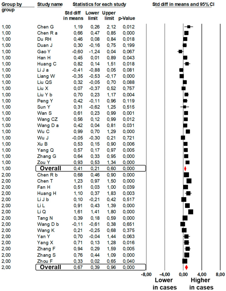
A total of 23 studies [6,8,25,26,31,32,35,38,40,41,44,45,46,54,61,63,64,67,68,70,73,76,81] showed significantly higher PT values in 1041 subjects with severe COVID-19 as compared to 3835 subjects with mild COVID-19 (SMD: 0.41; 95%CI: 0.21, 0.60, p < 0.001, Figure 1). The heterogeneity among studies was significant (I2: 84.3%, p < 0.001) and was not reduced by the exclusion of one study at a time.
Figure 1.
Forest plots of the difference in prothrombin time values between subjects with severe and those with mild COVID-19 (Group 1) and between non-survivors and survivors to COVID-19 (Group 2).
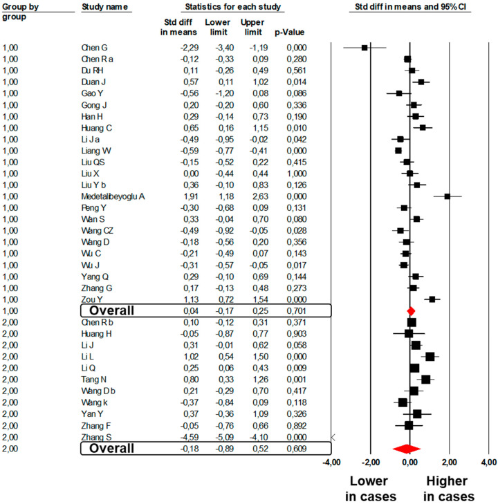
In contrast, no difference was found in 23 studies [6,8,25,26,31,32,35,36,38,40,41,44,45,46,51,54,63,64,67,68,73,76,81] reporting aPTT values between 1018 subjects with severe COVID-19 and 3976 subjects with mild disease (SMD: 0.04; 95%CI: −0.17, 0.25, p = 0.701; I2: 86.1%, p < 0.001, Figure 2).
Figure 2.
Forest plots of the difference in activated partial thromboplastin time values between subjects with severe and those with mild COVID-19 (Group 1) and between non-survivors and survivors to COVID-19 (Group 2).
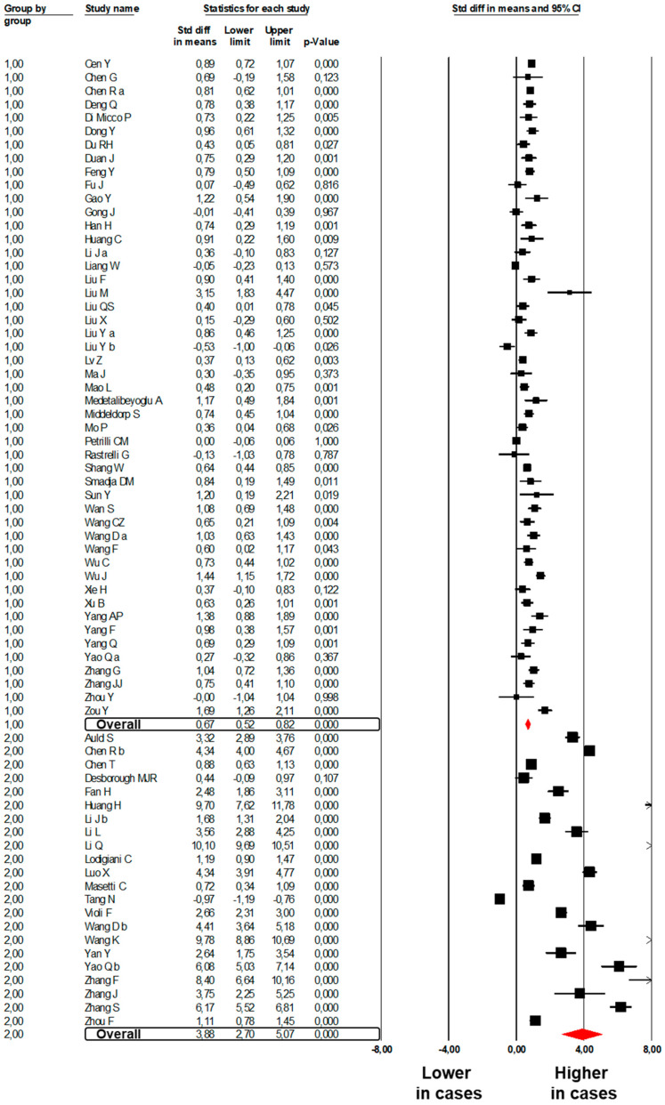
Forty-nine studies [6,8,24,25,26,27,28,30,31,32,33,34,35,36,38,41,42,43,44,45,46,47,48,49,50,51,52,53,55,57,58,59,61,63,64,65,67,68,69,70,71,72,73,74,76,77,80,81,87] showed higher D-Dimer levels in 5024 subjects with severe COVID-19 than in 8052 subjects with mild COVID-19 (SMD: 0.67; 95%CI: 0.52, 0.82, p < 0.001, Figure 3). Heterogeneity was significant (I2: 90.4%, p < 0.001) and was not reduced by the exclusion of one study at a time.
Figure 3.
Forest plots of the difference in D-Dimer values between subjects with severe and those with mild COVID-19 (Group 1) and between non-survivors and survivors to COVID-19 (Group 2).
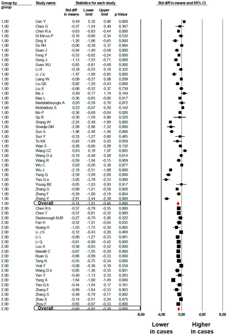
Significantly lower PLT count (SMD: −0.74; 95%CI: −1.01, −0.47, p < 0.001, Figure 4) was found between 1956 subjects with severe COVID-19 as compared to 6546 mild subjects enrolled in 38 studies [6,24,25,26,28,29,31,32,33,36,37,39,41,44,45,49,50,51,52,53,56,58,59,60,61,62,63,64,66,67,68,73,74,75,76,78,79,87]. The heterogeneity among studies was significant (I2: 95.5%, p < 0.001) and was not reduced after excluding one study at a time.
Figure 4.
Forest plots of the difference in platelet count between subjects with severe and those with mild COVID-19 (Group 1) and between non-survivors and survivors to COVID-19 (Group 2).
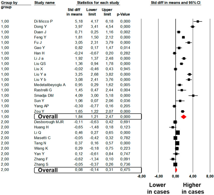
A total of 18 studies [28,30,32,33,34,35,38,40,44,45,46,47,51,57,59,61,71,81] showed significantly higher fibrinogen values in 469 subjects with severe as compared to 1434 subjects with mild COVID-19 (SMD: 1.84; 95%CI: 1.21, 2.47, p < 0.001, Figure 5). The heterogeneity among studies was significant (I2: 95.8%, p < 0.001) and was not reduced by the exclusion of one study at a time.
Figure 5.
Forest plots of the difference in fibrinogen values between subjects with severe and those with mild COVID-19 (Group 1) and between non-survivors and survivors to COVID-19 (Group 2).
3.3. Study Group 2 (Dead with COVID-19 vs. Survived to COVID-19)
A total of 15 studies [20,23,26,83,85,86,87,88,89,95,96,97,98,100,102] showed significantly higher PT values in 840 subjects dead with COVID-19 as compared to 3287 subjects survived to COVID-19 (SMD: 0.67; 95%CI: 0.39, 0.96, p < 0.001, Figure 1). The heterogeneity among studies was significant (I2: 90.2%, p < 0.001) and was not reduced by the exclusion of one study at a time.
No significant difference was found in 11 studies [20,26,86,87,88,89,95,96,97,98,100] reporting aPTT values between 475 subjects dead with COVID-19 and 2774 subjects survived to COVID-19 (SMD: −0.18; 95%CI: −0.89, 0.52 p = 0.609; I2: 97.3%, p < 0.001, Figure 2).
Twenty-two studies [20,26,74,82,83,84,85,86,87,88,89,90,91,92,94,95,96,97,98,99,100,102] showed higher D-Dimer levels in 1149 subjects dead with COVID-19 as compared to 4407 subjects survived to COVID-19 (SMD: 3.88; 95%CI: 2.70, 5.07, p < 0.001, Figure 3). The heterogeneity among studies was significant (I2: 99.4%, p < 0.001) and was not reduced by the exclusion of one study at a time.
A lower PLT count (SMD: −0.60, 95%CI: −0.82, −0.38; p < 0.001, Figure 4) was found in 1339 non-survivors to COVID-19 as compared to 5637 survivors enrolled in 21 studies [20,22,26,74,83,84,85,86,87,88,89,91,92,93,94,95,97,98,100,101,102]. The heterogeneity among studies was significant (I2: 90.3%, p < 0.001) and was not reduced by the exclusion of one study at a time.
A total of nine studies [20,84,86,89,92,96,97,98,100] showed comparable fibrinogen values in 429 subjects dead with COVID-19 as compared to 2472 subjects survived (SMD: 0.08; 95%CI: −0.14, 0.31, p = 0.475, Figure 5). The heterogeneity among studies was significant (I2: 67.1%, p = 0.002) and was not reduced by the exclusion of one study at a time.
3.4. Publication Bias
Visual inspection of funnel plots and the Egger’s test suggested the absence of publication bias and of small-study effect for studies evaluating PT, aPTT, and PLT. In contrast, a significant publication bias was found for D-Dimer and fibrinogen, confirmed by the Egger’s test (Egger’s p < 0.001 and p = 0.039, respectively). Of interest, the Duval and Tweedie’s trim and fill analysis substantially confirmed all results for D-Dimer (SMD: 2.08; 95%CI: 1.63, 2.53) and fibrinogen (SMD: 1.34; 95%CI: 0.91, 1.78) after trimming and imputing studies (Supplemental Figure S2).
3.5. Meta-Regression Analyses
Meta-regression models showed an impact of CRP levels on the difference in PT (Z-value: 2.27, p = 0.023) and fibrinogen (Z-value: 2.89, p = 0.004). Male sex was associated with a lower difference in D-Dimer (Z-value: −2.43, p = 0.015) and PLT levels (Z-value: 1.97, p = 0.048). A higher difference in fibrinogen was observed for increasing prevalence of hypertension (Z-value: 2.63, p = 0.009) (Supplemental Table S1).
4. Discussion
Results of the present meta-analysis consistently show that patients with severe COVID-19 exhibit higher PT, D-Dimer, and fibrinogen values, with a lower PLT count than subjects with mild disease. A significant difference for PT, D-Dimer, and PLT was also found in non-survivors to COVID-19, with similar fibrinogen levels as compared to survivors. Regression models further refined results, showing that differences in PT and fibrinogen levels directly correlated with the degree of inflammation, as expressed by CRP values. Moreover, prevalence of male sex significantly impacted the difference in D-Dimer levels and PLT count. Last, a higher difference in fibrinogen was observed for increasing prevalence of hypertension.
In the attempt to identify new biomarkers of disease severity, some meta-analytical studies have been performed so far on a similar topic, with contrasting results. Our findings are partially consistent with those of a previous meta-analysis [103], documenting significantly higher PT and D-Dimer levels in patients with severe COVID-19. However, differing from our findings, the authors documented no significant difference in PLT levels between severe and mild patients. Two further meta-analyses were specifically focused on PLT, showing that a low PLT count is associated with an increased risk of severe disease and mortality in patients with COVID-19 [104,105]. Overall, literature data about hemostatic parameters and COVID-19 have rapidly grown and these previous meta-analyses included a limited number of available studies. Here, we report the largest meta-analysis (84 studies) including multiple hemostatic parameters, with the aim to provide a comprehensive overview on the association between COVID-19 severity and hemostatic changes.
In the past decade, thrombocytopenia was documented in more than 50% of patients infected with SARS, being this laboratory finding identified as a significant predictor of mortality [106,107] with a high prognostic value [108]. Thrombocytopenia is common in several critical conditions, suggesting the presence of severe organ dysfunction and, in some cases, the development of intravascular coagulopathy [109]. Besides thrombocytopenia, our study suggests that both severe patients and non-survivors to COVID-19 exhibit significantly higher values of D-Dimer and prolonged PT.
The prognostic role of PT has been evaluated in several clinical settings, being identified as a predictor of death after myocardial infarction [110,111], after surgical intervention [112], in acute decompensated heart failure [113] or in neoplastic diseases [114]. PT is commonly used to evaluate the extrinsic and common pathways of coagulation, thus detecting low fibrinogen concentrations and deficiencies of factors II, V, VII, and X [115]. On the other hand, aPTT evaluates the function of all clotting factors with the only exception of factor VII [115]. As factor VII is the one with the shortest half-life, PT is the first hemostatic parameter affected in many clinical conditions [116].
In our meta-analysis, we documented a significant prolongation of PT, without significant modifications in aPTT. In this regard, some hypotheses can be done, such as the highly increased factor VIII activity [117] correcting an otherwise prolonged aPTT [118]. However, considering the hepatic origin of clotting factors and the short half-life of factor VII [119], we can also hypothesize that a prolonged PT with normal aPTT may be due to acute liver dysfunction. This is in line with the observed association between altered liver markers and COVID-19 severity [120]. Recent studies demonstrated that patients with severe COVID-19 have increased incidence of abnormal liver function, with significantly higher values of transaminases as compared to subjects with a mild disease [25,37]. In keeping with this, a recent meta-analysis confirmed the presence of hepatic impairments in SARS and MERS Coronaviridae infections [121]. Several hypotheses have been made to explain the presence of liver injury in severe COVID-19 patients, such as the fact that a high positive end-expiratory pressure may determine hepatic congestion through an increase in right atrial pressure [120]. However, the hypothesis of a concomitant liver damage from virally-induced cytotoxic T cells seems to be the most convincing [120].
The observation of higher PT values both in severe COVID-19 patients and non-survivors could also be interpreted from an alternative (or additional) point of view. In 2003, a total of 2.5% of SARS patients showed evidence of disseminated intravascular coagulation (DIC), being this condition frequently associated with death [122]. Accordingly, among 183 in-hospital patients with COVID-19, 71.4% of non-survivors had overt DIC as compared to only 0.6% of survivors [21]. The development of a consumption coagulopathy in severe COVID-19 may account also for the lower PLT count and the increased D-Dimer levels documented in our meta-analysis, in line with the laboratory criteria for overt DIC proposed by the International Society of Thrombosis and Hemostasis (ISTH) [123]. However, most patients with severe COVID-19 may not fulfill ISTH criteria for DIC, as coagulopathy seems to have its specific features in this clinical setting. For example, thrombocytopenia is less pronounced in severe COVID-19 patients as compared to sepsis-related DIC, while D-Dimer levels are exceptionally high [124]. Moreover, we documented higher fibrinogen levels in severe patients when compared with mild subjects, thus refuting the hypothesis of a consumption coagulopathy in this clinical setting. In line with previous evidence [117], this finding is further supported by the lack of changes in aPTT values both in severe and in COVID-19 non-survivors.
A further interesting finding is the direct correlation between CRP and PT values. CRP levels correlate with the intensity of the systemic inflammatory response and, since hemostasis and inflammation are strictly related [125], this supports the hypothesis of a systemic activation of the coagulation system in response to a dysregulation of the inflammatory markers. This is also in agreement with the increase in fibrinogen levels, mainly documented in most severe patients.
Taken together, the currently available evidence seems to suggest that SARS-CoV-2 infection can rapidly evolve into a severe condition with pulmonary and multiorgan complications, potentially resulting in a consistent hemostatic imbalance with hypercoagulable state. However, the exact mechanisms behind the observed hemostatic changes in severe COVID-19 patients are still unclear. It has been hypothesized that the coagulopathy in SARS-CoV-2 infection may be a combination of a low-grade DIC with its distinct features and a localized pulmonary thrombotic microangiopathy [124]. Alternatively, the hemostatic imbalance has been associated to the severe inflammatory state [117]. The high frequency of antiphospholipid antibodies documented among critically ill COVID-19 patients has also been related to the genesis of the hypercoagulable state [126,127]. In the meantime, a growing amount of literature data suggests that a high incidence of thromboembolic complications is expected among severe and critical patients with COVID-19, even in the presence of adequate thromboprophylaxis [128,129].
Further cohort studies are needed to clarify the pathogenesis and the prognostic value of the hemostatic changes associated to SARS-CoV-2 infection. This could allow for the identification of new markers of disease progression. Moreover, considering the unknown long-term outcomes of COVID-19, the growing amount of studies suggesting the presence of a residual chronic disability, and the need of post-discharge rehabilitation after the acute phase [130,131,132,133,134,135], it is essential to identify new biomarkers related to the disease for an adequate management of critical patients even after ICU (e.g., general ward, rehabilitation centers, and home). The hypothesis that persistent thromboembolic phenomena may contribute to the long-term outcomes of COVID-19 [136,137] and to the functional sequelae of critically ill patients after discharge [12,129] further supports the urgent need of studies addressing the mechanisms of such hemostatic imbalance in this clinical setting.
Some potential limitations of our study need to be discussed. First, studies included in our meta-analysis enrolled patients with different characteristics, since these studies had different inclusion and exclusion criteria. Moreover, considerable information is missing in included studies. Considering that meta-analyses are performed on pooled data, our meta-regression models allowed for the adjustment for some—but not all—potential confounders. Thus, although the multivariate approach was able to refine analyses by assessing the influence of most clinical and demographic variables on the observed results, our findings should be interpreted with great caution.
Second, most results of our meta-analysis were affected by significant heterogeneity. Although it was not possible to definitively identify the origin of such heterogeneity, the impact of major clinical and demographic variables on our results was evaluated through regression models. Moreover, we excluded the presence of publication bias by using different methods.
Finally, we performed our analyses by using SMD instead of mean difference, this method being designed to be used when the same outcome is evaluated in a variety of ways [138]. This allowed us to provide consistent meta-analytical information with an appropriate statistical methodology, but we were not able to provide a quantitative measure of the difference in the hemostatic parameters between cases and controls. This partially reduces the clinical relevance of our results.
In conclusion, our meta-analysis suggests that significant hemostatic changes are associated with COVID-19 severity. Strict monitoring of these parameters is needed to identify early modifications with the aim to stratify the death risk and to implement adequate therapeutic approaches. Considering the risk of fatal complications and poor long-term outcomes, further studies should better define the prognostic role of hemostatic parameters in COVID-19 patients.
Acknowledgments
Nothing to report.
Supplementary Materials
The following are available online at https://www.mdpi.com/2077-0383/9/7/2244/s1, Figure S1: Preferred Reporting Items for Systematic Reviews and Meta-Analyses (PRISMA) flow diagram, Figure S2: Funnel plots of effect size versus standard error for studies evaluating the difference in PT, aPTT, D-Dimer, platelet count and fibrinogen, Table S1: Meta-regression analyses. Impact of age, male gender, hypertension, diabetes, and C-reactive protein (CRP) on the difference in PT, D-Dimer, platelet count, and fibrinogen levels, Table S2: PubMed search (June 16, 2020).
Author Contributions
M.N.D.D.M. and I.C. conceived and designed the study, performed statistical analysis, interpreted results, and drafted the manuscript. G.A.S. performed statistical analysis. R.L., A.S., and M.M. acquired clinical data and reviewed the original draft of the manuscript. A.D.M. and P.A. supervised the project, drafted the manuscript and performed critical revision. All authors have read and agreed to the published version of the manuscript.
Funding
This research received no external funding.
Conflicts of Interest
The authors declare no conflict of interest.
References
- 1.Zhu N., Zhang D., Wang W., Li X., Yang B., Song J., Zhao X., Huang B., Shi W., Lu R., et al. A novel coronavirus from patients with pneumonia in China, 2019. N. Engl. J. Med. 2020;382:727–733. doi: 10.1056/NEJMoa2001017. [DOI] [PMC free article] [PubMed] [Google Scholar]
- 2.World Health Organization Coronavirus Disease 2019 (COVID-19). Situation Report-66. [(accessed on 26 March 2020)]; Available online: https://www.who.int/emergencies/diseases/novel-coronavirus-2019/situation-reports.
- 3.Chen N., Zhou M., Dong X., Qu J., Gong F., Han Y., Qiu Y., Wang J., Liu Y., Wei Y., et al. Epidemiological and clinical characteristics of 99 cases of 2019 novel coronavirus pneumonia in Wuhan, China: A descriptive study. Lancet. 2020;395:507–513. doi: 10.1016/S0140-6736(20)30211-7. [DOI] [PMC free article] [PubMed] [Google Scholar]
- 4.Team TNCPERE The epidemiological characteristics of an outbreak of 2019 novel coronavirus diseases (COVID-19) in China. China CDC Wkly. 2020;2:113–122. [PMC free article] [PubMed] [Google Scholar]
- 5.Wu P., Hao X., Lau E.H.Y., Wong J.Y., Leung K.S.M., Wu J.T., Cowling B.J., Leung G.M. Real-Time tentative assessment of the epidemiological characteristics of novel coronavirus infections in Wuhan, China, as at 22 January 2020. Eurosurveillance. 2020;25 doi: 10.2807/1560-7917.ES.2020.25.3.2000044. [DOI] [PMC free article] [PubMed] [Google Scholar]
- 6.Wan S., Xiang Y., Fang W., Zheng Y., Li B., Hu Y., Lang C., Huang D., Sun Q., Xiong Y., et al. Clinical features and treatment of COVID-19 patients in northeast Chongqing. J. Med. Virol. 2020;92:797–806. doi: 10.1002/jmv.25783. [DOI] [PMC free article] [PubMed] [Google Scholar]
- 7.Chan J.F., Yuan S., Kok K.H., To K.K., Chu H., Yang J., Xing F., Liu J., Yip C.C., Poon R.W., et al. A familial cluster of pneumonia associated with the 2019 novel coronavirus indicating person-to-person transmission: A study of a family cluster. Lancet. 2020;395:514–523. doi: 10.1016/S0140-6736(20)30154-9. [DOI] [PMC free article] [PubMed] [Google Scholar]
- 8.Huang C., Wang Y., Li X., Ren L., Zhao J., Hu Y., Zhang L., Fan G., Xu J., Gu X., et al. Clinical features of patients infected with 2019 novel coronavirus in Wuhan, China. Lancet. 2020;395:497–506. doi: 10.1016/S0140-6736(20)30183-5. [DOI] [PMC free article] [PubMed] [Google Scholar]
- 9.The Lancet Emerging understandings of 2019-nCoV. Lancet. 2020;395:311. doi: 10.1016/S0140-6736(20)30186-0. [DOI] [PMC free article] [PubMed] [Google Scholar]
- 10.Bastola A., Sah R., Rodriguez-Morales A.J., Lal B.K., Jha R., Ojha H.C., Shrestha B., Chu D.K.W., Poon L.L.M., Costello A., et al. The first 2019 novel coronavirus case in Nepal. Lancet Infect. Dis. 2020;20:279–280. doi: 10.1016/S1473-3099(20)30067-0. [DOI] [PMC free article] [PubMed] [Google Scholar]
- 11.Rivera-Lillo G., Torres-Castro R., Fregonezi G., Vilaro J., Puppo H. Challenge for rehabilitation after hospitalization for COVID-19. Arch. Phys. Med. Rehabil. 2020 doi: 10.1016/j.apmr.2020.04.013. [DOI] [PMC free article] [PubMed] [Google Scholar]
- 12.Ambrosino P., Papa A., Maniscalco M., Di Minno M.N.D. COVID-19 and functional disability: Current insights and rehabilitation strategies. Postgrad. Med. J. 2020 doi: 10.1136/postgradmedj-2020-138227. in press. [DOI] [PMC free article] [PubMed] [Google Scholar]
- 13.Yang L.L., Yang T. Pulmonary Rehabilitation for Patients with Coronavirus Disease 2019 (COVID-19) Chronic Dis. Transl. Med. 2020;6:79–86. doi: 10.1016/j.cdtm.2020.05.002. [DOI] [PMC free article] [PubMed] [Google Scholar]
- 14.Gando S., Levi M., Toh C.H. Disseminated intravascular coagulation. Nat. Rev. Dis. Prim. 2016;2:16037. doi: 10.1038/nrdp.2016.37. [DOI] [PubMed] [Google Scholar]
- 15.Shitrit D., Izbicki G., Shitrit A.B., Kramer M.R., Rudensky B., Sulkes J., Hersch M. Prognostic value of a new quantitative D-dimer test in critically ill patients 24 and 48 h following admission to the intensive care unit. Blood Coagul. Fibrinolysis. 2004;15:15–19. doi: 10.1097/00001721-200401000-00003. [DOI] [PubMed] [Google Scholar]
- 16.Moher D., Liberati A., Tetzlaff J., Altman D.G. Preferred reporting items for systematic reviews and meta-analyses: The PRISMA statement. PLoS Med. 2009;6:e1000097. doi: 10.1371/journal.pmed.1000097. [DOI] [PMC free article] [PubMed] [Google Scholar]
- 17.Higgins J.P., Thompson S.G., Deeks J.J., Altman D.G. Measuring inconsistency in meta-analyses. BMJ. 2003;327:557–560. doi: 10.1136/bmj.327.7414.557. [DOI] [PMC free article] [PubMed] [Google Scholar]
- 18.Sterne J.A., Egger M., Smith G.D. Systematic reviews in health care: Investigating and dealing with publication and other biases in meta-analysis. BMJ. 2001;323:101–105. doi: 10.1136/bmj.323.7304.101. [DOI] [PMC free article] [PubMed] [Google Scholar]
- 19.Duval S., Tweedie R. Trim and fill: A simple funnel-plot-based method of testing and adjusting for publication bias in meta-analysis. Biometrics. 2000;56:455–463. doi: 10.1111/j.0006-341X.2000.00455.x. [DOI] [PubMed] [Google Scholar]
- 20.Tang N., Bai H., Chen X., Gong J., Li D., Sun Z. Anticoagulant treatment is associated with decreased mortality in severe coronavirus disease 2019 patients with coagulopathy. J. Thromb. Haemost. 2020;18:1094–1099. doi: 10.1111/jth.14817. [DOI] [PMC free article] [PubMed] [Google Scholar]
- 21.Tang N., Li D., Wang X., Sun Z. Abnormal coagulation parameters are associated with poor prognosis in patients with novel coronavirus pneumonia. J. Thromb. Haemost. 2020;18:844–847. doi: 10.1111/jth.14768. [DOI] [PMC free article] [PubMed] [Google Scholar]
- 22.Yang X., Yang Q., Wang Y., Wu Y., Xu J., Yu Y., Shang Y. Thrombocytopenia and its association with mortality in patients with COVID-19. J. Thromb. Haemost. 2020;18:1469–1472. doi: 10.1111/jth.14848. [DOI] [PMC free article] [PubMed] [Google Scholar]
- 23.Yang X., Yu Y., Xu J., Shu H., Xia J., Liu H., Wu Y., Zhang L., Yu Z., Fang M., et al. Clinical course and outcomes of critically ill patients with SARS-CoV-2 pneumonia in Wuhan, China: A single-centered, retrospective, observational study. Lancet Respir. Med. 2020;8:475–481. doi: 10.1016/S2213-2600(20)30079-5. [DOI] [PMC free article] [PubMed] [Google Scholar]
- 24.Cen Y., Chen X., Shen Y., Zhang X.H., Lei Y., Xu C., Jiang W.R., Xu H.T., Chen Y., Zhu J., et al. Risk factors for disease progression in mild to moderate COVID-19 patients- a multi-center observational study. Clin. Microbiol. Infect. 2020 doi: 10.1016/j.cmi.2020.05.041. [DOI] [PMC free article] [PubMed] [Google Scholar]
- 25.Chen G., Wu D., Guo W., Cao Y., Huang D., Wang H., Wang T., Zhang X., Chen H., Yu H., et al. Clinical and immunologic features in severe and moderate Coronavirus Disease 2019. J. Clin. Investig. 2020 doi: 10.1172/JCI137244. [DOI] [PMC free article] [PubMed] [Google Scholar]
- 26.Chen R., Sang L., Jiang M., Yang Z., Jia N., Fu W., Xie J., Guan W., Liang W., Ni Z., et al. Longitudinal hematologic and immunologic variations associated with the progression of COVID-19 patients in China. J. Allergy Clin. Immunol. 2020;146:89–100. doi: 10.1016/j.jaci.2020.05.003. [DOI] [PMC free article] [PubMed] [Google Scholar]
- 27.Deng Q., Hu B., Zhang Y., Wang H., Zhou X., Hu W., Cheng Y., Yan J., Ping H., Zhou Q. Suspected myocardial injury in patients with COVID-19: Evidence from front-line clinical observation in Wuhan, China. Int. J. Cardiol. 2020;311:116–121. doi: 10.1016/j.ijcard.2020.03.087. [DOI] [PMC free article] [PubMed] [Google Scholar]
- 28.Di Micco P., Russo V., Carannante N., Imparato M., Rodolfi S., Cardillo G., Lodigiani C. Clotting factors in COVID-19: Epidemiological association and prognostic values in different clinical presentations in an Italian Cohort. J. Clin. Med. 2020;9:1371. doi: 10.3390/jcm9051371. [DOI] [PMC free article] [PubMed] [Google Scholar]
- 29.Ding X., Yu Y., Lu B., Huo J., Chen M., Kang Y., Lou J., Liu Z. Dynamic profile and clinical implications of hematological parameters in hospitalized patients with coronavirus disease 2019. Clin. Chem. Lab. Med. 2020;58:1365–1371. doi: 10.1515/cclm-2020-0411. [DOI] [PubMed] [Google Scholar]
- 30.Dong Y., Zhou H., Li M., Zhang Z., Guo W., Yu T., Gui Y., Wang Q., Zhao L., Luo S., et al. A novel simple scoring model for predicting severity of patients with SARS-CoV-2 infection. Transbound. Emerg. Dis. 2020 doi: 10.1111/tbed.13651. [DOI] [PMC free article] [PubMed] [Google Scholar]
- 31.Du R.H., Liu L.M., Yin W., Wang W., Guan L.L., Yuan M.L., Li Y.L., Hu Y., Li X.Y., Sun B., et al. Hospitalization and critical care of 109 decedents with COVID-19 Pneumonia in Wuhan, China. Ann. Am. Thorac. Soc. 2020 doi: 10.1513/AnnalsATS.202003-225OC. [DOI] [PMC free article] [PubMed] [Google Scholar]
- 32.Duan J., Wang X., Chi J., Chen H., Bai L., Hu Q., Han X., Hu W., Zhu L., Wang X., et al. Correlation between the variables collected at admission and progression to severe cases during hospitalization among patients with COVID-19 in Chongqing. J. Med. Virol. 2020 doi: 10.1002/jmv.26082. [DOI] [PMC free article] [PubMed] [Google Scholar]
- 33.Feng Y., Ling Y., Bai T., Xie Y., Huang J., Li J., Xiong W., Yang D., Chen R., Lu F., et al. COVID-19 with Different Severities: A Multicenter Study of Clinical Features. Am. J. Respir. Crit. Care Med. 2020;201:1380–1388. doi: 10.1164/rccm.202002-0445OC. [DOI] [PMC free article] [PubMed] [Google Scholar]
- 34.Fu J., Kong J., Wang W., Wu M., Yao L., Wang Z., Jin J., Wu D., Yu X. The clinical implication of dynamic neutrophil to lymphocyte ratio and D-dimer in COVID-19: A retrospective study in Suzhou China. Thromb. Res. 2020;192:3–8. doi: 10.1016/j.thromres.2020.05.006. [DOI] [PMC free article] [PubMed] [Google Scholar]
- 35.Gao Y., Li T., Han M., Li X., Wu D., Xu Y., Zhu Y., Liu Y., Wang X., Wang L. Diagnostic utility of clinical laboratory data determinations for patients with the severe COVID-19. J. Med. Virol. 2020;92:791–796. doi: 10.1002/jmv.25770. [DOI] [PMC free article] [PubMed] [Google Scholar]
- 36.Gong J., Ou J., Qiu X., Jie Y., Chen Y., Yuan L., Cao J., Tan M., Xu W., Zheng F., et al. A tool to early predict severe corona virus disease 2019 (COVID-19): A multicenter study using the risk nomogram in Wuhan and Guangdong, China. Clin. Infect. Dis. 2020 doi: 10.1093/cid/ciaa443. [DOI] [PMC free article] [PubMed] [Google Scholar]
- 37.Guan W.J., Ni Z.Y., Hu Y., Liang W.H., Ou C.Q., He J.X., Liu L., Shan H., Lei C.L., Hui D.S.C., et al. Clinical characteristics of coronavirus disease 2019 in China. N. Engl. J. Med. 2020;382:1708–1720. doi: 10.1056/NEJMoa2002032. [DOI] [PMC free article] [PubMed] [Google Scholar]
- 38.Han H., Yang L., Liu R., Liu F., Wu K.L., Li J., Liu X.H., Zhu C.L. Prominent changes in blood coagulation of patients with SARS-CoV-2 infection. Clin. Chem. Lab. Med. 2020;58:1116–1120. doi: 10.1515/cclm-2020-0188. [DOI] [PubMed] [Google Scholar]
- 39.Li H., Xiang X., Ren H., Xu L., Zhao L., Chen X., Long H., Wang Q., Wu Q. Serum Amyloid A is a biomarker of severe Coronavirus Disease and poor prognosis. J. Infect. 2020;80:646–655. doi: 10.1016/j.jinf.2020.03.035. [DOI] [PMC free article] [PubMed] [Google Scholar]
- 40.Li J., Li M., Zheng S., Li M., Zhang M., Sun M., Li X., Deng A., Cai Y., Zhang H. Plasma albumin levels predict risk for nonsurvivors in critically ill patients with COVID-19. Biomark. Med. 2020 doi: 10.2217/bmm-2020-0254. [DOI] [PMC free article] [PubMed] [Google Scholar]
- 41.Liang W., Liang H., Ou L., Chen B., Chen A., Li C., Li Y., Guan W., Sang L., Lu J., et al. Development and validation of a clinical risk score to predict the occurrence of critical Illness in hospitalized patients with COVID-19. JAMA Intern. Med. 2020 doi: 10.1001/jamainternmed.2020.2033. [DOI] [PMC free article] [PubMed] [Google Scholar]
- 42.Liu F., Zhang Q., Huang C., Shi C., Wang L., Shi N., Fang C., Shan F., Mei X., Shi J., et al. CT quantification of pneumonia lesions in early days predicts progression to severe illness in a cohort of COVID-19 patients. Theranostics. 2020;10:5613–5622. doi: 10.7150/thno.45985. [DOI] [PMC free article] [PubMed] [Google Scholar]
- 43.Liu M., He P., Liu H.G., Wang X.J., Li F.J., Chen S., Lin J., Chen P., Liu J.H., Li C.H. Clinical characteristics of 30 medical workers infected with new coronavirus pneumonia. Zhonghua Jie He He Hu Xi Za Zhi. 2020;43:209–214. doi: 10.3760/cma.j.issn.1001-0939.2020.03.014. [DOI] [PubMed] [Google Scholar]
- 44.Liu S., Luo H., Wang Y., Wang D., Ju S., Yang Y. Characteristics and associations with severity in COVID-19 patients: A multicenter cohort study from Jiangsu province, China. Lancet. 2020 doi: 10.2139/ssrn.3548753. [DOI] [Google Scholar]
- 45.Liu X., Li Z., Liu S., Sun J., Chen Z., Jiang M., Zhang Q., Wei Y., Wang X., Huang Y.Y., et al. Potential therapeutic effects of dipyridamole in the severely ill patients with COVID-19. Acta Pharm. Sin. B. 2020 doi: 10.1016/j.apsb.2020.04.008. [DOI] [PMC free article] [PubMed] [Google Scholar]
- 46.Liu Y., Liao W., Wan L., Xiang T., Zhang W. Correlation between relative nasopharyngeal virus RNA load and lymphocyte count disease severity in patients with COVID-19. Viral Immunol. 2020 doi: 10.1089/vim.2020.0062. [DOI] [PubMed] [Google Scholar]
- 47.Liu Y., Sun W., Li J., Chen L., Wang Y., Zhang L., Yu L. Clinical features and progression of acute respiratory distress syndrome in coronavirus disease 2019. medRxiv. 2020 doi: 10.1101/2020.02.17.20024166. [DOI] [Google Scholar]
- 48.Lv Z., Cheng S., Le J., Huang J., Feng L., Zhang B., Li Y. Clinical characteristics and co-infections of 354 hospitalized patients with COVID-19 in Wuhan, China: A retrospective cohort study. Microbes. Infect. 2020;22:195–199. doi: 10.1016/j.micinf.2020.05.007. [DOI] [PMC free article] [PubMed] [Google Scholar]
- 49.Ma J., Yin J., Qian Y., Wu Y. Clinical characteristics and prognosis in cancer patients with COVID-19: A single center’s retrospective study. J. Infect. 2020 doi: 10.1016/j.jinf.2020.04.006. [DOI] [PMC free article] [PubMed] [Google Scholar]
- 50.Mao L., Jin H., Wang M., Hu Y., Chen S., He Q., Chang J., Hong C., Zhou Y., Wang D., et al. Neurologic manifestations of hospitalized patients with coronavirus disease 2019 in Wuhan, China. JAMA Neurol. 2020;77:683–690. doi: 10.1001/jamaneurol.2020.1127. [DOI] [PMC free article] [PubMed] [Google Scholar]
- 51.Medetalibeyoglu A., Senkal N., Capar G., Kose M., Tukek T. Characteristics of the initial patients hospitalized for COVID-19: A single-center report. Turk. J. Med. Sci. 2020 doi: 10.3906/sag-2004-98. [DOI] [PMC free article] [PubMed] [Google Scholar]
- 52.Middeldorp S., Coppens M., van Haaps T.F., Foppen M., Vlaar A.P., Muller M.C.A., Bouman C.C.S., Beenen L.F.M., Kootte R.S., Heijmans J., et al. Incidence of venous thromboembolism in hospitalized patients with COVID-19. J. Thromb. Haemost. 2020 doi: 10.1111/jth.14888. [DOI] [PMC free article] [PubMed] [Google Scholar]
- 53.Mo P., Xing Y., Xiao Y., Deng L., Zhao Q., Wang H., Xiong Y., Cheng Z., Gao S., Liang K., et al. Clinical characteristics of refractory COVID-19 pneumonia in Wuhan, China. Clin. Infect. Dis. 2020 doi: 10.1093/cid/ciaa270. [DOI] [PMC free article] [PubMed] [Google Scholar]
- 54.Peng Y.D., Meng K., Guan H.Q., Leng L., Zhu R.R., Wang B.Y., He M.A., Cheng L.X., Huang K., Zeng Q.T. Clinical characteristics and outcomes of 112 cardiovascular disease patients infected by 2019-nCoV. Zhonghua Xin Xue Guan Bing Za Zhi. 2020;48:E004. doi: 10.3760/cma.j.cn112148-20200220-00105. [DOI] [PubMed] [Google Scholar]
- 55.Petrilli C.M., Jones S.A., Yang J., Rajagopalan H., O’Donnell L., Chernyak Y., Tobin K.A., Cerfolio R.J., Francois F., Horwitz L.I. Factors associated with hospital admission and critical illness among 5279 people with coronavirus disease 2019 in New York City: Prospective cohort study. BMJ. 2020;369:m1966. doi: 10.1136/bmj.m1966. [DOI] [PMC free article] [PubMed] [Google Scholar]
- 56.Qu R., Ling Y., Zhang Y.H., Wei L.Y., Chen X., Li X.M., Liu X.Y., Liu H.M., Guo Z., Ren H., et al. Platelet-To-Lymphocyte ratio is associated with prognosis in patients with coronavirus disease-19. J. Med. Virol. 2020 doi: 10.1002/jmv.25767. [DOI] [PMC free article] [PubMed] [Google Scholar]
- 57.Rastrelli G., Di Stasi V., Inglese F., Beccaria M., Garuti M., Di Costanzo D., Spreafico F., Greco G.F., Cervi G., Pecoriello A., et al. Low testosterone levels predict clinical adverse outcomes in SARS-CoV-2 pneumonia patients. Andrology. 2020 doi: 10.1111/andr.12821. [DOI] [PMC free article] [PubMed] [Google Scholar]
- 58.Shang W., Dong J., Ren Y., Tian M., Li W., Hu J., Li Y. The value of clinical parameters in predicting the severity of COVID-19. J. Med. Virol. 2020 doi: 10.1002/jmv.26031. [DOI] [PMC free article] [PubMed] [Google Scholar]
- 59.Smadja D.M., Guerin C.L., Chocron R., Yatim N., Boussier J., Gendron N., Khider L., Hadjadj J., Goudot G., Debuc B., et al. Angiopoietin-2 as a marker of endothelial activation is a good predictor factor for intensive care unit admission of COVID-19 patients. Angiogenesis. 2020 doi: 10.1007/s10456-020-09730-0. [DOI] [PMC free article] [PubMed] [Google Scholar]
- 60.Sun S., Cai X., Wang H., He G., Lin Y., Lu B., Chen C., Pan Y., Hu X. Abnormalities of peripheral blood system in patients with COVID-19 in Wenzhou, China. Clin. Chim. Acta. 2020;507:174–180. doi: 10.1016/j.cca.2020.04.024. [DOI] [PMC free article] [PubMed] [Google Scholar]
- 61.Sun Y., Dong Y., Wang L., Xie H., Li B., Chang C., Wang F.S. Characteristics and prognostic factors of disease severity in patients with COVID-19: The Beijing experience. J. Autoimmun. 2020:102473. doi: 10.1016/j.jaut.2020.102473. [DOI] [PMC free article] [PubMed] [Google Scholar]
- 62.To K.K., Tsang O.T., Leung W.S., Tam A.R., Wu T.C., Lung D.C., Yip C.C., Cai J.P., Chan J.M., Chik T.S., et al. Temporal profiles of viral load in posterior oropharyngeal saliva samples and serum antibody responses during infection by SARS-CoV-2: An observational cohort study. Lancet Infect. Dis. 2020;20:565–574. doi: 10.1016/S1473-3099(20)30196-1. [DOI] [PMC free article] [PubMed] [Google Scholar]
- 63.Wang C.Z., Hu S.L., Wang L., Li M., Li H.T. Early risk factors of the exacerbation of Coronavirus disease 2019 pneumonia. J. Med. Virol. 2020 doi: 10.1002/jmv.26071. [DOI] [PMC free article] [PubMed] [Google Scholar]
- 64.Wang D., Hu B., Hu C., Zhu F., Liu X., Zhang J., Wang B., Xiang H., Cheng Z., Xiong Y., et al. Clinical characteristics of 138 Hospitalized Patients with 2019 novel coronavirus-infected pneumonia in Wuhan, China. JAMA. 2020;323:1061–1069. doi: 10.1001/jama.2020.1585. [DOI] [PMC free article] [PubMed] [Google Scholar]
- 65.Wang F., Hou H., Luo Y., Tang G., Wu S., Huang M., Liu W., Zhu Y., Lin Q., Mao L., et al. The laboratory tests and host immunity of COVID-19 patients with different severity of illness. JCI Insight. 2020;5:e137799. doi: 10.1172/jci.insight.137799. [DOI] [PMC free article] [PubMed] [Google Scholar]
- 66.Wang R., Pan M., Zhang X., Han M., Fan X., Zhao F., Miao M., Xu J., Guan M., Deng X., et al. Epidemiological and clinical features of 125 Hospitalized Patients with COVID-19 in Fuyang, Anhui, China. Int. J. Infect. Dis. 2020;95:421–428. doi: 10.1016/j.ijid.2020.03.070. [DOI] [PMC free article] [PubMed] [Google Scholar]
- 67.Wu C., Chen X., Cai Y., Xia J., Zhou X., Xu S., Huang H., Zhang L., Du C., Zhang Y., et al. Risk factors associated with acute respiratory distress syndrome and death in patients with coronavirus disease 2019 Pneumonia in Wuhan, China. JAMA Intern. Med. 2020 doi: 10.1001/jamainternmed.2020.0994. [DOI] [PMC free article] [PubMed] [Google Scholar]
- 68.Wu J., Li W., Shi X., Chen Z., Jiang B., Liu J., Wang D., Liu C., Meng Y., Cui L., et al. Early antiviral treatment contributes to alleviate the severity and improve the prognosis of patients with novel coronavirus disease (COVID-19) J. Intern. Med. 2020;288:128–138. doi: 10.1111/joim.13063. [DOI] [PubMed] [Google Scholar]
- 69.Xie H., Zhao J., Lian N., Lin S., Xie Q., Zhuo H. Clinical characteristics of non-ICU hospitalized patients with coronavirus disease 2019 and liver injury: A retrospective study. Liver Int. 2020;40:1321–1326. doi: 10.1111/liv.14449. [DOI] [PMC free article] [PubMed] [Google Scholar]
- 70.Xu B., Fan C.Y., Wang A.L., Zou Y.L., Yu Y.H., He C., Xia W.G., Zhang J.X., Miao Q. Suppressed T cell-mediated immunity in patients with COVID-19: A clinical retrospective study in Wuhan, China. J. Infect. 2020;81:e51–e60. doi: 10.1016/j.jinf.2020.04.012. [DOI] [PMC free article] [PubMed] [Google Scholar]
- 71.Yang A.P., Li H.M., Tao W.Q., Yang X.J., Wang M., Yang W.J., Liu J.P. Infection with SARS-CoV-2 causes abnormal laboratory results of multiple organs in patients. Aging. 2020;12:10059–10069. doi: 10.18632/aging.103255. [DOI] [PMC free article] [PubMed] [Google Scholar]
- 72.Yang F., Shi S., Zhu J., Shi J., Dai K., Chen X. Clinical characteristics and outcomes of cancer patients with COVID-19. J. Med. Virol. 2020 doi: 10.1002/jmv.25972. [DOI] [PubMed] [Google Scholar]
- 73.Yang Q., Xie L., Zhang W., Zhao L., Wu H., Jiang J., Zou J., Liu J., Wu J., Chen Y., et al. Analysis of the clinical characteristics, drug treatments and prognoses of 136 patients with coronavirus disease 2019. J. Clin. Pharm. Ther. 2020 doi: 10.1111/jcpt.13170. [DOI] [PMC free article] [PubMed] [Google Scholar]
- 74.Yao Q., Wang P., Wang X., Qie G., Meng M., Tong X., Bai X., Ding M., Liu W., Liu K., et al. A retrospective study of risk factors for severe acute respiratory syndrome coronavirus 2 infections in hospitalized adult patients. Pol. Arch. Intern. Med. 2020;130:390–399. doi: 10.20452/pamw.15312. [DOI] [PubMed] [Google Scholar]
- 75.Young B.E., Ong S.W.X., Kalimuddin S., Low J.G., Tan S.Y., Loh J., Ng O.T., Marimuthu K., Ang L.W., Mak T.M., et al. Epidemiologic features and clinical course of patients infected with SARS-CoV-2 in Singapore. JAMA. 2020 doi: 10.1001/jama.2020.3204. [DOI] [PMC free article] [PubMed] [Google Scholar]
- 76.Zhang G., Hu C., Luo L., Fang F., Chen Y., Li J., Peng Z., Pan H. Clinical features and short-term outcomes of 221 patients with COVID-19 in Wuhan, China. J. Clin. Virol. 2020;127:104364. doi: 10.1016/j.jcv.2020.104364. [DOI] [PMC free article] [PubMed] [Google Scholar]
- 77.Zhang J.J., Dong X., Cao Y.Y., Yuan Y.D., Yang Y.B., Yan Y.Q., Akdis C.A., Gao Y.D. Clinical characteristics of 140 patients infected with SARS-CoV-2 in Wuhan, China. Allergy. 2020 doi: 10.1111/all.14238. [DOI] [PubMed] [Google Scholar]
- 78.Zheng F., Tang W., Li H., Huang Y.X., Xie Y.L., Zhou Z.G. Clinical characteristics of 161 cases of corona virus disease 2019 (COVID-19) in Changsha. Eur. Rev. Med. Pharmacol. Sci. 2020;24:3404–3410. doi: 10.26355/eurrev_202003_20711. [DOI] [PubMed] [Google Scholar]
- 79.Zheng Y., Zhang Y., Chi H., Chen S., Peng M., Luo L., Chen L., Li J., Shen B., Wang D. The hemocyte counts as a potential biomarker for predicting disease progression in COVID-19: A retrospective study. Clin. Chem. Lab. Med. 2020;58:1106–1115. doi: 10.1515/cclm-2020-0377. [DOI] [PubMed] [Google Scholar]
- 80.Zhou Y., Zhang Z., Tian J., Xiong S. Risk factors associated with disease progression in a cohort of patients infected with the 2019 novel coronavirus. Ann. Palliat. Med. 2020;9:428–436. doi: 10.21037/apm.2020.03.26. [DOI] [PubMed] [Google Scholar]
- 81.Zou Y., Guo H., Zhang Y., Zhang Z., Liu Y., Wang J., Lu H., Qian Z. Analysis of coagulation parameters in patients with COVID-19 in Shanghai, China. Biosci. Trends. 2020 doi: 10.5582/bst.2020.03086. [DOI] [PubMed] [Google Scholar]
- 82.Auld S.C., Caridi-Scheible M., Blum J.M., Robichaux C., Kraft C., Jacob J.T., Jabaley C.S., Carpenter D., Kaplow R., Hernandez-Romieu A.C., et al. ICU and ventilator mortality among critically Ill adults with coronavirus disease 2019. Crit. Care Med. 2020 doi: 10.1097/CCM.0000000000004457. [DOI] [PMC free article] [PubMed] [Google Scholar]
- 83.Chen T., Dai Z., Mo P., Li X., Ma Z., Song S., Chen X., Luo M., Liang K., Gao S., et al. Clinical characteristics and outcomes of older patients with coronavirus disease 2019 (COVID-19) in Wuhan, China (2019): A single-centered, retrospective study. J. Gerontol. A Biol. Sci. Med. Sci. 2020 doi: 10.1093/gerona/glaa089. [DOI] [PMC free article] [PubMed] [Google Scholar]
- 84.Desborough M.J.R., Doyle A.J., Griffiths A., Retter A., Breen K.A., Hunt B.J. Image-Proven thromboembolism in patients with severe COVID-19 in a tertiary critical care unit in the United Kingdom. Thromb. Res. 2020;193:1–4. doi: 10.1016/j.thromres.2020.05.049. [DOI] [PMC free article] [PubMed] [Google Scholar]
- 85.Fan H., Zhang L., Huang B., Zhu M., Zhou Y., Zhang H., Tao X., Cheng S., Yu W., Zhu L., et al. Cardiac injuries in patients with coronavirus disease 2019: Not to be ignored. Int. J. Infect. Dis. 2020;96:294–297. doi: 10.1016/j.ijid.2020.05.024. [DOI] [PMC free article] [PubMed] [Google Scholar]
- 86.Huang H., Zhang M., Chen C., Zhang H., Wei Y., Tian J., Shang J., Deng Y., Du A., Dai H. Clinical Characteristics of COVID-19 in patients with pre-existing ILD: A retrospective study in a single center in Wuhan, China. J. Med. Virol. 2020 doi: 10.1002/jmv.26174. [DOI] [PMC free article] [PubMed] [Google Scholar]
- 87.Li J., Long X., Luo H., Fang F., Lv X., Zhang D., Sun Y., Luo F., Li N., Zhang Q., et al. Clinical Characteristics of deceased patients infected with SARS-CoV-2 in Wuhan, China. Lancet. 2020 doi: 10.2139/ssrn.3546043. [DOI] [Google Scholar]
- 88.Li L., Yang L., Gui S., Pan F., Ye T., Liang B., Hu Y., Zheng C. Association of clinical and radiographic findings with the outcomes of 93 patients with COVID-19 in Wuhan, China. Theranostics. 2020;10:6113–6121. doi: 10.7150/thno.46569. [DOI] [PMC free article] [PubMed] [Google Scholar]
- 89.Li Q., Cao Y., Chen L., Wu D., Yu J., Wang H., He W., Chen L., Dong F., Chen W., et al. Hematological features of persons with COVID-19. Leukemia. 2020 doi: 10.1038/s41375-020-0910-1. [DOI] [PMC free article] [PubMed] [Google Scholar]
- 90.Lodigiani C., Iapichino G., Carenzo L., Cecconi M., Ferrazzi P., Sebastian T., Kucher N., Studt J.D., Sacco C., Alexia B., et al. Venous and arterial thromboembolic complications in COVID-19 patients admitted to an academic hospital in Milan, Italy. Thromb. Res. 2020;191:9–14. doi: 10.1016/j.thromres.2020.04.024. [DOI] [PMC free article] [PubMed] [Google Scholar]
- 91.Luo X., Zhou W., Yan X., Guo T., Wang B., Xia H., Ye L., Xiong J., Jiang Z., Liu Y., et al. Prognostic value of C-reactive protein in patients with COVID-19. Clin. Infect. Dis. 2020 doi: 10.1093/cid/ciaa641. [DOI] [PMC free article] [PubMed] [Google Scholar]
- 92.Masetti C., Generali E., Colapietro F., Voza A., Cecconi M., Messina A., Omodei P., Angelini C., Ciccarelli M., Badalamenti S., et al. High mortality in COVID-19 patients with mild respiratory disease. Eur. J. Clin. Investig. 2020 doi: 10.1111/eci.13314. [DOI] [PMC free article] [PubMed] [Google Scholar]
- 93.Ruan Q., Yang K., Wang W., Jiang L., Song J. Clinical predictors of mortality due to COVID-19 based on an analysis of data of 150 patients from Wuhan, China. Intensiv. Care Med. 2020 doi: 10.1007/s00134-020-05991-x. [DOI] [PMC free article] [PubMed] [Google Scholar]
- 94.Violi F., Cangemi R., Romiti G.F., Ceccarelli G., Oliva A., Alessandri F., Pirro M., Pignatelli P., Lichtner M., Carraro A., et al. Is albumin predictor of mortality in COVID-19? Antioxid. Redox Signal. 2020 doi: 10.1089/ars.2020.8142. [DOI] [PubMed] [Google Scholar]
- 95.Wang D., Yin Y., Hu C., Liu X., Zhang X., Zhou S., Jian M., Xu H., Prowle J., Hu B., et al. Clinical course and outcome of 107 patients infected with the novel coronavirus, SARS-CoV-2, discharged from two hospitals in Wuhan, China. Crit. Care. 2020;24:188. doi: 10.1186/s13054-020-02895-6. [DOI] [PMC free article] [PubMed] [Google Scholar]
- 96.Wang K., Zuo P., Liu Y., Zhang M., Zhao X., Xie S., Zhang H., Chen X., Liu C. Clinical and laboratory predictors of in-hospital mortality in patients with COVID-19: A cohort study in Wuhan, China. Clin. Infect. Dis. 2020 doi: 10.1093/cid/ciaa538. [DOI] [PMC free article] [PubMed] [Google Scholar]
- 97.Yan Y., Yang Y., Wang F., Ren H., Zhang S., Shi X., Yu X., Dong K. Clinical characteristics and outcomes of patients with severe covid-19 with diabetes. BMJ Open Diabetes Res. Care. 2020;8 doi: 10.1136/bmjdrc-2020-001343. [DOI] [PMC free article] [PubMed] [Google Scholar]
- 98.Zhang F., Xiong Y., Wei Y., Hu Y., Wang F., Li G., Liu K., Du R., Wang C.Y., Zhu W. Obesity predisposes to the risk of higher mortality in young COVID-19 patients. J. Med. Virol. 2020 doi: 10.1002/jmv.26039. [DOI] [PMC free article] [PubMed] [Google Scholar]
- 99.Zhang J., Liu P., Wang M., Wang J., Chen J., Yuan W., Li M., Xie Z., Dong W., Li H., et al. The clinical data from 19 critically ill patients with coronavirus disease 2019: A single-centered, retrospective, observational study. Z. Gesundh. Wiss. 2020:1–4. doi: 10.1007/s10389-020-01291-2. [DOI] [PMC free article] [PubMed] [Google Scholar]
- 100.Zhang S., Guo M., Duan L., Wu F., Wang Z., Xu J., Ma Y., Lv Z., Tan X., Zhang S., et al. Short term outcomes and risk factors for mortality in patients with COVID-19 in Wuhan, China: A retrospective study. Lancet Infect. Dis. 2020 doi: 10.2139/ssrn.3551390. [DOI] [Google Scholar]
- 101.Zhao X., Wang K., Zuo P., Liu Y., Zhang M., Xie S., Zhang H., Chen X., Liu C. Early decrease in blood platelet count is associated with poor prognosis in COVID-19 patients-indications for predictive, preventive, and personalized medical approach. EPMA J. 2020:1–7. doi: 10.1007/s13167-020-00208-z. [DOI] [PMC free article] [PubMed] [Google Scholar]
- 102.Zhou F., Yu T., Du R., Fan G., Liu Y., Liu Z., Xiang J., Wang Y., Song B., Gu X., et al. Clinical course and risk factors for mortality of adult inpatients with COVID-19 in Wuhan, China: A retrospective cohort study. Lancet. 2020;395:1054–1062. doi: 10.1016/S0140-6736(20)30566-3. [DOI] [PMC free article] [PubMed] [Google Scholar]
- 103.Xiong M., Liang X., Wei Y.D. Changes in blood coagulation in patients with severe coronavirus disease 2019 (COVID-19): A meta-analysis. Br. J. Haematol. 2020 doi: 10.1111/bjh.16725. [DOI] [PMC free article] [PubMed] [Google Scholar]
- 104.Jiang S.Q., Huang Q.F., Xie W.M., Lv C., Quan X.Q. The association between severe COVID-19 and low platelet count: Evidence from 31 observational studies involving 7613 participants. Br. J. Haematol. 2020 doi: 10.1111/bjh.16817. [DOI] [PubMed] [Google Scholar]
- 105.Lippi G., Plebani M., Henry B.M. Thrombocytopenia is associated with severe coronavirus disease 2019 (COVID-19) infections: A meta-analysis. Clin. Chim. Acta. 2020;506:145–148. doi: 10.1016/j.cca.2020.03.022. [DOI] [PMC free article] [PubMed] [Google Scholar]
- 106.He W.Q., Chen S.B., Liu X.Q., Li Y.M., Xiao Z.L., Zhong N.S. Death risk factors of severe acute respiratory syndrome with acute respiratory distress syndrome. Zhongguo Wei Zhong Bing Ji Jiu Yi Xue. 2003;15:336–337. [PubMed] [Google Scholar]
- 107.Yang M., Ng M.H., Li C.K. Thrombocytopenia in patients with severe acute respiratory syndrome (review) Hematology. 2005;10:101–105. doi: 10.1080/10245330400026170. [DOI] [PubMed] [Google Scholar]
- 108.Zou Z., Yang Y., Chen J., Xin S., Zhang W., Zhou X., Mao Y., Hu L., Liu D., Chang B., et al. Prognostic factors for severe acute respiratory syndrome: A clinical analysis of 165 cases. Clin. Infect. Dis. 2004;38:483–489. doi: 10.1086/380973. [DOI] [PMC free article] [PubMed] [Google Scholar]
- 109.Zarychanski R., Houston D.S. Assessing thrombocytopenia in the intensive care unit: The past, present, and future. Hematol. Am. Soc. Hematol. Educ. Program. 2017;2017:660–666. doi: 10.1182/asheducation-2017.1.660. [DOI] [PMC free article] [PubMed] [Google Scholar]
- 110.Meltzer L.E., Palmon F., Ferrigan M., Pekover J., Sauer H., Kitchell J.R. Prothrombin levels and fatality rates in acute myocardial infarction. JAMA. 1964;187:986–993. doi: 10.1001/jama.1964.03060260014003. [DOI] [PubMed] [Google Scholar]
- 111.Wang X., Chen R., Li Y., Miao F. Predictive value of prothrombin time for all-cause mortality in acute myocardial infarction patients. Conf. Proc. IEEE Eng. Med. Biol. Soc. 2018;2018:5366–5369. doi: 10.1109/EMBC.2018.8513654. [DOI] [PubMed] [Google Scholar]
- 112.Akamatsu N., Sugawara Y., Kanako J., Arita J., Sakamoto Y., Hasegawa K., Kokudo N. Low Platelet counts and prolonged prothrombin time early after operation predict the 90 days morbidity and mortality in living-donor liver transplantation. Ann. Surg. 2017;265:166–172. doi: 10.1097/SLA.0000000000001634. [DOI] [PubMed] [Google Scholar]
- 113.Okada A., Sugano Y., Nagai T., Takashio S., Honda S., Asaumi Y., Aiba T., Noguchi T., Kusano K.F., Ogawa H., et al. Prognostic value of prothrombin time international normalized ratio in acute decompensated heart failure-a combined marker of hepatic insufficiency and hemostatic abnormality. Circ. J. 2016;80:913–923. doi: 10.1253/circj.CJ-15-1326. [DOI] [PubMed] [Google Scholar]
- 114.Tas F., Kilic L., Serilmez M., Keskin S., Sen F., Duranyildiz D. Clinical and prognostic significance of coagulation assays in lung cancer. Respir. Med. 2013;107:451–457. doi: 10.1016/j.rmed.2012.11.007. [DOI] [PubMed] [Google Scholar]
- 115.Levy J.H., Szlam F., Wolberg A.S., Winkler A. Clinical use of the activated partial thromboplastin time and prothrombin time for screening: A review of the literature and current guidelines for testing. Clin. Lab. Med. 2014;34:453–477. doi: 10.1016/j.cll.2014.06.005. [DOI] [PubMed] [Google Scholar]
- 116.Morrissey J.H., Macik B.G., Neuenschwander P.F., Comp P.C. Quantitation of activated factor VII levels in plasma using a tissue factor mutant selectively deficient in promoting factor VII activation. Blood. 1993;81:734–744. doi: 10.1182/blood.V81.3.734.734. [DOI] [PubMed] [Google Scholar]
- 117.Panigada M., Bottino N., Tagliabue P., Grasselli G., Novembrino C., Chantarangkul V., Pesenti A., Peyvandi F., Tripodi A. Hypercoagulability of COVID-19 patients in intensive care unit: A report of thromboelastography findings and other parameters of hemostasis. J. Thromb. Haemost. 2020;18:1738–1742. doi: 10.1111/jth.14850. [DOI] [PMC free article] [PubMed] [Google Scholar]
- 118.Tripodi A., Chantarangkul V., Martinelli I., Bucciarelli P., Mannucci P.M. A shortened activated partial thromboplastin time is associated with the risk of venous thromboembolism. Blood. 2004;104:3631–3634. doi: 10.1182/blood-2004-03-1042. [DOI] [PubMed] [Google Scholar]
- 119.Kerr R., Newsome P., Germain L., Thomson E., Dawson P., Stirling D., Ludlam C.A. Effects of acute liver injury on blood coagulation. J. Thromb. Haemost. 2003;1:754–759. doi: 10.1046/j.1538-7836.2003.00194.x. [DOI] [PubMed] [Google Scholar]
- 120.Bangash M.N., Patel J., Parekh D. COVID-19 and the liver: Little cause for concern. Lancet Gastroenterol. Hepatol. 2020 doi: 10.1016/S2468-1253(20)30084-4. [DOI] [PMC free article] [PubMed] [Google Scholar]
- 121.Kukla M., Skonieczna-Zydecka K., Kotfis K., Maciejewska D., Loniewski I., Lara L.F., Pazgan-Simon M., Stachowska E., Kaczmarczyk M., Koulaouzidis A., et al. COVID-19, MERS and SARS with concomitant liver injury-systematic review of the existing literature. J. Clin. Med. 2020;9:1420. doi: 10.3390/jcm9051420. [DOI] [PMC free article] [PubMed] [Google Scholar]
- 122.Lillicrap D. Disseminated intravascular coagulation in patients with 2019-nCoV pneumonia. J. Thromb. Haemost. 2020;18:786–787. doi: 10.1111/jth.14781. [DOI] [PMC free article] [PubMed] [Google Scholar]
- 123.Jr Taylor F.B., Toh C.H., Hoots W.K., Wada H., Levi M. Towards definition, clinical and laboratory criteria, and a scoring system for disseminated intravascular coagulation. Thromb. Haemost. 2001;86:1327–1330. doi: 10.1055/s-0037-1616068. [DOI] [PubMed] [Google Scholar]
- 124.Levi M., Thachil J., Iba T., Levy J.H. Coagulation abnormalities and thrombosis in patients with COVID-19. Lancet Haematol. 2020 doi: 10.1016/S2352-3026(20)30145-9. [DOI] [PMC free article] [PubMed] [Google Scholar]
- 125.Hoppensteadt D., Tsuruta K., Hirman J., Kaul I., Osawa Y., Fareed J. Dysregulation of inflammatory and hemostatic markers in sepsis and suspected disseminated intravascular coagulation. Clin. Appl. Thromb. Hemost. 2015;21:120–127. doi: 10.1177/1076029613509476. [DOI] [PubMed] [Google Scholar]
- 126.Pineton de Chambrun M., Frere C., Miyara M., Amoura Z., Martin-Toutain I., Mathian A., Hekimian G., Combes A. High frequency of antiphospholipid antibodies in Critically-ill COVID-19 Patients: A link with hypercoagulability? J. Intern. Med. 2020 doi: 10.1111/joim.13126. [DOI] [PMC free article] [PubMed] [Google Scholar]
- 127.Zhang Y., Xiao M., Zhang S., Xia P., Cao W., Jiang W., Chen H., Ding X., Zhao H., Zhang H., et al. Coagulopathy and antiphospholipid antibodies in patients with Covid-19. N. Engl. J. Med. 2020;382:e38. doi: 10.1056/NEJMc2007575. [DOI] [PMC free article] [PubMed] [Google Scholar]
- 128.Di Minno A., Ambrosino P., Calcaterra I., Di Minno M.N.D. COVID-19 and venous thromboembolism: A meta-analysis of literature studies. Semin. Thromb. Hemost. 2020 doi: 10.1055/s-0040-1715456. in press. [DOI] [PMC free article] [PubMed] [Google Scholar]
- 129.Ambrosino P., Di Minno A., Maniscalco M., Di Minno M.N.D. COVID-19 and venous thromboembolism: Current insights and prophylactic strategies. Ann. Med. 2020 doi: 10.1080/07853890.2020.1791355. [DOI] [PMC free article] [PubMed] [Google Scholar]
- 130.Borg K., Stam H. Covid-19 and physical and rehabilitation medicine. J. Rehabil. Med. 2020 doi: 10.2340/16501977-2679. [DOI] [PubMed] [Google Scholar]
- 131.Brugliera L., Spina A., Castellazzi P., Cimino P., Tettamanti A., Houdayer E., Arcuri P., Alemanno F., Mortini P., Iannaccone S. Rehabilitation of COVID-19 patients. J. Rehabil. Med. 2020 doi: 10.2340/16501977-2678. [DOI] [PubMed] [Google Scholar]
- 132.Godeau E., Debeaumont D., Artaud-Macari E., Lagache L., Bouar G.L., Coquart J. Sequelae of acute respiratory distress syndrome: Interest of rehabilitation. Case Rep. Crit. Care. 2019;2019:7953141. doi: 10.1155/2019/7953141. [DOI] [PMC free article] [PubMed] [Google Scholar]
- 133.Simpson R., Robinson L. Rehabilitation following critical illness in people with COVID-19 infection. Am. J. Phys. Med. Rehabil. 2020 doi: 10.1097/PHM.0000000000001443. [DOI] [PMC free article] [PubMed] [Google Scholar]
- 134.Falvey J.R., Ferrante L.E. Flattening the disability curve: Rehabilitation and recovery after COVID-19 infection. Heart Lung J. Crit. Care. 2020 doi: 10.1016/j.hrtlng.2020.05.001. [DOI] [PMC free article] [PubMed] [Google Scholar]
- 135.Lopez M., Bell K., Annaswamy T., Juengst S., Ifejika N. COVID-19 guide for the rehabilitation clinician: A review of non-pulmonary manifestations and complications. Am. J. Phys. Med. Rehabil. 2020 doi: 10.1097/PHM.0000000000001479. [DOI] [PMC free article] [PubMed] [Google Scholar]
- 136.Spagnolo P., Balestro E., Aliberti S., Cocconcelli E., Biondini D., Casa G.D., Sverzellati N., Maher T.M. Pulmonary fibrosis secondary to COVID-19: A call to arms? Lancet Respir. Med. 2020 doi: 10.1016/S2213-2600(20)30222-8. [DOI] [PMC free article] [PubMed] [Google Scholar]
- 137.Boccia M., Aronne L., Celia B., Mazzeo G., Ceparano M., D’Agnano V., Parrella R., Valente T., Bianco A., Perrotta F. COVID-19 and coagulative axis: Review of emerging aspects in a novel disease. Monaldi Arch. Chest Dis. 2020;90 doi: 10.4081/monaldi.2020.1300. [DOI] [PubMed] [Google Scholar]
- 138.Takeshima N., Sozu T., Tajika A., Ogawa Y., Hayasaka Y., Furukawa T.A. Which is more generalizable, powerful and interpretable in meta-analyses, mean difference or standardized mean difference? BMC Med. Res. Methodol. 2014;14:30. doi: 10.1186/1471-2288-14-30. [DOI] [PMC free article] [PubMed] [Google Scholar]
Associated Data
This section collects any data citations, data availability statements, or supplementary materials included in this article.


