Abstract
Purpose
To detect and quantify conjunctival microangiopathy with optical coherence tomography angiography (OCTA).
Methods
Imaging was performed in the temporal and nasal quadrant of the conjunctiva using a Heidelberg Spectralis spectral domain-OCT in OCTA mode adding a 25D lens to the standard 30° fundus lens. Images were acquired within a 10° × 5° cube at the limbus. Binary images were analyzed using ImageJ (Fiji software version 2.0) and an average relative conjunctival vessel density was assessed.
Results
Thirty-two patients with diabetes mellitus type 1 and 2 and 42 healthy individuals were included. Vessel density in healthy individuals was 16.7 ± 5.2% in the nasal and 17.9 ± 6.4% in the temporal quadrant. In patients with diabetes without retinopathy, vessel density was 16.3 ± 6.7% in the nasal and 15.3 ± 7.3% in the temporal conjunctiva. In patients with diabetic retinopathy, vessel density was 13.7 ± 4.3% in the nasal and 15.2 ± 6.5% in the temporal conjunctiva. There were statistically significant higher values in both nasal and temporal measurements among healthy individuals than in patients with diabetic retinopathy (P = 0.03 and P = 0.01, respectively).
Conclusions
Patients with diabetic retinopathy exhibit reduced vessel density, which may suggest diabetic microangiopathy in the conjunctiva. Anterior segment OCTA may detect conjunctival microangiopathy in patients with visual axis opacifications, where retinal OCTA is not possible.
Translational Relevance
The findings of this study bridge the gap between experimental anterior segment OCTA imaging and clinical screening for diabetic complications.
Keywords: diabetes mellitus, optical coherence tomography angiography, anterior segment optical coherence tomography, scanning laser ophthalmoscope, diabetic retinopathy
Introduction
In patients with diabetes mellitus, diabetic retinopathy (DR) represents a common microvascular complication. In fact, DR is the most frequent cause of blindness in the working-age population of the industrially developed countries.1 The global prevalence of diabetes mellitus was 6.4% in 2010 and is estimated to increase to 7.7% in 2030. The pathophysiology of DR is complex and depends on various factors, most important hyperglycemia, hypertension, and duration of diabetes mellitus.2 The resulting microangiopathy causes capillary permeability and blood vessel obliteration leading to the proliferative retinal disease and diabetic maculopathy.3
Compared with the pathology of the retina, alterations of the conjunctival vessels owing to diabetes have not been investigated frequently, and quantitative results are limited. An overall decrease in vessel density of the bulbar conjunctiva of patients with diabetes using digital red-free images captured with a standardized slit-lamp photography technique has been observed and thus an association of decreased vessel area and severity of retinopathy proposed.4
One of the latest diagnostic tools in ophthalmology is optical coherence tomography angiography (OCTA) allowing fast imaging to detect streaming blood and thereby allowing to generate images of the underlying vasculature.5 It has been promoted as an alternative to fluorescein angiography or indocyanine green angiography. Compared with these classic invasive imaging modalities, OCTA is a dye-free imaging and therefore has no risks to suffer from side effects of the dye.6
One limitation of OCTA is the inability to detect vascular permeability. In contrast, it generates images of higher contrast and resolution, owing to the lack of diffuse hyperfluorescence resulting from the dye leakage.7
In this study, spectral domain OCTA was used to image the bulbar conjunctiva in patients with diabetes mellitus and a healthy control group.
Methods
This study was conducted at the Department of Ophthalmology, Inselspital, Bern University Hospital, University of Bern, Switzerland, from December 2017 to February 2018. This study is registered at ClinicalTrials.gov with the identifier NCT02811536.
Informed consent was obtained from every study participant. The study design was approved by the local ethics commission. This study is adherent to the requirements of the Declaration of Helsinki.
The following patients and participants were included into the following groups: (1) healthy individuals without systemic diabetes mellitus, (2) patients with systemic diabetes mellitus type 1 and 2 without any signs of DR, (3) patients with diabetes with mild, moderate and severe nonproliferative DR as well as patients suffering from proliferative DR, according to the Early Treatment Diabetic Retinopathy Study severity grade.8,9 The patients were assigned to each group after previous clinical examination including fundoscopy. Patients with anterior segment disorders (i.e., pingueculum, pterygia, neoplasia, conjunctivitis), history of ocular trauma or injury or receiving intravitreal injections with anti-vascular endothelial growth factor were excluded from our study.
Image Acquisition
All participants underwent ophthalmic imaging using the Heidelberg spectral domain OCTA system (Spectralis SD-OCT, Heidelberg Engineering, Heidelberg, Germany). This imaging device is designed to perform retinal imaging. In order to image the anterior segment including OCTA, a 25-diopter lens was added to the 30°lens of the spectral domain OCT.
All images were taken in the temporal and nasal quadrant of each eye, always by the same operator (HF). The focus was adjusted to the anterior segment. The images were taken in high resolution mode using a 10 × 5° cube (approximately 1370 × 680 pixels) with 512 A-scans per B-scan, 250 B-scans per volume scan, automatic real time set to nine images averaged per B-scan. Time of acquisition was approximately 30 to 45 seconds per quadrant.
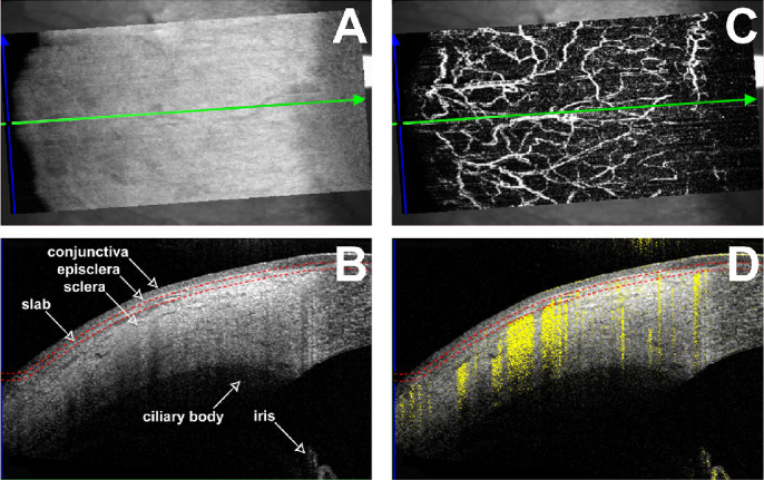
The software automatically creates en face OCTA images. In the software the contrast of each OCTA image was set to 1:5 to improve the image quality. So far, no specific algorithm exists for the segmentation of conjunctival vessels with OCTA imaging. Considering the OCTA projection artifacts as described,7 the slab for the projected OCTA image was chosen at a depth of 100 to 150 µm from the surface of the conjunctiva. The movement of the tear film on the bulbar conjunctiva causes the software to interpret it as flow (Fig. 1 D), leading to a misinterpretation of the en face OCTA image when choosing the slab within the conjunctiva. Therefore, a slab was chosen including the conjunctiva and the episclera/Tenon's capsule. We concluded this as the best choice to illustrate the conjunctival vascular bed of the anterior segment. The generated OCTA image was then exported as a portable network graphic file (Fig. 1). Reasons for exclusion of an image from our analysis were scan acquisition failure, insufficient fixation, or motion artifacts that made the image nongradable.
Figure 1.

Assessment of conjunctival vessels using anterior OCTA. (A) En face OCT of the temporal quadrant of a right eye corresponding to the conjunctival slab in B. Green and blue arrows refer to the position of the OCT scan. (B) OCT image in the position of the green arrow in A. The two red dotted lines show the depth of the OCTA slab (100 µm depth, 50 µm thickness). (C) En face OCTA revealing the vascular meshwork of the conjunctival slab. (D) OCTA scan in the position of the green arrow in A. The two red dotted lines show the depth of the OCTA slab (100 µm depth, 50 µm thickness). The reflectivity of vessels is shown in yellow within the OCT scan.
Imaging Analysis
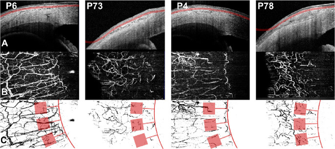
Using Fiji-software (ImageJ, National Institutes of Health, Bethesda, MD) files were converted into binary images in which all the pixels with values higher than a predefined value were marked as 1 (vessel pixels), the remaining as 0 (background pixels). Brightness was adjusted in a standardized way in order to highlight only bright vessels. Three regions of interest (ROI) in each image were selected. Every ROI consisted of a square of 200 × 200 pixels. The three ROI were aligned contiguous along the limbus at a distance of about 200 pixels (approximately 2–3 mm). For each ROI the percentage area of black pixels on the binary image was measured. This value represents the vessel area in this particular ROI. By averaging the value of three ROI, the average relative density of the conjunctival vessels for each image was calculated. (Fig. 2)
Figure. 2.

OCTA imaging of conjunctival vessels in healthy subjects (patient 69 [P69]), patients with diabetes without retinopathy (patient 73 [P73]) and patients with DR (patients 4 and 78 [P4, P78]). (A) Top row showing optical coherence tomography (OCT) scans with the corresponding angiography slabs (dotted red lines with transparent red) chosen at 100 µm depth, 50 µm thickness and corresponding en face OCTA images (B). (C) Binary image after standardization of the brightest vessels. The rim of the limbal vessels is demarcated with a red line. Red squares show the measured ROI.
Statistical Analysis
For statistical analysis, both nasal and temporal values were analyzed separately. An unpaired t-test was applied to compare healthy data with data of patients with DR and without DR. Multivariate regression analyses were used for the covariables found to be significant. A P-value of ≤ 0.05 was considered to be statistically significant. Data analysis was performed using Sigma Plot (Version 14, Systat Software GMBH, Erkrath, Germany) and GraphPad Prism version 5.02 for Windows (GraphPad Software, La Jolla, CA).
Results
Baseline Characteristics
Seventy-four individuals were included in this study. The mean age of all participating individuals was 47.82 years (range, 20–87 years). Forty-one patients were male, 33 patients were female. In group 1 (healthy individuals), 42 individuals were included; group 2 (patients with diabetes without retinopathy), 9 patients; and in group 3 (patients with diabetes with retinopathy), 23 patients (Table 1). Multivariate regression analysis accounting for age or gender using stepwise regression did not show a significant predictor for vessel density in our measurements. For each group, the mean vessel density of the temporal, as well of the nasal conjunctiva was calculated as described elsewhere in this article (Fig. 3). In group 1, there were 74 images of the temporal conjunctiva and 75 images of the nasal conjunctiva; in group 2, there were 12 images of the temporal conjunctiva and 11 images of the nasal conjunctiva; and in group 3, there were 36 images of the temporal conjunctiva and 26 images of the nasal conjunctiva were analyzed (Table 2).
Table 1.
Baseline Characteristics
| N | Age, Years (Range) | Male (n) | Female (n) | |
|---|---|---|---|---|
| Total included individuals | 74 | 47.82 (20–87) | 41 | 33 |
| Healthy individuals | 42 | 39.38 (20–77) | 23 | 19 |
| DM without RP | 9 | 64.11(43–81) | 5 | 4 |
| DM with RP | 23 | 58.13 (22–87) | 13 | 11 |
DM, diabetes mellitus; RP, retinopathy.
Figure. 3.

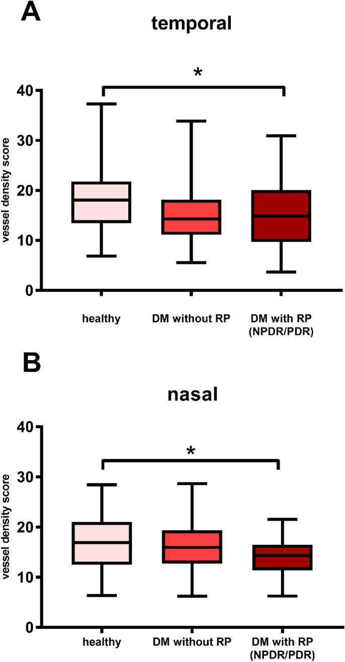
Box and whisker plots (minimum to maximum) plot of the relative vessel density score. The relative vessel density score is given in arbitrary units. Box and whiskers plots for the temporal (A) and nasal (B) measurements. There was a statistical difference in vessel density between group 1 and group 3 for the temporal (P = 0.0386), as well as the nasal (P = 0.0103) side (*). (Healthy, n = 42; diabetes mellitus without retinopathy [DM without RP], n = 9; diabetes mellitus with retinopathy [DM with RP], n = 23; NPDR, nonproliferative DR; PDR, proliferative DR).
Table 2.
Vessel Densities Score and P Values
| Temporal | Nasal | |||||
|---|---|---|---|---|---|---|
| Vessel Density Score | n | Average | SD | n | Average | SD |
| Group 1: Healthy | 74 | 17.9 | 6.4 | 75 | 16.7 | 5.2 |
| Group 2: DM without RP | 12 | 15.3 | 7.3 | 11 | 16.3 | 6.7 |
| Group 3: DM with RP | 36 | 15.2 | 6.5 | 26 | 13.7 | 4.3 |
| Temporal P Value | Nasal P Value | |||||
| Group 1 vs. group 2 | 0.1942 | 0.8043 | ||||
| Group 2 vs. group 3 | 0.9737 | 0.1744 | ||||
| Group 1 vs. group 3 | 0.0386 | 0.0103 | ||||
Vessel density scores are given in arbitrary units. Healthy = group 1; DM without RP = group 2; DM with RP = group 3. P values of unpaired t-test between all groups group are listed below. DM, diabetes mellitus; RP, retinopathy; SD, standard deviation.
Metrics in Healthy Eyes (Group 1)
The average vessel-density of the temporal conjunctiva in healthy eyes was measured to be 17.94% ± 6.4%. In the nasal conjunctiva, mean vessel density was 16.68 ± 5.2%. The unpaired t-test revealed no statistically significant difference between the temporal and the nasal quadrant of healthy eyes regarding the conjunctival vessel density (P = 0.07).
Metrics of the Eyes of Patients with Diabetes without Retinopathy (Group 2)
For group 2, the average vessel density of the temporal conjunctiva was 15.28 ± 7.3%, the nasal conjunctiva had an average vessel density of 16.25 ± 5.2%. Even though the measured average vessel density of patients with diabetes not suffering from retinopathy was slightly lower compared with healthy eyes, there was no statistically significant difference between groups 1 and 2, neither for the temporal, or for the nasal conjunctiva (P = 0.1942 and P = 0.8043, respectively).
Metrics of Eyes with DR (Group 3)
Measurements in group 3 resulted in a mean vessel density of 15.21 ± 6.5% for the temporal conjunctiva and 13.73 ± 4.3% in the nasal conjunctiva. Unpaired t-test on mean conjunctival vessel densities in healthy subjects (group 1) were significantly higher compared with vessel densities in patients with diabetes and DR (group 3) for both the temporal (P = 0.0386) and nasal conjunctiva (P = 0.0103) (Fig. 3, Table 2).
Discussion
Although OCT and OCTA are already established retinal imaging modalities, studies applying OCTA protocols to the anterior segment of the eye are relatively uncommon. One study quantified iris vasculature using anterior segment OCTA in patients with acute anterior uveitis.10 The quantification of the iris vessels was achieved by using an automated algorithm through a combination of different filters and intensity threshold to remove nonspecific, noncontiguous voxels. This algorithm allowed the quantitative visibility of the progressive decrease of the vessel caliper and dilation with the therapeutic control of the inflammation. In our study, we applied a similar algorithm to identify the conjunctival vessels. Although iris vessel analysis would be an interesting read out in patients with diabetes mellitus as well, it is prone to errors owing to constant movement of the iris musculature.
Other studies have investigated conjunctival vessels in patients with diabetes.11–13 The vessel analysis algorithm in infrared images of the conjunctiva revealed a decrease in tortuosity and loss of capillaries in patients with longer disease duration. It is known that disease duration plays a key role on the microangiopathic effects of diabetes on the conjunctival vasculature.4,14 It has been suggested that conjunctival hypoxia may explain the higher risk of infectious conjunctivitis in patients with diabetes compared with individuals without diabetes, because acute infectious conjunctivitis is more common among patients with diabetes mellitus.15
In healthy individuals, good repeatability and interobserver agreement was found in a previous OCTA study of the corneal limbus vascularization. Although a statistical significant difference in temporal and nasal quadrant vascularization was found in this previous report,16 no such difference was observed in our study. This may be explained by the choice of the size of the ROI and/or the area of the measurement (corneal limbus vs. conjunctiva). Nevertheless, we analyzed nasal and temporal quadrants separately.
Because OCTA is depth resolved, an additional advantage using this technology for anterior segment pathologic features is the simultaneous assessment of the abnormal vasculature, as well as the exact depth of the lesion.16 Furthermore, different vascular layers are individually evaluated by obtaining different depths of the tissues. Analysis of infrared imaging or color photography of the conjunctiva may be biased by submerged vessels, therefore leading to increased vessel density. As such OCTA has a clear advantage compared with conventional imaging, for posterior as well as anterior segment imaging. In patients with diabetes, swept source OCTA revealed a higher detection rate of intraretinal microvascular abnormalities compared with Early Treatment Diabetic Retinopathy Study imaging. In fact, a more severe state of DR than suggested by color fundus grading was found.17
As OCTA images are generated through motion contrast of flowing blood, they generate high resolution stationary image of the vascular bed.18–28 The depth resolution of the OCT enables the segmentation of different layers, which is a vast advantage to fluorescein angiography. Nevertheless real time dynamics (i.e., leakage) are only visible by injection of a dye. A disadvantage of the OCTA are artifacts that complicate the evaluation of the image. Projection artifacts from superficial layers create false artefactual images of vessels seen deeper than they actually inhibit.7 Therefore, conclusions from OCTA should only be made by experienced physicians.
Our study has several limitations. The OCTA is currently predominantly used to image the retina. Thus, imaging of the anterior segment with OCTA remains exploratory. The majority of the vessels visible in the anterior segment OCTA are precapillary vessels. Nevertheless, we found a significantly lower vessel density in patients with DR compared with healthy individuals and patients without DR. There are several possible explanations for these findings. The most obvious would be that the precapillary vessel density diminishes with the general rarefication of the conjunctival vessels. Another explanation is that the lower vessel density seen in the precapillary vessels is due to decreased blood velocity and flow, because of changes in blood rheology.29,30 Conjunctival vessels show a high variability. We suspected to find a difference in vessel density between nasal and temporal measurements, but no significant difference was found. Nevertheless, we analyzed both nasal and temporal quadrants separately to avoid a possible difference in vessel density. Artifacts causing poor image quality are a major issue. Most segmentation and movement artifacts involve areas of the conjunctiva in the image periphery and such ROI within the image center should be chosen for analysis. In our study, we only included ROI within 2 to 3 mm from the anatomic limbus. Furthermore, images were carefully evaluated for artifacts causing interference with our measurements. A challenge to increase image quality is eye tracking. Although the devices use markers of the retinal anatomy such as the optic nerve or the macular vessels to track the eye, for anterior imaging, the pupil or the conjunctival vessels could be used in anterior segment devices. Another limitation lies in the small sample size of our cohort in patients with diabetes without retinopathy and a potential bias owing to different age range between healthy individuals and diabetics. As such, the lack of effect sizes of interest such as between groups 1 and 2 should be interpreted with caution. The reason for the small sample size in this group was the exclusion of patients receiving anti-vascular endothelial growth factor owing to diabetic macular edema. Furthermore, in our cohort we did not find an age dependency of conjunctival vessel density. Nevertheless, our study is the first to investigate the conjunctival slab in healthy and patients with diabetes with OCTA.
In conclusion, OCTA imaging of the anterior segment remains exploratory. However, we show a significant decrease in vessel density in patients with diabetes with DR, compared with healthy individuals.
Acknowledgments
Disclosure: K. Schuerch, None; H. Frech, None; M. Zinkernagel, Novartis (C, I), Bayer (C, F), Heidelberg Engineering (F), Boeringer Ingelheim (F)
References
- 1. Congdon N, O'Colmain B, Klaver CC, et al.. Causes and prevalence of visual impairment among adults in the United States. Arch Ophthalmol. 2004; 122: 477–485. [DOI] [PubMed] [Google Scholar]
- 2. Yau JW, Rogers SL, Kawasaki R, et al.. Global prevalence and major risk factors of diabetic retinopathy. Diabetes Care. 2012; 35: 556–564. [DOI] [PMC free article] [PubMed] [Google Scholar]
- 3. Ebneter A, Zinkernagel MS.. Novelties in diabetic retinopathy. Endocrine Dev. 2016; 31: 84–96. [DOI] [PubMed] [Google Scholar]
- 4. Owen CG, Newsom RS, Rudnicka AR, Ellis TJ, Woodward EG. Vascular response of the bulbar conjunctiva to diabetes and elevated blood pressure. Ophthalmology. 2005; 112: 1801–1808. [DOI] [PubMed] [Google Scholar]
- 5. Munk MR, Giannakaki-Zimmermann H, Berger L, et al.. OCT-angiography: A qualitative and quantitative comparison of 4 OCT-A devices. PloS One. 2017; 12: e0177059. [DOI] [PMC free article] [PubMed] [Google Scholar]
- 6. Lipson BK, Yannuzzi LA.. Complications of intravenous fluorescein injections. Int Ophthalmol Clin. 1989; 29: 200–205. [DOI] [PubMed] [Google Scholar]
- 7. Spaide RF, Fujimoto JG, Waheed NK. Image artifacts in optical coherence tomography angiography. Retina. 2015; 35: 2163–2180. [DOI] [PMC free article] [PubMed] [Google Scholar]
- 8. Diabetic retinopathy study. Report Number 6. Design, methods, and baseline results. Report Number 7. A modification of the Airlie House classification of diabetic retinopathy. Prepared by the Diabetic Retinopathy. Invest Ophthalmol Vis Sci. 1981; 21: 1–226. [PubMed] [Google Scholar]
- 9. Grading diabetic retinopathy from stereoscopic color fundus photographs–an extension of the modified Airlie House classification. ETDRS report number 10. Early Treatment Diabetic Retinopathy Study Research Group. Ophthalmology. 1991; 98: 786–806. [PubMed] [Google Scholar]
- 10. Pichi F, Sarraf D, Arepalli S, et al.. The application of optical coherence tomography angiography in uveitis and inflammatory eye diseases. Prog Retin Eye Res. 2017; 59: 178–201. [DOI] [PubMed] [Google Scholar]
- 11. Dexel T, Bohme H, Schmidt P, Haslbeck M, Mehnert H. Investigation of conjunctival vessels in diabetics with and without retinopathy by means of a microcirculation-index. Bibl Anat. 1977; 16: 442–444. [PubMed] [Google Scholar]
- 12. Ditzel J, Sagild U.. Morphologic and hemodynamic changes in the smaller blood vessels in diabetes mellitus. II. The degenerative and hemodynamic changes in the bulbar conjunctiva of normotensive diabetic patients. N Engl J Med. 1954; 250: 587–594. [DOI] [PubMed] [Google Scholar]
- 13. Landau J, Davis E.. The small blood-vessels of the conjunctiva and nailbed in diabetes mellitus. Lancet. 1960; 2: 731–734. [DOI] [PubMed] [Google Scholar]
- 14. Owen CG, Newsom RS, Rudnicka AR, Barman SA, Woodward EG, Ellis TJ. Diabetes and the tortuosity of vessels of the bulbar conjunctiva. Ophthalmology. 2008; 115: e27–e32. [DOI] [PubMed] [Google Scholar]
- 15. Kruse A, Thomsen RW, Hundborg HH, Knudsen LL, Sorensen HT, Schonheyder HC. Diabetes and risk of acute infectious conjunctivitis–a population-based case-control study. Diabet Med. 2006; 23: 393–397. [DOI] [PubMed] [Google Scholar]
- 16. Ang M, Sim DA, Keane PA, et al.. Optical coherence tomography angiography for anterior segment vasculature imaging. Ophthalmology. 2015; 122: 1740–1747. [DOI] [PubMed] [Google Scholar]
- 17. Schaal KB, Munk MR, Wyssmueller I, Berger LE, Zinkernagel MS, Wolf S. Vascular abnormalities in diabetic retinopathy assessed with swept-source optical coherence tomography angiography widefield imaging. Retina. 2019; 39: 79–87. [DOI] [PubMed] [Google Scholar]
- 18. Fingler J, Schwartz D, Yang C, Fraser SE. Mobility and transverse flow visualization using phase variance contrast with spectral domain optical coherence tomography. Opt Exp. 2007; 15: 12636–12653. [DOI] [PubMed] [Google Scholar]
- 19. Fingler J, Readhead C, Schwartz DM, Fraser SE. Phase-contrast OCT imaging of transverse flows in the mouse retina and choroid. Invest Ophthalmol Vis Sci. 2008; 49: 5055–5059. [DOI] [PubMed] [Google Scholar]
- 20. Yu L, Chen Z.. Doppler variance imaging for three-dimensional retina and choroid angiography. J Biomed Opt. 2010; 15: 016029. [DOI] [PMC free article] [PubMed] [Google Scholar]
- 21. Liu G, Qi W, Yu L, Chen Z. Real-time bulk-motion-correction free Doppler variance optical coherence tomography for choroidal capillary vasculature imaging. Opt Exp. 2011; 19: 3657–3666. [DOI] [PMC free article] [PubMed] [Google Scholar]
- 22. Jia Y, Tan O, Tokayer J, et al.. Split-spectrum amplitude-decorrelation angiography with optical coherence tomography. Opt Exp. 2012; 20: 4710–4725. [DOI] [PMC free article] [PubMed] [Google Scholar]
- 23. Mariampillai A, Leung MK, Jarvi M, et al.. Optimized speckle variance OCT imaging of microvasculature. Opt Lett. 2010; 35: 1257–1259. [DOI] [PubMed] [Google Scholar]
- 24. Mariampillai A, Standish BA, Moriyama EH, et al.. Speckle variance detection of microvasculature using swept-source optical coherence tomography. Opt Lett. 2008; 33: 1530–1532. [DOI] [PubMed] [Google Scholar]
- 25. Kim DY, Fingler J, Werner JS, Schwartz DM, Fraser SE, Zawadzki RJ. In vivo volumetric imaging of human retinal circulation with phase-variance optical coherence tomography. Biomed Opt Exp. 2011; 2: 1504–1513. [DOI] [PMC free article] [PubMed] [Google Scholar]
- 26. An L, Shen TT, Wang RK. Using ultrahigh sensitive optical microangiography to achieve comprehensive depth resolved microvasculature mapping for human retina. J Biomed Opt. 2011; 16: 106013. [DOI] [PMC free article] [PubMed] [Google Scholar]
- 27. Conroy L, DaCosta RS, Vitkin IA. Quantifying tissue microvasculature with speckle variance optical coherence tomography. Opt Lett. 2012; 37: 3180–3182. [DOI] [PubMed] [Google Scholar]
- 28. Mahmud MS, Cadotte DW, Vuong B, et al.. Review of speckle and phase variance optical coherence tomography to visualize microvascular networks. J Biomed Opt. 2013; 18: 50901. [DOI] [PubMed] [Google Scholar]
- 29. Mendivil A, Cuartero V, Mendivil MP. Ocular blood flow velocities in patients with proliferative diabetic retinopathy and healthy volunteers: a prospective study. Br J Ophthalmol. 1995; 79: 413–416. [DOI] [PMC free article] [PubMed] [Google Scholar]
- 30. Ciulla TA, Harris A, Latkany P, et al.. Ocular perfusion abnormalities in diabetes. Acta Ophthalmol Scand. 2002; 80: 468–477. [DOI] [PubMed] [Google Scholar]


