Figure 4.
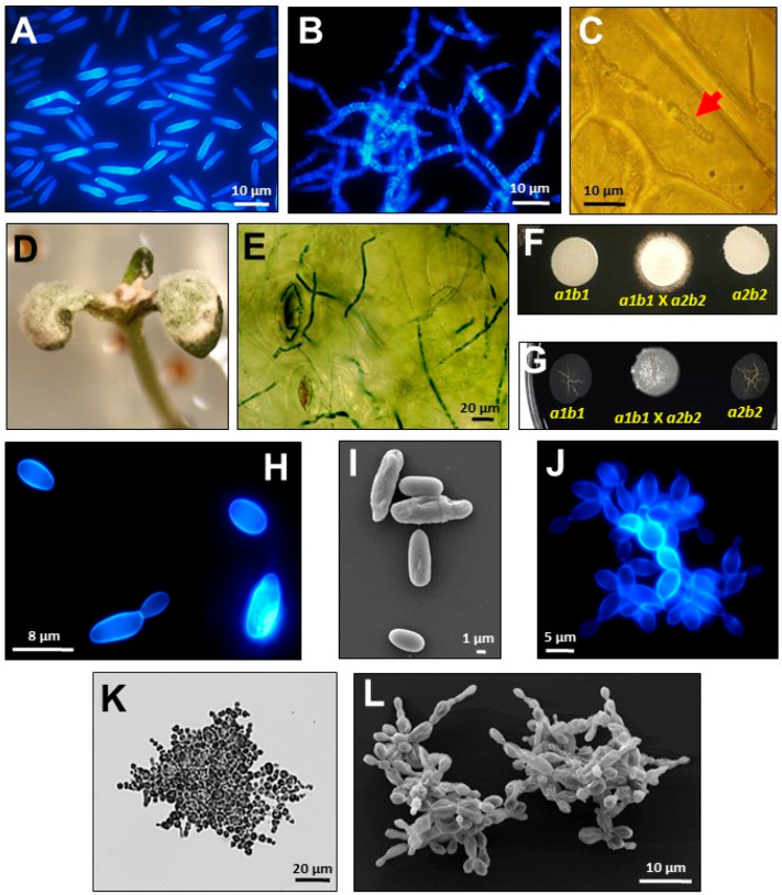
Multicellular shapes developed by Ustilaginomycetes under different growth or development conditions. (A–F), Images of U. maydis. (G–L) Images of S. reilianum. (A,B) Epifluorescence microphotographs of the fungus showing yeast-like unicellular growth, or multicellular growth as mycelium, in minimal medium pH 7 or pH 3, respectively. (C) Multicellular growth of the fungus as mycelium (red arrow) during the colonization of maize plant cells. (D,E) Multicellular growth of the fungus during Arabidopsis infection. (F,G) Multicellular growth of fungi as white-fuzzy colonies after mating of sexually compatible sporidia on plates with charcoal containing minimal medium. (H,I) Epifluorescence and scanning electron microphotographs of the fungus growing unicellularly like yeasts. (J–L) Epifluorescence, bright field, and scanning electron microphotographs of the multicellular clusters of the fungus. In all the epifluorescence microphotographs, the fungi were stained with calcofluor white (Sigma-aldrich, 18909). In (E,K), the fungi were stained with cotton blue-lactophenol (Sigma-Aldrich, 61335, St. Louis, MO, USA).

