



Abstract
X-linked adrenoleukodystrophy (ALD) is a neurodegenerative peroxisomal disorder with variable clinical phenotypes. Childhood cerebral ALD (CCALD) is at the most severe end of the disease spectrum. In CCALD, the clinical manifestations include increasing deficits in behavior, vision, hearing, coordination, and motor function, as well as seizures. Without treatment, CCALD often results in apparent vegetative state within 1 to 2 years of appearance of initial signs and symptoms. We present the case of a boy with classic inflammatory CCALD who exhibited spontaneous attenuation in disease progression. While extremely rare, spontaneous arrest of disease progression may occur in boys with inflammatory CCALD.
Keywords: cerebral adrenoleukodystrophy, inflammatory, spontaneous arrest
Introduction
X-linked adrenoleukodystrophy (ALD; OMIM, phenotype MIM number: 300100) is a genetic disorder, resulting from pathogenic mutations in ABCD1 (ATP-binding cassette, subfamily D, member 1), which is the coding gene for the ALD protein (ALDP). The ALDP is an ATP-binding peroxisomal transporter and dysfunction of ALDP results in the accumulation of very long chain fatty acids (VLCFAs) in body tissue. Phenotypic expression of X-ALD is highly variable, with the most frequent variants being childhood cerebral ALD (CCALD) and adrenomyeloneuropathy (AMN).1,2 Typically, CCALD affects boys between the ages of 4 and 10 years and manifests with rapidly progressive cerebral demyelination. Early development of affected boys is normal before the onset of CCALD; however, without treatment, initial behavioral difficulties resembling attention deficit and hyperactivity disorder eventually advance to significant neurologic impairment, including cortical blindness, loss of communication, and progressive spasticity within a mean of 1.9 years.3
This neurologic progression results from an inflammatory demyelination occurring in the cerebral white matter, characterized on magnetic resonance imaging (MRI) as symmetric T2-weighted hyperintensity, typically initiating in the posterior corpus callosum. The other common MRI finding is a garland of enhancement near the leading edge of white matter disease associated with the disruption of the blood brain barrier and active demyelination. The presence of contrast enhancement has been demonstrated to predict active progression and disability in affected boys.
In affected males, reports of attenuated disease are observed in approximately 10% of individuals; however, spontaneous cessation of active disease progression as determined by positive contrast enhancement has been rarely reported.4 Korenke et al reported three boys with CCALD who showed an arrest of their initial neurological deterioration for subsequent periods of 5 to 12 years.5 Moreover, repeated neuroimaging did not show progression of demyelination in their case series. However, as only one patient underwent MRI with contrast as an initial neuroimaging modality and demonstrated resolution of gadolinium enhancement in repeated MRI, it is not clear whether the other two patients also had inflammatory CCALD. Here we report the second untreated case of inflammatory CCALD that shows disease attenuation both clinically and radiographically.
Case Report
The patient is an Asian boy who was diagnosed with CCALD when he experienced a new-onset seizure at 8 years of age. His elder brother died of complications of CCALD at the age of 6 years. The plasma very long chain fatty acid profile in the patient was abnormal: C26:0 3.09 nmol/mL (reference ≤ 1.30 nmol/mL); C26:0/C22:0 0.073 (reference ≤ 0.01); and C24:0/C22:0 1.91 (reference ≤ 1.39). He was subsequently referred to the University of Minnesota for evaluation of hematopoietic stem cell transplantation (HSCT). Upon initial evaluation at age 9 years, a neurologic examination revealed an alert and awake child with dysarthric speech. He was able to respond to all questions in very short sentences with long latency. His language aptitude was clearly below his grade level. His visual acuity was 20/30 on the right and 20/40 on the left as evaluated by a Snellen near card. He had normal visual tracking and fixations. His optokinetic nystagmus was intact. Fundoscopic exam revealed pale optic discs with sharp margins, and his hearing was intact. He demonstrated normal muscle strength and tone without hyperreflexia. The Babinski response was negative bilaterally. His fine motor coordination was mildly abnormal, and his gait was narrow-based and stable. His cerebral ALD neurologic function score was 3 for the presence of nonfebrile seizures, aphasia/apraxia, and running difficulties. The brain MRI revealed extensive areas of demyelination with contrast enhancement (►Fig. 1). Abnormal T2 hyperintensity was noted in the posterior parieto-occipital region which extended to the anterior periventricular temporal lobe white matter. Posterior body and splenium of the corpus callosum, as well as posterior dorsal midbrain, were also involved. There was contrast enhancement surrounding the regions of abnormal T2 hyperintensity with corresponding restricted diffusion. Loes score was 15 and gadolinium intensity score (GIS) was 3. The patient was found to have adrenal insufficiency at the time of diagnosis and was treated with glucocorticoids. His seizures were managed by oxcarbazepine and levetiracetam. It was determined that his cerebral disease was too advanced for consideration of HSCT. His family was counseled that he was expected to progress to a vegetative state, followed eventually by death within 1 to 2 years of the current presentation in line with the natural course of the disease. The patient was subsequently lost to follow-up.
Fig. 1.
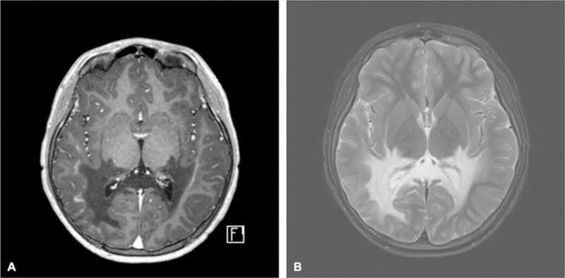
Brain MRI at the age of 9 years. (A) T2-weighted image shows a symmetric and confluent demyelinating lesion within the parieto-occipital lobes. (B) T1-weighted image post gadolinium administration demonstrates contrast enhancement at the leading edge of demyelinating lesion. MRI, magnetic resonance imaging.
At the age of 12 years, he presented to the emergency department of our hospital after having had a seizure at school. The patient’s neurologic status had not considerably progressed compared with that at his initial visit. He remained attentive and cooperative during the neurologic exam. He was able to follow two-step commands. His speech was dysarthric but he was able to answer questions in full sentences. He was noted to have left gaze preference and bidirectional gaze evoked horizontal nystagmus, more prominent on the leftward gaze. His muscle strength and tones were normal. He was slightly hyperreflexic on the left side. His gait was narrow-based and stable but he was not able to do tandem. The Babinski response was positive bilaterally, and the cerebral ALD neurologic function score remained as 3. A repeat MRI brain scan revealed the progression of moderate atrophy in the right parieto-occipital region as well as bilateral posterior temporal lobes (►Fig. 2). However, the previously noted contrast enhancement along the leading edge of demyelination was resolved. The Loes score was 23 and GIS was 0. ►Table 1 summarizes the patient’s clinical and radiographic disease progression.
Fig. 2.

Brain MRI at the age of 12 years. (A) T2-weighted image shows demyelinating lesion and cerebral volume loss. (B) T1-weighted image post gadolinium administration demonstrates resolution of contrast enhancement.
Table 1.
Progression of disease over 3 years
25-point scoring system used to evaluate gross clinical neurologic status of the cerebral ALD cohort. A score of 0 denotes absence of clinical signs of cerebral disease. ALD, adrenoleukodystrophy.
34-point ALD MRI severity scale developed by Loes et al.6ALD, adrenoleukodystrophy; MRI, magnetic resonance imaging.
A four-point scale of gadolinium intensity relative to the choroid plexus used to predict neurologic outcome in children with cerebral ALD.7ALD, adrenoleukodystrophy.
Discussion
Inflammatory cerebral ALD develops in approximately 35% of males with ALD aged < 12 years and in a smaller percentage of affected patients aged 12 years.2 Changes on MRI in the brain precede clinical manifestations by several years. Previous work has shown a strong association between the presence of contrast enhancement on T1-weighted MRIs and disease progression.6,7 Miller et al developed a four-point scale of gadolinium intensity relative to the choroid plexus.7 In boys with CCALD, the enhancement score itself predicts neurologic outcomes following treatment and such data are used to guide treatment decisions for clinicians and families.
Although there are several hypotheses proposed to explain the initiation of the demyelinating process, the sequential mechanisms responsible for the full-blown inflammatory demyelination in CCALD remain poorly understood. Accumulation of VLCFAs in myelin could lead to the progressive destabilization of myelin sheaths and subsequent demyelination.8 In 10 to 15% of patients that develop cerebral demyelination (as detected by nonenhancing T2-weighted abnormalities), the demyelinating process appears to halt spontaneously.9 In these cases, contrast enhancement is not seen, suggesting that a disruption of the blood–brain barrier did not occur. However, once initial cerebral demyelination converts into the rapidly progressive inflammatory phase, as evidenced by contrast enhancement on MRI, the lesion progresses rapidly and the patient’s neurologic status deteriorates accordingly. It has been suggested previously that microglial dysfunction contributes to neuroinflammation and thus, alters the neurovascular unit.10 Elevated levels of proinflammatory chemokines have been observed in the cerebrospinal fluid of CCALD patients and correlate with the severity of MRI findings.11 Within the active lesion, oxidative stress has also been observed in activated astrocytes and macrophages.12 There are case reports demonstrating that moderate or severe head trauma can initiate the conversion to rapidly progressive inflammatory demyelination,13,14 emphasizing the crucial role of environmental factors in progression of the disease.
Most patients with cerebral ADL die within a decade after diagnosis if they are not treated with HSCT.15 Allogenic HSCT is more likely to be effective if performed at an early stage of neurodegeneration and with limited cerebral involvement.16–18 Lentiviral vector mediated hematopoietic stem cell gene therapy (HSCGT) has also been investigated as a potential alternative to allogenic HSCT. In a single-group, open-label phase 2 and 3 safety and efficacy study, Lenti-D HSCGT was demonstrated to be a safe and effective alternative to HSCT in boys with early-stage CCALD.19 This multicenter study (STARBEAM ALD-102) is still ongoing to fully assess the duration of response and long-term safety.
The present report is the second case demonstrating a spontaneous attenuation of inflammatory demyelinating cerebral disease, and subsequent apparent stabilization of neurologic function, neither of which are predicted based on the known natural history of the disease. The ultimate course of this patient is unknown; however, it should be acknowledged that while extremely rare, spontaneous attenuation of disease progression at a functional level and resolution of gadolinium enhancement may occur in boys with inflammatory advanced CCALD.
We caution that in spite of this, clinicians should remain advised upon the detection of CCALD to rapidly refer these affected boys to experienced transplant centers for evaluation and treatment consideration. Treatment options for advanced patients should be offered with great caution as their condition often deteriorates even more quickly after myeloablative conditioning. A conservative strategy, as employed for treatment in our patient, may also result in a better quality of life over time in rare cases.
Acknowledgments
Conflict of Interest
Dr. Raymond has served as a consultant for Bluebird bio (Cambridge, MA), Minoryx (Barceona), and Viking Therapeutics (San Diego, CA). Dr. Miller is a full-time empolyee of Sangamo Therapeutics, Inc.
References
- 1.Moser HW, Moser AB, Smith KD, et al. Adrenoleukodystrophy: phenotypic variability and implications for therapy. J Inherit Metab Dis 1992;15(04):645–664 [DOI] [PubMed] [Google Scholar]
- 2.Raymond GV, Moser AB, Fatemi A X–Linked adrenoleukodystrophy. GeneReviews Available at: https://www.ncbi.nlm.nih.gov/books/NBK1315/. Accessed November 5, 2018 [Google Scholar]
- 3.Moser HW, Naidu S, Kumar AJ, Rosenbaum AE. The adrenoleukodystrophies. Crit Rev Neurobiol 1987;3(01):29–88 [PubMed] [Google Scholar]
- 4.Berger J, Forss-Petter S, Eichler FS. Pathophysiology of X-linked adrenoleukodystrophy. Biochimie 2014;98:135–142 [DOI] [PMC free article] [PubMed] [Google Scholar]
- 5.Korenke GC, Pouwels PJ, Frahm J, et al. Arrested cerebral adrenoleukodystrophy: a clinical and proton magnetic resonance spectroscopy study in three patients. Pediatr Neurol 1996;15(02): 103–107 [DOI] [PubMed] [Google Scholar]
- 6.Loes DJ, Hite S, Moser H, et al. Adrenoleukodystrophy: a scoring method for brain MR observations. Am J Neuroradiol 1994;15 (09):1761–1766 [PMC free article] [PubMed] [Google Scholar]
- 7.Miller WP, Mantovani LF, Muzic J, et al. Intensity of MRI gadolinium enhancement in cerebral adrenoleukodystrophy: a biomarker for inflammation and predictor of outcome following transplantation in higher risk patients. Am J Neuroradiol 2016;37(02):367–372 [DOI] [PMC free article] [PubMed] [Google Scholar]
- 8.Loes DJ, Fatemi A, Melhem ER, et al. Analysis of MRI patterns aids prediction of progression in X-linked adrenoleukodystrophy. Neurology 2003;61(03):369–374 [DOI] [PubMed] [Google Scholar]
- 9.Melhem ER, Loes DJ, Georgiades CS, Raymond GV, Moser HW. X-linked adrenoleukodystrophy: the role of contrast-enhanced MR imaging in predicting disease progression. Am J Neuroradiol 2000;21(05):839–844 [PMC free article] [PubMed] [Google Scholar]
- 10.Ho JK, Moser H, Kishimoto Y, Hamilton JA. Interactions of a very long chain fatty acid with model membranes and serum albumin. Implications for the pathogenesis of adrenoleukodystrophy. J Clin Invest 1995;96(03):1455–1463 [DOI] [PMC free article] [PubMed] [Google Scholar]
- 11.Lund TC, Stadem PS, Panoskaltsis-Mortari A, et al. Elevated cerebral spinal fluid cytokine levels in boys with cerebral adrenoleukodystrophy correlates with MRI severity. PLoS One 2012;7 (02):e32218. [DOI] [PMC free article] [PubMed] [Google Scholar]
- 12.Powers JM, Pei Z, Heinzer AK, et al. Adrenoleukodystrophy: oxidative stress of mice and men. J Neuropathol Exp Neurol 2005;64(12):1067–1079 [DOI] [PubMed] [Google Scholar]
- 13.Raymond GV, Seidman R, Monteith TS, et al. Head trauma can initiate the onset of adrenoleukodystrophy. J Neurol Sci 2010; 290(1,2):70–74 [DOI] [PubMed] [Google Scholar]
- 14.Weller M, Liedtke W, Petersen D, Opitz H, Poremba M. Very-late-onset adrenoleukodystrophy: possible precipitation of demyelination by cerebral contusion. Neurology 1992;42(02):367–370 [DOI] [PubMed] [Google Scholar]
- 15.Mahmood A, Raymond GV, Dubey P, Peters C, Moser HW. Survival analysis of haematopoietic cell transplantation for childhood cerebral X-linked adrenoleukodystrophy: a comparison study. Lancet Neurol 2007;6(08):687–692 [DOI] [PubMed] [Google Scholar]
- 16.Baumann M, Korenke GC, Weddige-Diedrichs A, et al. Haemato-poietic stem cell transplantation in 12 patients with cerebral X-linked adrenoleukodystrophy. Eur J Pediatr 2003;162(01):6–14 [DOI] [PubMed] [Google Scholar]
- 17.Peters C, Charnas LR, Tan Y, et al. Cerebral X-linked adrenoleukodystrophy: the international hematopoietic cell transplantation experience from 1982 to 1999. Blood 2004;104(03):881–888 [DOI] [PubMed] [Google Scholar]
- 18.Miller WP, Rothman SM, Nascene D, et al. Outcomes after allogeneic hematopoietic cell transplantation for childhood cerebral adrenoleukodystrophy: the largest single-institution cohort report. Blood 2011;118(07):1971–1978 [DOI] [PubMed] [Google Scholar]
- 19.Eichler F, Duncan C, Musolino PL, et al. Hematopoietic stem-cell gene therapy for cerebral adrenoleukodystrophy. N Engl J Med 2017;377(17):1630–1638 [DOI] [PMC free article] [PubMed] [Google Scholar]