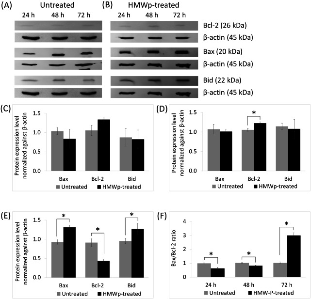
Figure 2. Effects of HMWp on apoptosis-related protein expression in MCF7 cells.
Western blot analysis of Bcl-2 family proteins (Bcl-2, Bax, and Bid) from (A) untreated control and (B) HMWp treated MCF7 cells at 24 h, 48 h and 72 h. The untreated control and HMWp-treated samples were derived from the same experiment and that gels/blots were processed in parallel. β-actin was served as internal control. Relative protein expressions of Bcl-2, Bax and Bid in MCF7 cells at (C) 24 h, (D) 48 h and (E) 72 h, were determined by normalization to β-actin. (F) Bax/Bcl-2 ratio in untreated control and HMWp-treated MCF7 cells. Treatment with HMWp for 72 h increased the Bax/Bcl-2 ratio compared with untreated control. Data are expressed as means ± SD from three independent experiments. * indicates significant difference in expression level/fold change between the untreated control and treated cells (SPSS, independent samples t-test, p < 0.05).

