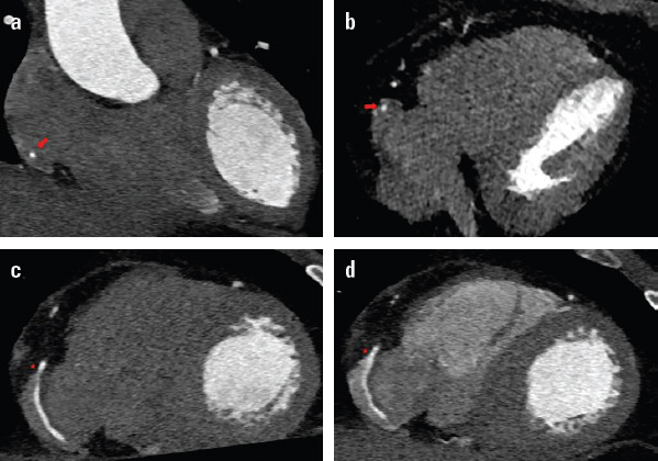
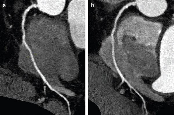
Coronary congenital anomalies are rare. Most are benign, but some can cause myocardial damage or sudden death. With the advancement of imaging studies, its diagnosis is increasing. We present a series of three cases in which the right coronary artery has an intra-auricular path. We examined a middle-aged man and two women with no cardiovascular history. They consulted a doctor for chest pain. Conventional ergometry and transthoracic echocardiography were performed and revealed normal results. Due to persistence of the symptoms, a computed tomography of the coronary arteries is requested. There were no significant atherosclerotic lesions found; however, an abnormality was found. Anatomical characteristics were evaluated using conventional axial images and curved multiplanar reconstructions, with retrospective acquisition and single injection of contrast (Fig. 1 and 2). The abnormality is diagnosed by evaluating that any segment of the artery is completely surrounded by blood in the right atrium. The artery originates from the right Valsalva sinus, with the junction of the proximal and middle thirds being affected. It crosses the inferolateral portion inside the right atrium with an average length of 27.7 mm in three cases and a maximum depth of 5 mm. Our cases do not associate with other coronary anomalies. The symptoms (if any) are unknown. In a 2-year follow-up, no major cardiovascular events or cardiovascular death appeared. It is important to be aware that this anomaly is not visualized by coronary angiography. Apart from the lack of knowledge about its prognosis, its clinical importance may lie in the risk of accidental injury during endovascular procedures or in cardiac surgery.
Figure 1.

. (a, b) Coronal image with cardiac synchronization and contrast only in left cavities which demonstrates the middle/distal third of the right coronary artery inside the right atrium (arrow). Plane images 4C oblique images following the axis of the artery. The intra-atrial path of the artery with contrast completely surrounding it. (c) With contrast in one phase. (d) With contrast in two phases to opacify the right cavities
Figure 2.

Images with multiplanar reconstruction following the axis of the artery, demonstrating the intracavitary path of the artery. (a) With contrast in one phase. (b) With two-phase contrast to opacify the right cavities
Footnotes
Informed consent: About the consent: Since the series was collected retrospectively, it was not possible to request written consent from the patients, in any case there is no identifying data on them in the images.


