Abstract
Patient: Male, 8-year-old
Final Diagnosis: COVID-19
Symptoms: Abdominal pain • respiratory distress • status epilepticus • vomiting
Medication: —
Clinical Procedure: —
Specialty: Infectious Diseases • Pediatrics and Neonatology
Objective:
Unusual clinical course
Background:
As the severe acute respiratory syndrome coronavirus 2 (SARS CoV2) spreads around the world infecting people of all ages, clinicians and researchers are working to gather data on the presentation of coronavirus disease (COVID-19). Further study is necessary to better diagnose and treat COVID-19 patients.
Case Report:
We describe the case of an 8-year-old boy admitted with status epilepticus, who also tested positive for COVID-19, while afebrile, with no initial respiratory symptoms. Benzodiazepines were given per treatment guidelines, abating the seizure activity. He subsequently developed respiratory distress and desaturation requiring temporary emergent intubation. All clinical symptoms resolved within a few hours. Results of a computed tomography (CT) scan of the brain were within normal limits. Results of a 24-h electroencephalogram (EEG) were abnormal, indicative of diffuse cerebral dysfunction. As a result of intubation and findings of bilateral infiltrates on chest x-ray, a COVID-19 test was administered and the result was positive.
Conclusions:
For proper diagnosis and treatment, patients and clinicians should be aware that COVID-19 may not present in the typical fashion of respiratory distress and fever. The present case suggests a rare neurological presentation of COVID-19.
MeSH Keywords: COVID-19, Pediatrics, SARS Virus, Status Epilepticus
Background
The rapid spread of the coronavirus disease (COVID-19), originating in Wuhan, China, has resulted in a pandemic affecting millions of people worldwide. The presentation of COVID-19 can range from asymptomatic to severe respiratory distress [1]. Data from China and the United States suggest children have a milder disease course than adults. While most adults present with fever, cough, and shortness of breath, children can present atypically [2]. Here, we present a pediatric case of COVID-19 with the initial presentation of status epilepticus.
Case Report
On April 22, 2020, an 8-year-old boy with a history of attention deficit hyperactivity disorder (ADHD), motor tics, and a recent seizure presented to the Emergency Department (ED) in status epilepticus. His mother reported that he woke up early in the morning of presentation complaining of nausea and abdominal pain. She noted his head was turned to the left and his eyes were looking upward. He had 3 episodes of vomiting that were non-bilious and non-bloody while his head was still turned left. While being transported to the ED, he was reportedly in and out of consciousness. The mother denies any shaking movement in the body, drooling, urination, or defecation during that time. She also denied he had fever, cough, diarrhea, shortness of breath, recent travel, or known COVID-19 exposure.
Results of a routine EEG at a neurology visit 2.5 weeks prior to this presentation were abnormal, with intermittent spikes and waves. Neurology had been monitoring the boy due to ADHD and recent complaints of motor tics. The diagnosis of possible pediatric autoimmune neuropsychiatric disorders associated with streptococcal infections (PANDAS) was made and he was placed on a 10-day course of amoxicillin. His mother was advised to administer only amoxicillin for the first 5 days and then to administer amoxicillin along with cefdinir for the next 5 days, both of which can lower the seizure threshold. An MRI scheduled for May 12 was postponed due to the COVID pandemic.
In the ED, a left-sided focal seizure with rhythmic movement of the left arm and blinking of the left eye was observed, which the parents stated had been occurring for 30 minutes. Vital signs were stable and the patient was afebrile. His body weight was 45.0 kg. Lorazepam 2 mg intravenous (IV) with a loading dose of 2250 mg (50mg/kg) of levetiracetam IV was given and the seizure seemed to abate. The patient began to have agonal breathing and desaturations and was emergently intubated. The need for intubation and the chest x-ray prompted a COVID-19 test.
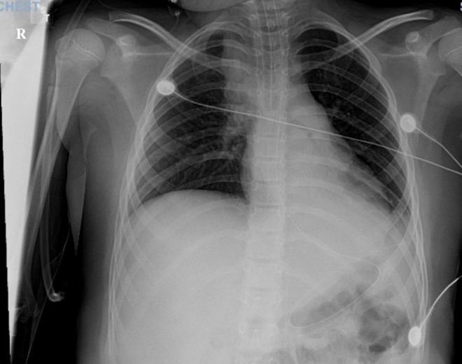
The chest X-ray revealed bilateral infiltrates (Figure 1), for which a dose of ceftriaxone 2000 mg IV was given. The results of a brain CT scan without contrast were within normal limits. Laboratory results revealed normal chemistries and bloodwork, including a normal coagulation panel. Blood culture and urine cultures displayed no growth. The result of reverse transcription-polymerase chain reaction testing (RT-PCR) for SARS-CoV-2 was positive from the St. Joseph’s Health Microbiology Laboratory, and all other respiratory pathogen results were negative. As a result of clinical experience and recommendations at that time, a 5-day course of oral hydroxychloroquine 200 mg was initiated.
Figure 1. A chest X-ray revealing right upper-lobe infiltration.

Soon after arrival at the Pediatric Intensive Care Unit (PICU), he was extubated and was stable on room air with no further seizure activity. Methylprednisolone 40 mg IV push and magnesium 1 g IV were given daily for respiratory support, as well as daily oral preparations of cholecalciferol 50 mcg and ascorbic acid 500 mg for additional nutritional support. Daily therapeutic enoxaparin 40 mg subcutaneous was given during hospitalization. His temperature remained normal during his entire hospital stay.
Laboratory results on day 2 displayed a change from admission in absolute neutrophil count, from 5.65×10³/mm3 to 8.56×10³/mm3, and a decrease in absolute lymphocyte count, from 5.5×10³/mm3 to 0.96×10³/mm3, with a neutrophil-to-lymphocyte ratio of 8.92. The 24-h EEG result was abnormal, indicative of diffuse cerebral dysfunction of non-specific etiology. The patient remained clinically stable and was discharged home to quarantine and to finish a course of hydroxychloroquine with outpatient MRI and neurology follow-up.
Discussion
Due to the rapid spread of the coronavirus and with most cases occurring in adults, the impact on children is difficult to assess. While fever, cough, and shortness of breath are among the most commonly reported symptoms in adults, a significantly lower percentage of children have these 3 symptoms. The lack of data on pediatric COVID-19 cases makes diagnosing COVID-19 more challenging in the children [2]. The presentation of a child in status epilepticus, while afebrile, with no signs of initial respiratory concerns, draws attention to the need for data regarding the most typical pediatric presentation of COVID-19 disease.
In the disease course of this particular COVID-19 patient, it is unclear whether the respiratory distress and desaturation requiring short-term intubation were results of the COVID-19 infection or a sequelae of status epilepticus. Our patient had no notable respiratory symptoms for the remainder of his hospital stay, and none of the home contacts ever exhibited any signs of fever or respiratory symptoms.
As the clinical spectrum of pediatric presentation of COVID-19 has yet to be established, clinicians should be aware of the potential for COVID-19 to present with other organ involvement, such as neurologic symptoms. Our patient presented with predominant neurologic symptoms, and subsequent chest x-ray evaluation and COVID-19 testing revealed COVID-19 infection. Due to the initial presentation of status epilepticus, COVID-19 was not initially considered. Given our patient’s recent antibiotic use and neurologic history, there were multiple potential causes for the presentation of status epilepticus, which also made COVID-19 initially appear less likely. If intubation and therefore chest x-ray after seizure had not been required, he may have never been tested for COVID-19. As seen in this case, when a child presents with other potential causes for neurological symptoms, it is important for clinicians to consider COVID-19 as a potential diagnosis, even in a patient with mild respiratory symptoms.
This case of status epilepticus as the predominant presentation of COVID-19 also raises the question of the neuroinvasive ability of SARS-CoV-2. Many viruses, including influenza, adenovirus, and respiratory syncytial virus, have been known to induce seizures [3]. Middle-Eastern respiratory syndrome coronavirus (MERS-CoV) causes dysfunction in several organ systems, including the respiratory, renal, cardiovascular, and central nervous systems. Some reported cases of MERS-CoV presented with coma, ataxia, and focal motor deficits [4]. Evidence supporting the ability of some coronaviruses to invade the brainstem suggests the possibility that SARS CoV2 has a similar ability to invade the central nervous system [5]. Coronaviruses can enter cells that express angiotensin-converting enzyme 2 (ACE2), which has been found to be expressed by neurons and glial cells [6].
Research involving 214 hospitalized COVID-19 patients in Wuhan, China, revealed 36.4% had neurologic manifestations such as acute cerebrovascular disease, impaired consciousness, and skeletal muscle injury [7]. Other COVID-19 patients have been reported to have neurologic symptoms of headache, nausea, and vomiting [5]. In the present patient, no tests of cerebrospinal fluid were done to definitively show if the virus directly infected the brain, but his initial predominant presentation of status epilepticus supports the idea that SARSCoV-2 can induce seizures through a neurotropic mechanism. Additionally, there have been reports of adult cases of COVID-19 with the predominant symptom of status epilepticus, which further suggests pediatric patients can present similarly [8].
As the investigation into the characteristics of COVID-19 develops, some studies have assessed the potential benefit of monitoring the neutrophil-to-lymphocyte ratio (NLR). A retrospective analysis of COVID-19 patients at a hospital in Wuhan, China suggested the NLR is an independent risk factor for mortality in hospitalized COVID-19 patients [9]. Analysis of the immune response in hospitalized COVID-19 patients showed most had an increase in NLR and T lymphopenia, which were more evident in severe cases [9,10]. Our patient was admitted with a normal NLR, but on the 2nd day of hospitalization he had an increased NLR of 8.9 and lymphopenia. Studies on the NLR have been limited to adults and the relevance of the NLR as a prognostic indicator for children has yet to be determined. Although our patient had an increased NLR and a positive clinical outcome, research suggests it could be of benefit to monitor NLR and lymphocyte count when diagnosing and treating COVID-19.
As the world continues to deal with this pandemic, clinicians must be aware of the possibility of neurologic presentations of COVID-19 to avoid missing or delaying diagnosis. It is also important to note that the absence of initial respiratory symptoms in COVID-19 patients does not rule out the possibility of sudden respiratory distress. Although the data suggest children have a milder disease course than adults, it is important to develop a better characterization of pediatric presentation for diagnostic purposes and to better understand the transmission dynamics in children [1,2,11].
Conclusions
This case report demonstrates that a predominant neurological presentation of status epilepticus can occur in patients with COVID-19. More information on the characteristics of the clinical course of COVID-19 is needed to better diagnosis and treat COVID-19, especially in children.
References:
- 1.Guan WJ, Ni ZY, Hu YL, et al. China Medical Treatment Expert Group for clinical characteristics of coronavirus disease 2019 in China. N Engl J Med. 2020;382(18):1708–20. doi: 10.1056/NEJMoa2002032. [DOI] [PMC free article] [PubMed] [Google Scholar]
- 2.CDC COVID-19 Response Team Coronavirus disease 2019 in children – United States, February 12–April 2, 2020. Morb Mortal Wkly Rep. 2020;69(14):422–26. doi: 10.15585/mmwr.mm6914e4. [DOI] [PMC free article] [PubMed] [Google Scholar]
- 3.Chung B, Wong V. Relationship between five common viruses and febrile seizure in children. Arch Dis Child. 2007;92(7):589–93. doi: 10.1136/adc.2006.110221. [DOI] [PMC free article] [PubMed] [Google Scholar]
- 4.Arabi YM, Harthi A, Hussein J, et al. Severe neurologic syndrome associated with Middle East respiratory syndrome corona virus (MERS-CoV) Infection. 2015;43(4):495–501. doi: 10.1007/s15010-015-0720-y. [DOI] [PMC free article] [PubMed] [Google Scholar]
- 5.Li YC, Bai WZ, Hashikawa T. The neuroinvasive potential of SARS-CoV2 may play a role in the respiratory failure of COVID-19 patients. J Med Virol. 2020;92(6):552–555. doi: 10.1002/jmv.25728. [DOI] [PMC free article] [PubMed] [Google Scholar]
- 6.Baig AM, Khaleeq A, Ali U, Syeda H. Evidence of the COVID-19 virus targeting the CNS: Tissue distribution, host-virus interaction, and proposed neurotropic mechanisms. ACS Chem Neurosci. 2020;11(7):995–98. doi: 10.1021/acschemneuro.0c00122. [DOI] [PubMed] [Google Scholar]
- 7.Mao L, Jin H, Wang M, et al. Neurologic manifestations of hospitalized patients with coronavirus disease 2019 in Wuhan, China. JAMA Neurol. 2020;77(6):1–9. doi: 10.1001/jamaneurol.2020.1127. [DOI] [PMC free article] [PubMed] [Google Scholar]
- 8.Vollono C, Rollo E, Romozzi M, et al. Focal status epilepticus as unique clinical feature of COVID-19: A case report. Seizure. 2020;78:109–12. doi: 10.1016/j.seizure.2020.04.009. [DOI] [PMC free article] [PubMed] [Google Scholar]
- 9.L Liu Y, Du X, Chen J, et al. Neutrophil-to-lymphocyte ratio as an independent risk factor for mortality in hospitalized patients with COVID-19. J Infect. 2020;81(1):e6–12. doi: 10.1016/j.jinf.2020.04.002. [DOI] [PMC free article] [PubMed] [Google Scholar]
- 10.Qin C, Zhou L, Hu Z, et al. Dysregulation of immune response in patients with COVID-19 in Wuhan, China. Clin Infect Dis. 2020 doi: 10.1093/cid/ciaa248. [Online ahead of print] [DOI] [PMC free article] [PubMed] [Google Scholar]
- 11.Dong Y, Mo X, Hu Y, et al. Epidemiology of COVID-19 among children in China. Pediatrics. 2020;145(6):e20200702. doi: 10.1542/peds.2020-0702. [DOI] [PubMed] [Google Scholar]


