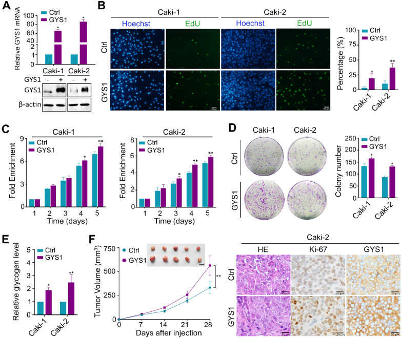
Figure 3.
GYS1 promotes ccRCC proliferation in vitro and in vivo. (A) GYS1 was overexpressed by transfection with GYS1 exogenous vector in Caki-1 and Caki-2 cells. The mRNA and protein levels of GYS1 were determined by qRT-PCR and western blotting. (B) EdU assays showed the replication of DNA in cells induced by GYS1. Green staining denotes duplicated cells and blue denotes the cell nucleus. (C) Cell activity was detected by CCK8 assay over five consecutive days. Relative absorbance was measured at OD450. Fold-enrichment was normalized to the absorbance on day 1. (D) Colony formation assays to determine the effect of GYS1 on cell growth. The number of colonies was counted using ImageJ software (NIH, Bethesda, MD, USA). (E) The glycogen content in cell lines with GYS1 overexpression were detected by a quantitative glycogen detection kit. (F) Xenograft mice experiment to evaluate tumor growth in vivo. Mice were sacrificed at 28 days after inoculation with Caki-2 cells. The volume of tumors in each group was calculated. Representative images of HE staining and IHC are shown. Statistical data are represented as the mean ± SD. *P < 0.05, **P < 0.01.

